Living cells on the move
Abstract
Spectacular collective phenomena such as jamming, turbulence, wetting and waves emerge when living cells migrate in groups.
Much like birds fly in flocks and fish swim in swarms, cells in our body move in groups. Collective cell migration enables embryonic development, wound healing and cancer cell invasion. These phenomena involve complex biochemical regulation, but their dynamics can ultimately be predicted by emerging physical principles of living matter.
When and where do cells migrate in groups?
When seen at the microscale, our body is a busy maze filled with actively moving cells. Cellular movements are slow, rarely exceeding m/min, but crucial to embryonic development, the immune response, tissue self-renewal and wound healing. Through the same mechanisms that sustain these physiological functions, cell movements drive devastating diseases such as acute inflammation and cancer. These can in fact be considered diseases of cell movement because arresting cells in a controlled manner would be sufficient to prevent their spread. Take cancer as an example; whereas its origin is well-known to be genetic, if we could selectively stop the movement of tumor cells, we would prevent them from escaping primary tumors and reach distant organs to metastasize. Understanding cell migration is therefore crucial to improve current strategies to fight disease.
 \phantomsubcaption\phantomsubcaption
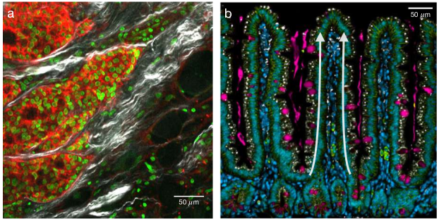
Cell migration comes in different flavors. Some cell types move as isolated self-propelled particles. For example, to chase and destroy pathogens, immune cells move individually through tissue pores. Similarly, in some types of cancer, single cells dissociate from tumors and travel solo through the surrounding tissue, eventually reaching blood vessels and metastasizing at distant organs. By contrast, in other physiological functions and pathological conditions, cells move as collectives. Collective cell movements are dominant in embryo development, where they enable the precise positioning of tissues and organ progenitors. The later coordination of growth and motion shapes organs well into postnatal life. Collective cell migration also drives wound healing in tissues such as the skin. Here, a continuous sheet of tightly adhered cells moves cohesively over the damaged area to restore a functional tissue. Cancer invasion also involves the migration of cell sheets, strands and clusters (Fig. 1). Within these groups, cells with different function can self-organize in spatial compartments to behave like an aberrant organ. This added functionality is thought to provide malignant tumors with distinct invasive strategies that improve their chances of spreading and metastasizing. Finally, collective cell migration is also involved in maintaining the inner surface of the gut, which is the fastest self-renewing tissue in mammals. It renews entirely every 3-5 days, which implies a daily loss of tens of grams of cells. Self-renewal proceeds thanks to the division of stem cells that reside at the bottom of tissue invaginations called crypts. The progeny of these stem cells then migrates collectively from the crypt to the top of finger-like protrusions called villi, where they are shed into the intestinal lumen and discarded (Fig. 1).
Collective cell migration is regulated by a myriad of molecular processes, from genetic programs to sensing and signaling pathways. Yet, these molecular processes act upon a limited number of physical quantities to determine cell movement. Therefore, coarse-grained physical approaches may provide crucial insight into biological questions. Furthermore, collective cell behaviors also inspire new physical theories of living systems. In this article, we highlight progress in this direction.
Cell assemblies as living matter
What physical principles underlie collective cell motion? In traditional condensed matter, interactions between electrons and between atomic nuclei give rise to fascinating collective phenomena such as magnetism and superconductivity. Analogously, cell-cell interactions lead to emergent collective phenomena in migrating cell groups. However, when treating cell colonies as materials, we must take into account some key features of living matter, as we illustrate throughout the article.
First, the primary constituents of living tissues are cells and extracellular networks of protein fibers such as collagen. The interactions between these mesoscale constituents are orders of magnitude weaker than interatomic interactions in conventional solids. So, with notable exceptions like bone, most biological tissues are soft materials, which can easily deform and flow.
Second, cells are machines with internal engines. Specialized proteins known as molecular motors harness the energy of chemical reactions to generate forces and produce mechanical work. These energy-transducing molecular processes ultimately power cell migration, allowing cells to move autonomously, without externally applied forces. This continuous supply of energy drives living tissues out of thermodynamic equilibrium. Importantly, the driving is local; it occurs at the level of individual constituents, i.e. single cells. In other words, cells are active constituents, and living tissues are a paradigmatic example of active matter — an exploding new field in nonequilibrium statistical physics.
But cells are not only mechanically active: they also sense their environment, process information, and respond by adapting their behavior. For example, stem cells plated on substrates of different stiffness differentiate into distinct cell types, from brain to bone cells. So living tissues are adaptive: they respond in programmed ways to environmental cues such as external forces, the mechanical properties of the extracellular matrix, and concentrations of nutrients and signaling molecules. Consequently, cell-cell and cell-environment interactions are often very complex. Unlike atoms and electrons in conventional condensed matter, cellular interactions cannot in general be fully described via an interaction potential with a fixed functional form. Thus, a key challenge in this field is to find effective ways to capture complex cell behaviors in terms of simple, physically-motivated interactions 1.
To flow or not to flow
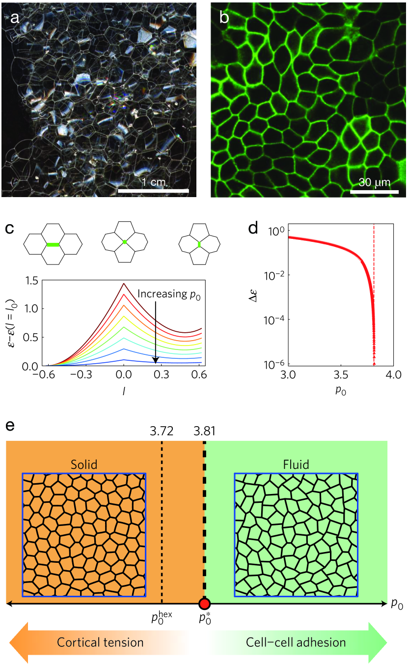
One way to think about interactions between deformable epithelial cells comes from the physics of foams. In foams, gas bubbles arrange in polygonal packings, with the liquid phase filling the interstitial spaces and providing surface tension to bubble interfaces (Fig. 2). Similarly, cells in epithelial monolayers also acquire polygonal shapes, with roughly straight edges subject to active tension generated inside cells (Fig. 2). This description of tissues as polygonal cell packings traces back to work by Hisao Honda and collaborators in 1980, and it was later popularized by work in the group of Frank Jülicher and collaborators 3. Because cells are deformable, edge lengths vary dynamically. These variations change the energy of the cellular network, which we can write in terms of areas and perimeters of cells as
| (1) |
This energy assumes that cells resist changes in their area and perimeter around the preferred values and with elastic moduli and , respectively. The preferred perimeter depends on cell-cell interactions and cellular activity, with cell-cell adhesion promoting longer edges and cellular tension favoring shorter edges. The preferred perimeter and area define a dimensionless parameter which informs about the preferred cell shape. Higher corresponds to more elongated cells, and smaller corresponds to more isotropic shapes, with less perimeter for the same area.
For a given , edge lengths vary until the system reaches its ground state, minimizing the energy in Eq. 1. In this process, an edge can shrink until it eventually disappears, leading to the formation of a new cell-cell interface (Fig. 2). These events, known as T1 transitions, allow cells to change neighbors, driving topological rearrangements of the cellular network. The ability to reorganize its constituents determines whether a material is solid or fluid. So, if cell rearrangements are difficult, the cellular network resists shear deformations; the tissue is solid. In contrast, if cells can rearrange easily, the network yields upon shear; the tissue is fluid. At small , i.e. for roundish cells, Eq. 1 reveals that there is an energy barrier preventing T1 rearrangements (Fig. 2). However, as we increase and cells become more elongated, the energy barrier decreases (Fig. 2). At a critical value of , the barrier vanishes (Fig. 2); cells can rearrange freely 2.
This simple model thus predicts a solid-fluid transition in tissues driven by changes in cell shape, encoded in (Fig. 2). This is a striking prediction, which showcases the bizarre mechanical properties of materials with deformable constituents. In conventional condensed matter, solids melt by either increasing temperature or reducing the packing fraction, i.e. decreasing pressure. Tissues, however, can melt at a fixed temperature and at the maximum packing fraction, , i.e. without gaps between cells 2. Tissues can become fluid by increasing the cell perimeter-to-area ratio, for example by either decreasing intracellular tension or, counterintuitively, increasing cell-cell adhesion (Fig. 2). The more cells adhere to one another, the more they elongate, and the easier it is for them to rearrange.
Shortly after its prediction 2, this solid-fluid transition driven by changes in cell shape was experimentally verified in layers of human bronchial epithelial cells 4. Moreover, this study showed that cells from healthy individuals tended to be caged by their neighbors and form a solid tissue, whereas cells from asthmatic individuals tended to remain unjammed, forming a fluid tissue. Therefore, these experiments suggested that the fluid-solid transition in tissues is involved in disease, opening the door to new treatments based on preventing this phase transition. Fluid-solid transitions also occur during development, enabling tissues to first turn fluid in order to remodel and acquire their shapes, and then solidify and mature. The emerging picture is that, in different biological contexts, cells can tune their shape and use the physical principles governing phase transitions in foams to decide whether to flow or not to flow.
Aligning with neighbors
The action starts once tissues become fluid and cells can move. Cells then engage in collective flows of different types, depending on how cells align with neighbors to coordinate their individual motions. Again, these cell-cell interactions depend strongly on cell shape.
Cells come in many different shapes: some are roughly spherical, some have rod-like shapes, and some cells develop a head-tail asymmetry to migrate persistently in one direction. This direction can be represented by a vector known as cell polarity. In groups, cells can align their individual polarities, forming phases of matter with orientational order. These alignment interactions and the resulting oriented phases can be described using concepts from the physics of magnetism and liquid crystals. For example, cells in a group can spontaneously break symmetry and align in a common direction. This type of alignment is known as polar order. To capture its emergence, one can introduce ferromagnetic-like interactions between individual cell polarities. At a coarse-grained level, collective cell polarity can be thought to result from an effective free energy with the Mexican-hat shape familiar from the Landau theory of phase transitions. This example thus illustrates how tools and concepts from other fields of physics are being borrowed to rationalize cell alignment.
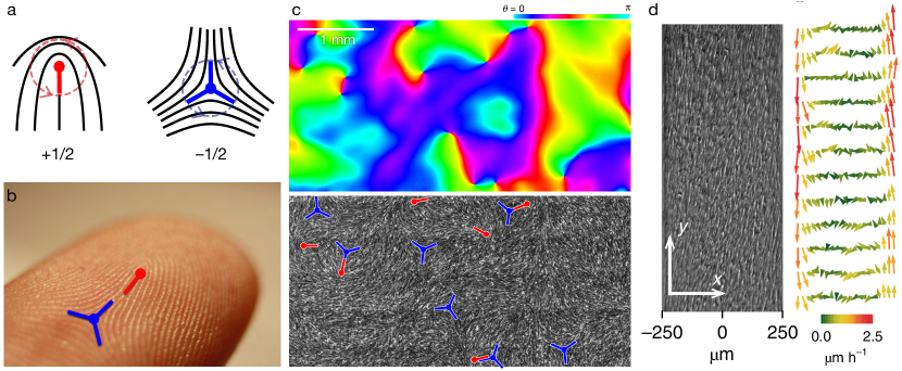
In other situations, cells align along one axis but choose no preferred direction of motion. This type of alignment is known as nematic order, taking its name from nematic liquid crystals, used in LCD screens. Prominent features of liquid crystals are singular points known as topological defects, where alignment is locally lost. You can find topological defects literally on your own hands: they are the points where your fingerprint ridges meet (Figs. 3 and 3). Recently, researchers have discovered topological defects in several cell assemblies, from bacterial colonies to epithelial tissues, confirming that they can be described as liquid crystals (Fig. 3). Interestingly, topological defects can play important biological roles. For example, in epithelial monolayers, defects promote cell death and extrusion 6. In colonies of the motile soil bacterium M. xanthus, defects promote the formation of multicellular aggregates known as fruiting bodies, which allow the bacterial population to survive starvation 7.
Flowing on their own
Describing orientational order is not enough to account for collective cell flows. To this end, physicists describe cell assemblies as active matter 1; 8. For example, when cells align with polar order, they can start migrating in the direction of alignment 9. This type of collective motion is known as flocking. The active-matter theory of this phenomenon was developed 25 years ago inspired by the mesmerizing flights of bird flocks 10. Today, the principles of flocking are applied to many other systems, from synthetic active colloids to bacterial swarms.
To describe nematic cell colonies, researchers use the theory of active liquid crystals, which generalizes the hydrodynamics of liquid crystals to include active (cell-generated) stresses. Among many other phenomena, this theory explains the cell flows observed around topological defects. Again, beyond cell colonies, the theory successfully describes many other systems, from biopolymer gels to shaken granular materials. Employing general theories of active matter to describe cell migration is particularly useful because it reveals connections to apparently-unrelated systems. Progressively, this approach is uncovering a classification of active systems based on symmetries and generic behaviors, in the spirit of universality classes in statistical mechanics.
 \phantomsubcaption\phantomsubcaption\phantomsubcaption\phantomsubcaption
For example, the theory of active liquid crystals was originally inspired by behaviors in bacterial suspensions and in the cell cytoskeleton. Soon after formulating the theory, researchers predicted that active stresses generate an instability whereby these fluids start flowing spontaneously, without having to apply external forces 14. To drive flows, active stresses have to overcome alignment forces in the liquid crystal, which happens only at sufficiently large scales. Therefore, the theory predicted that a strip of active fluid would flow only if it was wide enough 14.
More than a decade later, these predictions were tested in cell monolayers 5, showcasing the generic character of the theory. Whereas cells confined in narrow stripes did not flow, cells in stripes wider than a critical width developed a collective shear flow as predicted by the theory (Fig. 3). In very large tissues, cell flows become chaotic, creating disordered patterns of swirls known as active turbulence 15. Confinement can therefore prevent and organize spontaneous cell flows. This regulatory role of confinement may be relevant in embryonic development and tumor invasion, in which cell groups often migrate in tracks defined by the surrounding tissue (Fig. 1). Overall, these studies showcase how cells can leverage the physics of active fluids to engage in collective flow patterns, and how confinement controls whether and how cell groups flow on their own.
To spread or not to spread
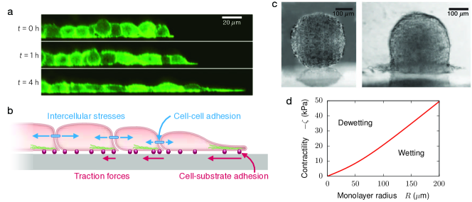
What happens if we release confinement and expose a cell monolayer to free space? Cells at the monolayer edge are able to sense that they have neighboring cells on one side but not on the other. In ways that remain to be fully understood, edge cells respond to this asymmetric environment by polarizing toward free space (Fig. 4). Specifically, these cells extend protrusions known as lamellipodia, with which they exert directed and persistent traction forces on the underlying substrate to migrate toward open ground. Because cells in the monolayer are adhered to one another, the migrating edge cells pull on cells in the second row, which then also polarize, migrate, and pull on inner cells, setting the monolayer under tension 16 (Fig. 4). At the molecular level, this supracellular coordination is mediated by a protein known as merlin, which transduces intercellular forces into cell polarization. In this mechanically-coordinated way, the entire cell monolayer spreads on the substrate, becoming progressively thinner 11 (Fig. 4). Combined with other mechanisms, this type of collective cell migration helps to close gaps in epithelial tissues, as in wound healing.
But tissues not always spread on substrates. Under certain conditions, a cell monolayer may instead retract from the substrate, eventually collapsing into a droplet-like cell aggregate 13 (Fig. 4). Tissue spreading and retraction are therefore reminiscent of the wetting and dewetting of liquid droplets. The degree of wetting depends on the balance between cohesive forces within the liquid and adhesive forces with the substrate. By analogy, early models proposed that tissue wetting was dictated by a competition between cell-cell () and cell-substrate () adhesion energies 17; 12, encoded in a spreading parameter . When , cell-cell adhesion dominates and the cell aggregate retracts from the substrate (dewetting, Fig. 4 left), whereas when , cell-substrate adhesion dominates and the aggregate spreads (wetting, Fig. 4 right). This simple conceptual framework was sufficient to interpret the behavior of cell aggregates of varying cell-cell and cell-substrate adhesion 17; 12. However, this analogy to passive liquids does not explicitly account for the active nature of cells.
Recent work addressed this limitation by treating the cell monolayer as a droplet of active liquid 13. Using this approach, one obtains the spreading parameter directly in terms of active cellular forces. Supported by experiments, the model predicts that the wetting transition results from the competition between two types of active forces: cell-substrate traction forces that promote spreading versus cell-cell pulling forces that promote retraction. This active wetting framework makes another key prediction: the spreading parameter depends on the droplet radius. Cell monolayers larger than a critical radius wet the substrate, whereas smaller monolayers dewet from it (Fig. 4). This prediction is striking because it has no counterpart in the classic wetting picture, in which the spreading parameter depends solely on surface tensions. For regular liquid droplets, size does not matter. In contrast, tissue wetting is size-dependent. This prediction was verified in experiments, providing evidence for the active nature of tissue wetting 13.
Besides its relevance in physics, the existence of a critical size for tissue wetting might explain drastic changes in tissue morphology during embryonic development and cancer progression. For example, a disturbing possibility is that a growing tumor might become able to spread onto the surrounding tissue once it reaches a critical size. Overall, this work exemplifies how the quest to understand collective cell migration motivates the development of new physics, leading to the discovery of phenomena like active wetting. Finally, this physics approach offers clues on how cell aggregates may tune active forces to control whether to spread or not to spread.
Mechanical waves without inertia
Tissue spreading revealed yet another striking collective phenomenon: Mechanical waves start spontaneously at the leading edge and propagate across the cell monolayer 11 (Fig. 5). These waves are slow, with speeds m/h, and wavelengths that span several cell diameters. Similar to longitudinal sound waves, tissue waves stretch and compress cells as they travel. Most strikingly, the waves are self-sustained; they travel long distances ( mm) unattenuated. This observation is surprising because cell motion is so slow that inertia is negligible. Therefore, these waves cannot be sustained by the common back-and-forth between kinetic and potential energy familiar from the harmonic oscillator. Moreover, there are many sources of dissipation in tissues, including cell-cell and cell-substrate friction, which could potentially damp the waves. Thus, the very existence of mechanical waves in tissues implies an active driving mechanism that compensates damping and generates an effective inertia.
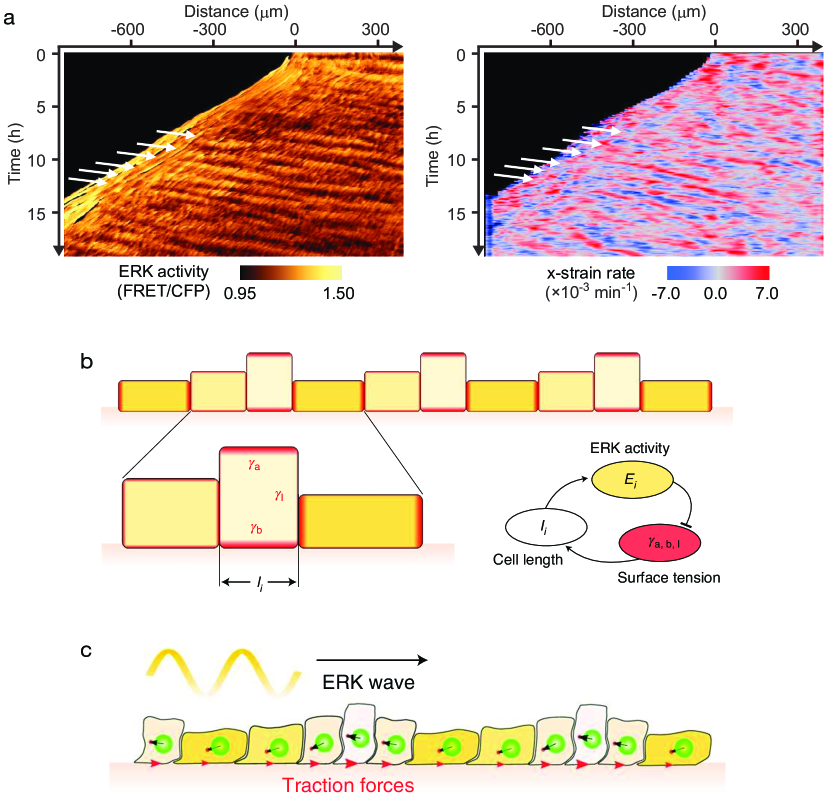
The quest to understand these waves led to a plethora of physical models, ultimately revealing a palette of possible mechanisms 1. Recently, the situation has been clarified by experiments revealing that mechanical waves are accompanied by waves of ERK, an extracellular signaling molecule that affects cellular activity (Fig. 5). Following these observations, researchers developed a theory based on the feedback between the biochemical regulator and cell mechanics (Fig. 5), which produces coupled chemical and mechanical waves. Assuming that cells polarize in response to stress gradients in the monolayer, the theory also explains propagation away from the leading edge in spreading tissues 18 (Fig. 5).
These results show that cells can exploit mechanochemical feedbacks to transmit local information over long distances. This tissue-scale communication is relevant for wound healing, enabling distant cells to coordinate their migration toward the wound. Similar principles operate in morphogenesis, enabling coordinated cell deformations to precisely shape tissues without requiring local genetic control of cellular forces. From a physics perspective, this research highlights that the cells’ ability to generate, sense, and respond to signals, both chemical and mechanical, can give rise to emergent phenomena as counter-intuitive as mechanical waves without inertia.
Outlook
The physics of active living matter is increasingly successful at explaining the dynamics of collective cell migration. This core biological process is being understood through concepts such as orientational order, flow, turbulence, jamming, wetting and wave propagation. The mechanistic origin and physical properties of these phenomena in cells, however, differ fundamentally from those in non-living matter. Whereas new active matter theories progressively manage to explain the broad phenomenology of collective cell migration in terms of a small number of physical variables, how cells tune these variables through thousands of genes and biochemical reactions remains a major open question.
Acknowledgments
We apologize to the many colleagues whose work could not be cited owing to space constraints. R.A. acknowledges support from the Human Frontier Science Program (LT000475/2018-C).
References
- (1) Alert, R. & Trepat, X. Physical Models of Collective Cell Migration. Annu. Rev. Condens. Matter Phys. 11, 77–101 (2020).
- (2) Bi, D., Lopez, J. H., Schwarz, J. M. & Manning, M. L. A density-independent rigidity transition in biological tissues. Nat. Phys. 11, 1074–1079 (2015).
- (3) Farhadifar, R., Röper, J.-C., Aigouy, B., Eaton, S. & Jülicher, F. The Influence of Cell Mechanics, Cell-Cell Interactions, and Proliferation on Epithelial Packing. Curr. Biol. 17, 2095–2104 (2007).
- (4) Park, J.-A. et al. Unjamming and cell shape in the asthmatic airway epithelium. Nat. Mater. 14, 1040–8 (2015).
- (5) Duclos, G. et al. Spontaneous shear flow in confined cellular nematics. Nat. Phys. 14, 728–732 (2018).
- (6) Saw, T. B. et al. Topological defects in epithelia govern cell death and extrusion. Nature 544, 212–216 (2017).
- (7) Copenhagen, K., Alert, R., Wingreen, N. S. & Shaevitz, J. W. Topological defects promote layer formation in Myxococcus xanthus colonies. Nat. Phys. 17, 211–215 (2021).
- (8) Marchetti, M. C. et al. Hydrodynamics of soft active matter. Rev. Mod. Phys. 85, 1143–1189 (2013).
- (9) Malinverno, C. et al. Endocytic reawakening of motility in jammed epithelia. Nat. Mater. 16, 587–596 (2017).
- (10) Vicsek, T., Czirók, A., Ben-Jacob, E., Cohen, I. & Shochet, O. Novel Type of Phase Transition in a System of Self-Driven Particles. Phys. Rev. Lett. 75, 1226–1229 (1995).
- (11) Serra-Picamal, X. et al. Mechanical waves during tissue expansion. Nat. Phys. 8, 628–634 (2012).
- (12) Gonzalez-Rodriguez, D., Guevorkian, K., Douezan, S. & Brochard-Wyart, F. Soft Matter Models of Developing Tissues and Tumors. Science 338, 910–917 (2012).
- (13) Pérez-González, C. et al. Active wetting of epithelial tissues. Nat. Phys. 15, 79–88 (2019).
- (14) Voituriez, R., Joanny, J. F. & Prost, J. Spontaneous flow transition in active polar gels. Europhys. Lett. 70, 404–410 (2005).
- (15) Blanch-Mercader, C. et al. Turbulent Dynamics of Epithelial Cell Cultures. Phys. Rev. Lett. 120, 208101 (2018).
- (16) Trepat, X. et al. Physical forces during collective cell migration. Nat. Phys. 5, 426–430 (2009).
- (17) Douezan, S. et al. Spreading dynamics and wetting transition of cellular aggregates. Proc. Natl. Acad. Sci. U. S. A. 108, 7315–7320 (2011).
- (18) Boocock, D., Hino, N., Ruzickova, N., Hirashima, T. & Hannezo, E. Theory of mechanochemical patterning and optimal migration in cell monolayers. Nat. Phys. 17, 267–274 (2021).