Optical read-out of the Néel vector in metallic antiferromagnet Mn2Au
Abstract
Metallic antiferromagnets with broken inversion symmetry on the two sublattices, strong spin-orbit coupling and high Néel temperatures offer new opportunities for applications in spintronics. Especially Mn2Au, with high Néel temperature and conductivity, is particularly interesting for real-world applications. Here, manipulation of the orientation of the staggered magnetization, i.e. the Néel vector, by current pulses has been recently demonstrated, with the read-out limited to studies of anisotropic magnetoresistance or X-ray magnetic linear dichroism. Here, we report on the in-plane reflectivity anisotropy of Mn2Au (001) films, which were Néel vector aligned in pulsed magnetic fields. In the near-infrared, the anisotropy is 0.6%, with higher reflectivity for the light polarized along the Néel vector. The observed magnetic linear dichroism is about four times larger than the anisotropic magnetoresistance. This suggests the dichroism in Mn2Au is a result of the strong spin-orbit interactions giving rise to anisotropy of interband optical transitions, in-line with recent studies of electronic band-structure. The considerable magnetic linear dichroism in the near-infrared could be used for ultrafast optical read-out of the Néel vector in Mn2Au.
pacs:
74.40.Gh, 78.47.J-, 78.47.D-, 74.72.-hI Introduction
There is an increasing interest in antiferromagnetic (AFM) materials as active elements in future spintronics devices, including data storage applications [1, 2, 3, 4, 5]. This is motivated by the absence of net magnetization and the related stray fields, which limit the minimum distance between two bits, and thus leads to higher storage density compared to ferromagnets. Moreover, the inherently fast spin dynamics in AFMs, which takes place in the THz range [6, 7, 8, 9, 10], is orders of magnitude faster then typical spin dynamics in ferromagnetic materials (GHz range) [11]. The absence of net magnetization, however, makes the manipulation and read-out of magnetic order, which is given by the staggered magnetization, i.e. the Néel vector, generally difficult.
Recently, two fully compensated metallic antiferromagnets CuMnAs and Mn2Au have been in the research focus. Here, the specific crystal and magnetic structure in combination with large spin-orbit coupling enable current driven manipulation of the Néel vector [12]. In particular, it was suggested that application of electric current pulses of the order of A cm-2 can rotate the Néel vector due to the bulk Néel spin-orbit torques (NSOT) [12]. Such current-driven switching was indeed demonstrated in metallic AFMs CuMnAs [13, 14, 15] and Mn2Au [16, 17, 18], with pulse durations down to picoseconds, achieved by driving currents with pulsed THz radiation [19]. Note that the high Néel temperature ( K) and high electrical conductivity [20, 21] make Mn2Au particularly interesting for real-world applications. In Mn2Au the current pulses were shown to reorient only a fraction (up to 30 ) of the domains [17], implying further studies of the switching mechanisms as well as novel approaches are required.
The absence of net magnetization makes the efficient read-out of the direction of the Néel vector challenging. Indeed, the electrical read-out via the anisotropic magnetoresistance (AMR) and the planar Hall effect is well established [13, 16]. However, recent studies show that in Mn2Au the AMR does not exceed 0.15 % [22]. Moreover, the read-out time in the AMR detection scheme is inherently limited by the electronics to timescales orders of magnitude larger than the intrinsic switching timescales [19]. Thus, using optical methods, which would enable fast read-out on the femtosecond scale, should be explored [23, 24]. While linear magneto-optic effects are commonly used to investigate magnetization and its dynamics in ferro- and ferri-magnets [25, 26], as well as in non-collinear antiferromagnets [27], the presence of the quadratic magneto-optical effects (Cotton-Mouton/Voigt effect) was demonstrated in several insulating AFMs [28, 29, 30, 7]. To probe the small changes in the refractive index due to the quadratic magneto-optical effects, polarimetry studies are commonly performed in the transmission geometry, since the changes in the optical phase accumulate over the optical path length within the material [28, 29, 30]. For metallic collinear AFM, with optical penetration depths on the order of tens of nanometers, the corresponding changes are small. Thus, for CuMnAs another approach has been recently demonstrated [31, 32]. Here, the thermomodulation aspect of the optical pump-probe technique has been used to detect small photoinduced changes in rotation of the polarization of the optical probe beam. Since such changes depend on the polarization of light with respect to the Néel vector, probe polarization dependence is used to determine the direction of the Néel vector [31, 32]. Indeed, similar approach has been recently used to study dynamics in metallic AFM Fe2As [33] and insulating CoO [34]. Finally, we note, that several recent reports demonstrated large magneto-optical contrast also in ultrathin NiO [35, 36, 37] and CoO [38] films. However, in this case the origin of the magneto-optical contrast is in large magnetostriction.
In Mn2Au, the recent angular-resolved photoemission study of Néel vector aligned Mn2Au films demonstrated the breaking of the C4z symmetry, a consequence of antiferromagnetic order and strong spin-orbit interaction [39]. In fact, a pronounced in-plane anisotropy of the electronic band structure is observed up to the binding energy of a few eV [39]. The existence of flat bands at the 1.5 eV binding energy, and their calculated anisotropy may present means to detect a substantial magnetic linear dichroism in the near-infrared (NIR) range.
Here, we report on near-infrared magnetic linear dichroism (MLD) measurements on Mn2Au thin films, performed in reflectance geometry at room temperature. To determine the magnitude of the MLD we investigate Mn2Au films whose staggered magnetization (Néel vector) was aligned using pulsed magnetic field [22]. The MLD in the NIR range is found to be %, about four times larger than the observed anisotropic magnetoresistance in Mn2Au [22]. The comparatively large MLD suggests it originates from the anisotropy in the interband absorption as a result of C4z symmetry breaking.
II Results and discussion
II.1 Mn2Au thin films
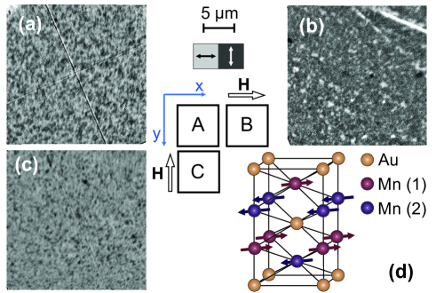
Mn2Au has a body-centered tetragonal crystal structure, whose unit cell is depicted in Fig. 1(d). It is an easy plane (001) AFM, with a strong out-of plane hard axis and a weak in-plane magnetic anisotropy. The spin orientations in adjacent layers are presented by arrows, with the Néel vector pointing along the easy [110] directions [20, 21, 40].
The -axis epitaxial Mn2Au thin film is grown on r-cut (102) Al2O3 substrate, with the lateral size of 1010 mm2 and thickness of 530 m by the radio–frequency magnetron sputtering at 600 oC - see Ref. [21,37] for fabrication details. To ensure epitaxial growth, a 45 nm thick Mn2Au film is deposited on a 20 nm thick (001) Ta buffer layer. To protect the surface, a 2 nm Al layer is deposited on Mn2Au, forming an aluminum-oxide capping layer. Mn2Au grows epitaxially with [110] and [10] axes parallel to the substrate edges, which are along the and directions of r-cut Al2O3. As elaborated in Appendix B, the -axis of Mn2Au films grown on r-cut Al2O3 is tilted by 2-3o towards the direction.
As previously demonstrated [41, 42, 22, 39], the application of magnetic field, exceeding 30-50 T results in the aligning the Néel vector along one of the easy axes, perpendicular to the applied magnetic field [41]. While such a ”polarization” of the film may be incomplete, i.e. there is still a small fraction of orthogonally polarized domains, it is quasi permanent [39, 41, 22].
To perform systematic measurements, the as-grown sample is cut into 55 mm2 pieces. The three pieces investigated are labeled with letters -. Sample is in the as-prepared state, i.e. in a multidomain configuration, and is used as a reference. Samples and are Néel vector aligned [39, 41, 22] in the pulsed magnetic field of 60 T along the different, yet crystallographically equivalent {110} directions, as shown in the middle panel of Fig. 1. Throughout this work the default film orientation is such that and .
II.2 X-ray magnetic linear dichroism in polarized Mn2Au films
Prior to optical studies the AFM domain patterns of all samples are first imaged by X-ray magnetic linear dichroism photoemission electron microscopy (XMLD-PEEM). The XMLD-PEEM imaging of the AFM domains is performed at room temperature at the SIM beamline of the Swiss Light Source. The sample is illuminated by linearly polarized X-rays with the polarization in the sample plane and the angle of incidence of 16∘. The XMLD contrast was obtained by using the two-energy mode, as described in detail in Ref.[39]. Here the energies E1 = 639 eV and E2 = 638.2 eV were used, which correspond to the maximum and minimum of the XMLD [42].
The XMLD-PEEM images of all samples are shown in Fig. 1, demonstrating that samples and have orthogonally oriented Néel vector. In particular, each of the polarized samples has mostly one type of AFM domains, with narrow worm-like domains of the other type, similarly to earlier reports [41, 42]. The sample , on the other hand, has about equal density of the domains of both types. Hence, sample is used as a reference in optical dichroism measurements.
II.3 Magnetic linear dichroism in the near-infrared
General symmetry considerations
In a tetragonal easy-plane system like Mn2Au a symmetry-based phenomenological model of the quadratic magneto-optical effect generally implies two possible contributions to the MLD: an isotropic and an anisotropic [28]. The isotropic contribution does not depend on the direction of the Néel vector within the plane, and can be observed when light is propagating along the two-fold symmetry axis of the crystal (here, we are considering the crystal symmetry only). The anisotropic MLD does depend on the direction of the Néel vector [29] and is present when the light propagates along the fourfold symmetry axis. It is the anisotropic component, that can be used to determine the Néel vector orientation in a collinear AFM.
The corresponding dielectric tensor of an AFM with the tetragonal crystal structure and the Néel vector perpendicular to the fourfold symmetry axis (i.e. within the easy plane), can be written as [28]:
| (1) | |||
Here and are the in-plane and out-of-plane dielectric constants in the absence of magnetic order, is the Néel vector and are the phenomenological coefficients related to the MLD. Given the two possible directions of the Néel vector in Mn2Au, we choose the axis to be along the fourfold [001] axis, while and coincide with [110] and [10] crystallographic axes. The difference between and is governed by , yet includes also the component proportional to , i.e. the so called isotropic component to the MLD (the term proportional to is likely negligible compared to ). The difference may lead to the reflectivity-anisotropy in non-normal incidence configuration (see Appendix B).
Experimental approach
The goal of the experiment is to determine the anisotropic magnetic contribution to dichroism, given by , which is proportional to phenomenological constants and and depends on the (in-plane) Néel vector orientation. Such a MLD is expected to be in the range of 10-3 and could be overshadowed by extrinsic non-magnetic effects such as strain. In particular, in the case of an additional lattice distortion in the -plane due to strain, (and as well) would be different for the and directions, leading to reflectivity-anisotropy in the -plane unrelated to the magnetic order (see the discussion in Appendix B). Furthermore, such extrinsic contributions may vary from sample to sample due to slight variation of substrate properties such as miscut. Thus, to unambiguously determine the MLD, measurements on different magnetically aligned parts of the same film are performed.
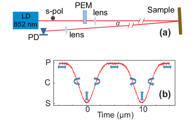
Optical dichroism is studied using a setup shown in Fig. 2(a) - for details see Appendix A. The measurements are performed at room temperature. We use the 852 nm laser diode and a Photo-Elastic modulator (PEM) to modulate the polarization of incoming light between the two orthogonal linear polarizations: s-polarized (vertical) and p-polarized (horizontal). The Mn2Au films were mounted such that their easy axes are along the horizontal/vertical directions, which correspond to and directions of . The light reflected from the sample is focused onto a photodiode and the AC signal is recorded using a digital oscilloscope. Measurements at multiple positions on each of the three samples are performed (the laser spot diameter on the sample was about 500 ) and averaged to minimize systematic errors.
Disentanglement of the MLD and structural dichroism
In our experimental geometry, with / corresponding to horizontal/vertical light polarizations, the MLD, given by in Eq. (1), should result in a difference between reflectivities of the s- and p-polarized light. However, the weak modulation of the intensity of the reflected light is also expected due to effects related to the operation of the PEM, as well as due to structural/strain anisotropy. Thus, the modulation of light intensity on the photo-diode, (the reflectivity, , of Mn2Au at 852 nm is 0.52 +/- 0.01), can be expressed as:
| (2) |
Here is the contribution to the signal related to the PEM operation and slight misalignment of optical components (present also when measuring uncoated gold mirror as a reference). is the amplitude related to the MLD of the sample, where - . Finally, is the amplitude of the dichroism caused by structural effects, such as strain. Here, we assume that in the first approximation the structural/strain dichroism is decoupled from , i.e. the strain does not affect the Néel vector. Thus, we assume is independent on the Néel vector direction, but depends on the sample orientation (see also Appendix B). Finally, is a periodic function describing the polarization state of light after passing through the PEM - see Fig. 2(a). Since identical experimental geometry is used for all samples, is the same in all measurements. Moreover, given the samples - were cut from the same film, should be identical to all samples, when mounted along the same direction with respect to the as-grown sample. Conversely, should change sign when the sample is rotated by 90o around the C4 axis, i.e. . When considering the MLD, given the multidomain state on the length scale of the laser spot size of 500 m. Finally, taking into account orthogonal direction of the Néel vector in the two magnetically aligned samples,it follows that . Under these assumptions the structural and magnetic contribution can be disentangled by the following referenced measurements:
| (3) | |||
| (4) |
Near-infrared magnetic linear dichroism in Mn2Au
Fig. 3 presents the results of dichroism measurements, which are performed in sequence, under identical conditions. The dashed black line, representing the evolution of the polarization state - see Fig. 2(b) - is added to all panels as a guide to the eye. Fig. 3(a) presents the raw data, , taken on samples , and . The complicated shape of the raw signal is largely a result of the optical elements of the setup, mainly the PEM. The PEM acts as a wave plate, where is modulated between and . This results in a sequence of polarization states s-p-s-p-s within one modulation period of the PEM (20 s). Note that between s- and p- polarizations, the light is circularly/elliptically polarized. Due to imperfections and asymmetry between the optical properties of the squeezed and elongated PEM crystal a modulation of the signal at 50 kHz is also observed.

To obtain the MLD component we first subtract the signal of the reference sample (as-prepared, ) from signals obtained on the magnetically polarized samples and , following Eq.(3). Moreover, as the MLD should give rise to a periodic modulation in reflectivity at the second harmonic of the PEM frequency, we fold the data recorded over two periods into the 10 s time window. The resulting variations are shown in Fig. 3(b). The differential signals do not have a complicated shape of the raw data as in Fig. 3(a), demonstrating that subtraction efficiently cancels out all system related modulations. Indeed, the shape of the differential signal follows the shape of the reference curve (dashed line). Furthermore, the absence of modulation of the differential signal at the 50 kHz PEM frequency clearly confirms the absence of circular dichroism. Fig. 3(c) presents the result obtained by the same approach for samples rotated by 90o around the C4 axis. The fact that the differential signals and in Fig. 3(c) are phase-shifted by underscores the MLD nature of the differential signals.
The MLD-induced change in reflectivity is given by the differences between the maxima and minima of the traces in Fig. 3 (b-c), multiplied by a factor of two (the signals shown in Fig. 3(b) and (c) are relative to the unpolarized reference sample ). The reflectivity is found to be higher for the light polarized parallel to the staggered magnetization. The average value of the MLD induced reflectivity change from all data combined is . We note, however, that it follows from Fig. 3(b) and (c) that the MLD extracted from and (), is larger than the MLD extracted from and (). This may suggest that the underlying microstructural strain favors the orientation of the Néel vector parallel to the -axis tilt (parallel to the axis of the substrate), resulting in a partial polarization of the as grown reference sample along the axis. Indeed, preliminary study using scanning electron microscopy with polarization analysis seem to support this observation [43].
Fig. 3(d) presents results of the measurements of structural dichroism. Following Eq.(4), for differences and the signal due to the MLD should cancel out and the remaining variation of reflectivity is due to the structural/strain effects. The extracted , with the reflectivity being larger for light polarized perpendicular to the direction of the -axis tilt, i.e. parallel to the axis of the r-cut sapphire (see also Appendix B). Thus, the structural dichroism is about twice smaller than the MLD in Mn2Au films on r-cut sapphire. Noteworthy, the value of extracted from is again noticeably lower that the value extracted from . This is consistent with the above suggestion that the as-grown sample is partially polarized along the axis of the substrate, resulting in a lower value of the extracted structural dichroism when monitoring .
III Conclusions
We demonstrate a pronounced magnetic linear dichroism in the NIR range in a collinear metallic antiferromagnet Mn2Au at room temperature. The observed MLD of exceeds the value of AMR in Mn2Au [22], recorded at liquid Helium temperatures, by about a factor of four. While no study on temperature dependence of the AMR in Mn2Au has been performed thus far, one may expect that the increased electron-phonon and electron-magnon scattering give rise to a suppression of AMR at higher temperatures, as typically observed. Thus, the ratio between the MLD and AMR may in fact be even higher at room temperature.
Recent broadband terahertz study of several ferromagnets demonstrated AMR signals up to 30 THz [44], with the effect slowly decreasing with increasing frequency. The large difference between the observed MLD in the near-infrared and the AMR in Mn2Au [22] implies the MLD in the near-infrared to be a result of the polarization dependent interband absorption. Indeed, the recent angular-resolved photoemission spectroscopy study does show that the Néel vector orientation leads to a pronounced valence band asymmetry up to binding energies of several eV [39]. Thus, further systematic studies of MLD as a function of photon energy may reveal spectral ranges with even higher MLD.
In view of the recent studies, demonstrating a large magneto-optical contrast in ultrathin NiO [35, 36, 37] and CoO [38] films, it is tempting to estimate the polarization rotation angle, which corresponds to the observed MLD-induced reflectivity variation of . Assuming the observed reflectivity change is dominated by the variation of the (interband) absorption coefficient, we estimate the polarization rotation angle for the light polarized at 45 away from the Néel vector to be mdeg, about a factor of 2 larger than the reported value in CoO thin films [38] and more than a factor of 5 larger than the reported value in NiO thin films [35].
As a simple non-perturbative table-top experiment, the presented approach can be extended to imaging mode, enabling detection of Néel vector orientation with micrometer spatial resolution. Moreover, in view of potential applications of Mn2Au, e.g. as a spintronic memory device, utilizing sequences of femtosecond optical pulses can provide read-out speeds matching the expected ultrafast switching times in antiferromagnetic memory devices.
Acknowledgements.
This work was funded by the Deutsche Forschungsgemeinschaft (DFG, German Research Foundation) Grant No. TRR 173 268565370 (project A05). This work received support from Horizon 2020 Framework Program of the European Commission under grant agreement No. 863155 (S-NEBULA). V.G. and M.F. acknowledge the financial support from the Graduate School of Excellence ”Materials Science in Mainz” (DFG GSC 266 49741853). We acknowledge the Paul Scherrer Institut, Villigen, Switzerland for the beamtime allocation under proposal 20200977 at the SIM beamline of the SLS. The authors thank the SIM beamline staff for the technical support. We acknowledge valuable discussions with H.-J. Elmers, H. Gomonay, P. Grigorev and L. Šmejkal.Appendix A: Experimental setup for optical dichroism
Our experimental setup is sketched in Fig. 2(a). For the non-cubic material, the experimental reflectivity-anisotropy can be a result of a finite angle of incidence. For the same reason, i.e. the difference in reflectivities of - and -polarized light, any dielectric mirrors need to be avoided in the beam path. Therefore, it is not possible to use normal incidence and to separate the reflected light using a semi-transparent dielectric mirror. To minimize such effects, the angle of incidence of is used. Moreover, we use minimal number of the optical elements, with the only reflecting surface being the surface of the sample, as shown in Fig. 2(a).
We use the 852 nm laser diode and a Photo-Elastic modulator (PEM) from Hinds Instruments, which operates at the frequency of 50 kHz. The PEM is set to retardation mode for 852 nm, resulting in a modulation of light polarization between -polarized (vertical) and -polarized (horizontal) at 100 kHz, the second harmonic of the PEM - see Fig. 2(b).
The laser intensity was 5 mW while the diameter of the laser spot on the sample was 500 m. The laser heating in thin films is governed by the absorbed light intensity and the thermal properties of the substrate (sapphire in our case). The temperature increase of the illuminated region can be estimated using a simple steady-state heat diffusion model [45, 46]. Using the reflectivity of 0.52, thermal conductivity of sapphire at room temperature of 35 W/mK [47] we estimated, using Eq.(3) of Ref.[46], the steady state heating to be K. Thus, the laser heating can be neglected.
Appendix B: The possible origins of structural dichroism
While our data clearly demonstrate the presence of a substantial MLD in Mn2Au, there are two possible origins giving rise to the structural component of the measured reflectivity anisotropy - see Fig. 3(d). To address these, we first need to consider the specifics of the epitaxial film growth on r-cut sapphire using the Ta (001) buffer layer [21, 40].
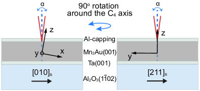
As elaborated elsewhere [17, 48], the -axis of the Mn2Au films grown on (102) Al2O3 is tilted with respect to the surface normal by about 2-3o (the tilt shows small variation from sample to sample). It turns out the the -axis of the film tilts towards the axis of the substrate.
Given the tilted growth, the observed structural dichroism could be a result of the experimental geometry - the effect is illustrated in Fig. 4. Namely, due to the tilt of the film’s c-axis with respect to the surface normal, the rotation of sample by (or equivalently by rotating the light polarization) changes the projection of the vector of light onto the c-axis. The difference between and can thus give rise to reflectivity modulation. Unfortunately, due to the lack of data on (anisotropic) optical properties of Mn2Au, this contribution cannot be estimated.
On the other hand, the observed dichroism can also be a result of the underlying strain as a result of the anisotropic thermal expansion of sapphire [49, 50]; such an effect can not be neglected on the low-symmetry cut of the Al2O3. Indeed, investigations of sample morphology using atomic force microscopy suggest breaking of the four-fold symmetry [40]. Considering the underlying strain to be responsible for the non-magnetic components of dichroism, its amplitude could roughly be estimated from . Assuming a linear relation between and the relative difference in the / lattice spacings the latter should be of the order of 0.1 %. Using the XRD measurements we are, however, unable to resolve such a weak asymmetry between the [110] and [10] lattice spacings parameters, because of the rather large mosaicity of [21, 40].
References
- Zutic et al. [2004] I. Zutic, J. Fabian, and S. Das Sarma, Spintronics: Fundamentals and applications, Rev. Mod. Phys. 76, 323 (2004).
- MacDonald and Tsoi [2011] A. H. MacDonald and M. Tsoi, Antiferromagnetic metal spintronics, Phil. Trans. R. Soc. A. 369, 3098 (2011).
- Gomonay and Loktev [2014] E. V. Gomonay and V. M. Loktev, Spintronics of antiferromagnetic systems (review article), Low Temperature Physics 40, 17 (2014).
- Jungwirth et al. [2016] T. Jungwirth, X. Marti, P. Wadley, and J. Wunderlich, Antiferromagnetic spintronics, Nature Nanotech 11, 231 (2016).
- Baltz et al. [2018] V. Baltz, A. Manchon, M. Tsoi, T. Moriyama, T. Ono, and Y. Tserkovnyak, Antiferromagnetic spintronics, Rev. Mod. Phys. 90, 015005 (2018).
- Kampfrath et al. [2011] T. Kampfrath, A. Sell, G. Klatt, A. Pashkin, S. Mährlein, T. Dekorsy, M. Wolf, M. Fiebig, A. Leitenstorfer, and R. Huber, Coherent terahertz control of antiferromagnetic spin waves, Nature Photonics 5, 31 (2011).
- Reid et al. [2015] A. H. M. Reid, T. Rasing, R. V. Pisarev, H. A. Dürr, and M. C. Hoffmann, Terahertz-driven magnetism dynamics in the orthoferrite DyFeO3, Applied Physics Letters 106, 082403 (2015).
- Yamaguchi et al. [2010] K. Yamaguchi, M. Nakajima, and T. Suemoto, Coherent control of spin precession motion with impulsive magnetic fields of half-cycle terahertz radiation, Phys. Rev. Lett. 105, 237201 (2010).
- Kim et al. [2014] T. H. Kim, S. Y. Hamh, J. W. Han, C. Kang, C.-S. Kee, S. Jung, J. Park, Y. Tokunaga, Y. Tokura, and J. S. Lee, Coherently controlled spin precession in canted antiferromagnetic YFeO3 using terahertz magnetic field, Applied Physics Express 7, 093007 (2014).
- Arana et al. [2017] M. Arana, F. Estrada, D. S. Maior, J. B. S. Mendes, L. E. Fernandez-Outon, W. A. A. Macedo, V. M. T. S. Barthem, D. Givord, A. Azevedo, and S. M. Rezende, Observation of magnons in Mn2Au films by inelastic brillouin and raman light scattering, Applied Physics Letters 111, 192409 (2017).
- Camley et al. [2015] R. E. Camley, Z. Celinski, and R. L. Stamps, Magnetism of surfaces, interfaces, and nanoscale materials, Handbook of Surface Science 5, 2 (2015).
- Železnỳ et al. [2014] J. Železnỳ, H. Gao, K. Vỳbornỳ, J. Zemen, J. Mašek, A. Manchon, J. Wunderlich, J. Sinova, and T. Jungwirth, Relativistic néel-order fields induced by electrical current in antiferromagnets, Physical review letters 113, 157201 (2014).
- Wadley et al. [2016] P. Wadley, B. Howells, J. Železnỳ, C. Andrews, V. Hills, R. P. Campion, V. Novák, K. Olejník, F. Maccherozzi, S. Dhesi, et al., Electrical switching of an antiferromagnet, Science 351, 587 (2016).
- Grzybowski et al. [2017] M. J. Grzybowski, P. Wadley, K. W. Edmonds, R. Beardsley, V. Hills, R. P. Campion, B. L. Gallagher, J. S. Chauhan, V. Novak, T. Jungwirth, F. Maccherozzi, and S. S. Dhesi, Imaging current-induced switching of antiferromagnetic domains in CuMnAs, Phys. Rev. Lett. 118, 057701 (2017).
- Wadley et al. [2018] P. Wadley, S. Reimers, M. J. Grzybowski, C. Andrews, M. Wang, J. S. Chauhan, B. L. Gallagher, R. P. Campion, K. W. Edmonds, S. S. Dhesi, F. Maccherozzi, V. Novak, J. Wunderlich, and T. Jungwirth, Current polarity-dependent manipulation of antiferromagnetic domains, Nature Nanotechnology 13, 362 (2018).
- Bodnar et al. [2018] S. Y. Bodnar, L. Šmejkal, I. Turek, T. Jungwirth, O. Gomonay, J. Sinova, A. A. Sapozhnik, H. J. Elmers, M. Kläui, and M. Jourdan, Writing and reading antiferromagnetic Mn2Au by Néel spin-orbit torques and large anisotropic magnetoresistance, Nat. Commun. 9, 348 (2018).
- Bodnar et al. [2019] S. Y. Bodnar, M. Filianina, S.P. Bommanaboyena, T. Forrest, F. Maccherozzi, A. Sapozhnik, Y. Skourski, M. Kläui, and M. Jourdan, Imaging of current induced néel vector switching in antiferromagnetic Mn2Au, Physical Review B 99, 140409 (2019).
- Meinert et al. [2018] M. Meinert, D. Graulich, and T. Matalla-Wagner, Electrical switching of antiferromagnetic Mn2Au and the role of thermal activation, Phys. Rev. Applied 9, 064040 (2018).
- Olejník et al. [2018] K. Olejník, T. Seifert, Z. Kašpar, V. Novák, P. Wadley, R. P. Campion, M. Baumgartner, P. Gambardella, P. Němec, J. Wunderlich, J. Sinova, P. Kužel, M. Müller, T. Kampfrath, and T. Jungwirth, Terahertz electrical writing speed in an antiferromagnetic memory, Science Advances 4, 10.1126/sciadv.aar3566 (2018).
- Barthem et al. [2013] V. Barthem, C. Colin, H. Mayaffre, M.-H. Julien, and D. Givord, Revealing the properties of Mn2Au for antiferromagnetic spintronics, Nature Communications 4, 1 (2013).
- Jourdan et al. [2015] M. Jourdan, H. Bräuning, A. Sapozhnik, H. Elmers, H. Zabel, and M. Kläui, Epitaxial Mn2Au thin films for antiferromagnetic spintronics, Journal of Physics D: Applied Physics 48, 385001 (2015).
- Bodnar et al. [2020] S. Y. Bodnar, Y. Skourski, O. Gomonay, J. Sinova, M. Kläui, and M. Jourdan, Magnetoresistance effects in the metallic antiferromagnet Mn2Au, Phys. Rev. Applied 14, 014004 (2020).
- Němec et al. [2018] P. Němec, M. Fiebig, T. Kampfrath, and A. V. Kimel, Antiferromagnetic opto-spintronics, Nature Physics 14, 229 (2018).
- Siddiqui et al. [2020] S. A. Siddiqui, J. Sklenar, K. Kang, M. J. Gilbert, A. Schleife, N. Mason, and A. Hoffmann, Metallic antiferromagnets, Journal of Applied Physics 128, 040904 (2020).
- Erskine and Stern [1973] J. L. Erskine and E. A. Stern, Magneto-optic Kerr effect in Ni, Co, and Fe, Phys. Rev. Lett. 30, 1329 (1973).
- Kirilyuk et al. [2010] A. Kirilyuk, A. V. Kimel, and T. Rasing, Ultrafast optical manipulation of magnetic order, Rev. Mod. Phys. 82, 2731 (2010).
- Higo et al. [2018] T. Higo, H. Man, D. B. Gopman, L. Wu, T. Koretsune, O. M. van t Erve, Y. P. Kabanov, D. Rees, Y. Li, M.-T. Suzuki, et al., Large magneto-optical Kerr effect and imaging of magnetic octupole domains in an antiferromagnetic metal, Nature Photonics 12, 73 (2018).
- Borovik-Romanov et al. [1973] A. S. Borovik-Romanov, A. A. Kreines, A. A. Pankov, and T. M. A, Magnetic birefringence of light in antiferromagnetic transition-metal fluorides, Sov. Phys.-JETP 64, 1762 (1973).
- Krichevtsov and Pisarev [1983] B. B. Krichevtsov and R. V. Pisarev, Anisotropy of magnetic linear dichroism in cubic magnetic substances, Sov. Phys.-JETP 84, 865 (1983).
- Ferré and Gehring [1984] J. Ferré and G. A. Gehring, Linear optical birefringence of magnetic crystals, Rep. Prog. Phys. 47, 513 (1984).
- Saidl et al. [2017] V. Saidl, P. Němec, P. Wadley, V. Hills, R. Campion, V. Novák, K. Edmonds, F. Maccherozzi, S. Dhesi, B. Gallagher, et al., Optical determination of the néel vector in a cumnas thin-film antiferromagnet, Nature Photonics 11, 91 (2017).
- Surỳnek et al. [2020] M. Surỳnek, V. Saidl, Z. Kašpar, V. Novák, R. Campion, P. Wadley, and P. Němec, Investigation of magnetic anisotropy and heat dissipation in thin films of compensated antiferromagnet CuMnAs by pump–probe experiment, Journal of Applied Physics 127, 233904 (2020).
- Yang et al. [2019] K. Yang, K. Kang, Z. Diao, A. Ramanathan, M. H. Karigerasi, D. P. Shoemaker, A. Schleife, and D. G. Cahill, Magneto-optic response of the metallic antiferromagnet Fe2As to ultrafast temperature excursions, Phys. Rev. Materials 3, 124408 (2019).
- Zheng et al. [2018] Z. Zheng, J. Y. Shi, Q. Li, T. Gu, H. Xia, L. Q. Shen, F. Jin, H. C. Yuan, Y. Z. Wu, L. Y. Chen, and H. B. Zhao, Magneto-optical probe of ultrafast spin dynamics in antiferromagnetic CoO thin films, Phys. Rev. B 98, 134409 (2018).
- Xu et al. [2019] J. Xu, C. Zhou, M. Jia, D. Shi, C. Liu, H. Chen, G. Chen, G. Zhang, Y. Liang, J. Li, W. Zhang, and Y. Wu, Imaging antiferromagnetic domains in nickel oxide thin films by optical birefringence effect, Phys. Rev. B 100, 134413 (2019).
- Schreiber et al. [2020] F. Schreiber, L. Baldrati, C. Schmitt, R. Ramos, E. Saitoh, R. Lebrun, and M. Kläui, Concurrent magneto-optical imaging and magneto-transport readout of electrical switching of insulating antiferromagnetic thin films, Applied Physics Letters 117, 082401 (2020).
- Meer et al. [2021] H. Meer, F. Schreiber, C. Schmitt, R. Ramos, E. Saitoh, O. Gomonay, J. Sinova, L. Baldrati, and M. Kläui, Direct imaging of current-induced antiferromagnetic switching revealing a pure thermomagnetoelastic switching mechanism in NiO, Nano Letters 21, 114 (2021).
- Xu et al. [2020] J. Xu, H. Chen, C. Zhou, D. Shi, G. Chen, and Y. Wu, Optical imaging of antiferromagnetic domains in ultrathin CoO(001) films, New Journal of Physics 22, 083033 (2020).
- Elmers et al. [2020] H.-J. Elmers, S. Chernov, S. D’Souza, S. Bommanaboyena, S. Y. Bodnar, K. Medjanik, S. Babenkov, O. Fedchenko, D. Vasilyev, S. Agustsson, et al., Néel vector induced manipulation of valence states in the collinear antiferromagnet Mn2Au, ACS nano 14, 17554 (2020).
- Bommanaboyena et al. [2020] S. Bommanaboyena, T. Bergfeldt, R. Heller, M. Kläui, and M. Jourdan, High quality epitaxial Mn2Au (001) thin films grown by molecular beam epitaxy, Journal of Applied Physics 127, 243901 (2020).
- Sapozhnik et al. [2018] A. Sapozhnik, M. Filianina, S. Y. Bodnar, A. Lamirand, M.-A. Mawass, Y. Skourski, H.-J. Elmers, H. Zabel, M. Kläui, and M. Jourdan, Direct imaging of antiferromagnetic domains in Mn2Au manipulated by high magnetic fields, Physical Review B 97, 134429 (2018).
- Sapozhnik et al. [2017] A. A. Sapozhnik, R. Abrudan, Y. Skourski, M. Jourdan, H. Zabel, M. Kläui, and H.-J. Elmers, Manipulation of antiferromagnetic domain distribution in Mn2Au by ultrahigh magnetic fields and by strain, physica status solidi (RRL) - Rapid Research Letters 11, 1600438 (2017).
- Bommanaboyena et al. [2021] S. Bommanaboyena, D. Backes, L. Veiga, S. Dhesi, Y. Niu, B. Sarpi, T. Denneulin, A. Kovacs, T. Mashoff, O. Gomonay, et al., Readout of a antiferromagnetic spintronics systems by strong exchange coupling of Mn2Au and permalloy, arXiv preprint arXiv:2106.02333 (2021).
- Nadvorník et al. [2021] L. Nadvorník, M. Borchert, L. Brandt, R. Schlitz, K. A. de Mare, K. Vỳbornỳ, I. Mertig, G. Jakob, M. Kläui, S. T. Goennenwein, et al., Broadband terahertz probes of anisotropic magnetoresistance disentangle extrinsic and intrinsic contributions, Phys. Rev. X 11, 021030 (2021).
- Carslaw and Jaeger [1992] H. S. Carslaw and J. C. Jaeger, Conduction of heat in solids (Clarendon press, 1992).
- Mihailovic and Demsar [1999] D. Mihailovic and J. Demsar, Time-resolved optical studies of quasiparticle dynamics in high-temperature superconductors: Experiments and theory, in Spectroscopy of Superconducting Materials (American Chemical Society, 1999) Chap. 16, pp. 230–244.
- Burghartz and Schulz [1994] S. Burghartz and B. Schulz, Thermophysical properties of sapphire, AlN and MgAl2O4 down to 70 K, Journal of nuclear materials 212, 1065 (1994).
- Bodnar [2020] S. Bodnar, Manipulation of Néel Vector in Antiferromagnetic Mn2Au by Electric Current and Magnetic Field Pulses, Ph.D. thesis, Johannes Gutenberg-Universität Mainz (2020).
- Yim and Paff [1974] W. M. Yim and R. J. Paff, Thermal expansion of AlN, sapphire, and silicon, Journal of Applied Physics 45, 1456 (1974).
- Roder et al. [2006] C. Roder, S. Einfeldt, S. Figge, T. Paskova, D. Hommel, P. P. Paskov, B. Monemar, U. Behn, B. A. Haskell, P. T. Fini, and S. Nakamura, Stress and wafer bending of a-plane GaN layers on r-plane sapphire substrates, Journal of Applied Physics 100, 103511 (2006).