Three-level multilevel simulation of the neuroreceptor function of NMDA
Abstract
We present a new multiscale method to study the N-Methyl-D-Aspartate (NMDA) neuroreceptor starting from the reconstruction of its crystallographic structure. Thanks to the combination of homology modeling, Molecular Dynamics and Lattice Boltzmann simulations, we analyse the allosteric transition of NDMA upon ligand binding and compute the receptor response to ionic passage across the membrane.
I Introduction
Modern biological sciences are incessantly searching to unveil the molecular mechanisms of biological activity, their dis-functionalities and the ensuing diseases so as propose and develop possible remedies. Computer models have a major say in this endeavour, as they can often access regions of parameter space which are beyond reach of direct experimentation. In particular, molecular dynamics simulations have a long and time-honored history in the study of biomolecular assemblies culminating with the 2013 Nobel Prize in Chemistry nobelchemistry.
The field shows no sign of flagging; quite on the contrary it is witnessing burgeoning progress in multiple directions, and particular in terms of capabilities to tackle larger systems over longer temporal scales. Remarkable examples in point are the recent simulations of biological systems is the 100-million atom-scale model of an entire cell organelle, a photosynthetic chromatophore vesicle from a purple bacterium rochaix2019dynamic or the study of the N-Methyl-D-Aspartate (NMDA) neuroreceptor by the DE Shaw research group song2018mechanism. These outstanding achievements were obtained by exploiting the method of Molecular Dynamics (MD), whereby all atoms in the simulation are taken into account, with a massive computational effort, often undertaken on special hardware.
Such achievements unquestionably push the frontiers of simulation further ahead and pave the way to disruptive progress in many areas of biology and medicine. In particular, the possibility of simulating large systems is key to neuroscience, where the response of neuro-receptors to binding with natural or synthetic ligands can reveal the allosteric transitions in the membrane protein and how the channel pore function, with the modulation of the ionic passage. NMDA receptors, in particular, play a critical role in brain development and function, including learning and memory formation trimble2002molecular; burnell2018positive. Dysfunctional NMDA is implicated in various neurological disorders, such as Alzheimer’s disease, depression, stroke, epilepsy and schizophrenia. Therefore, computational capability to tackle large-scale protein transitions and ion passage has the potential to study many neurological diseases and design new drugs through medicinal chemistry methods. Some initial studies have indeed tackled the study of NMDA by a brute force MD approach that, although showing some degree of success, has made clear the difficulty in covering the complete times scale of the allosteric and functional response dai2013nmda; zheng2017probing; song2018mechanism; palmai2018does.
Under physiological conditions, the opening of the receptor ion channel requires concurrent binding of glycine and glutamate and relief of magnesium block at the ion channel pore by membrane depolarization. The resulting calcium flux triggers a cascade of signal transduction necessary for synaptic plasticity.
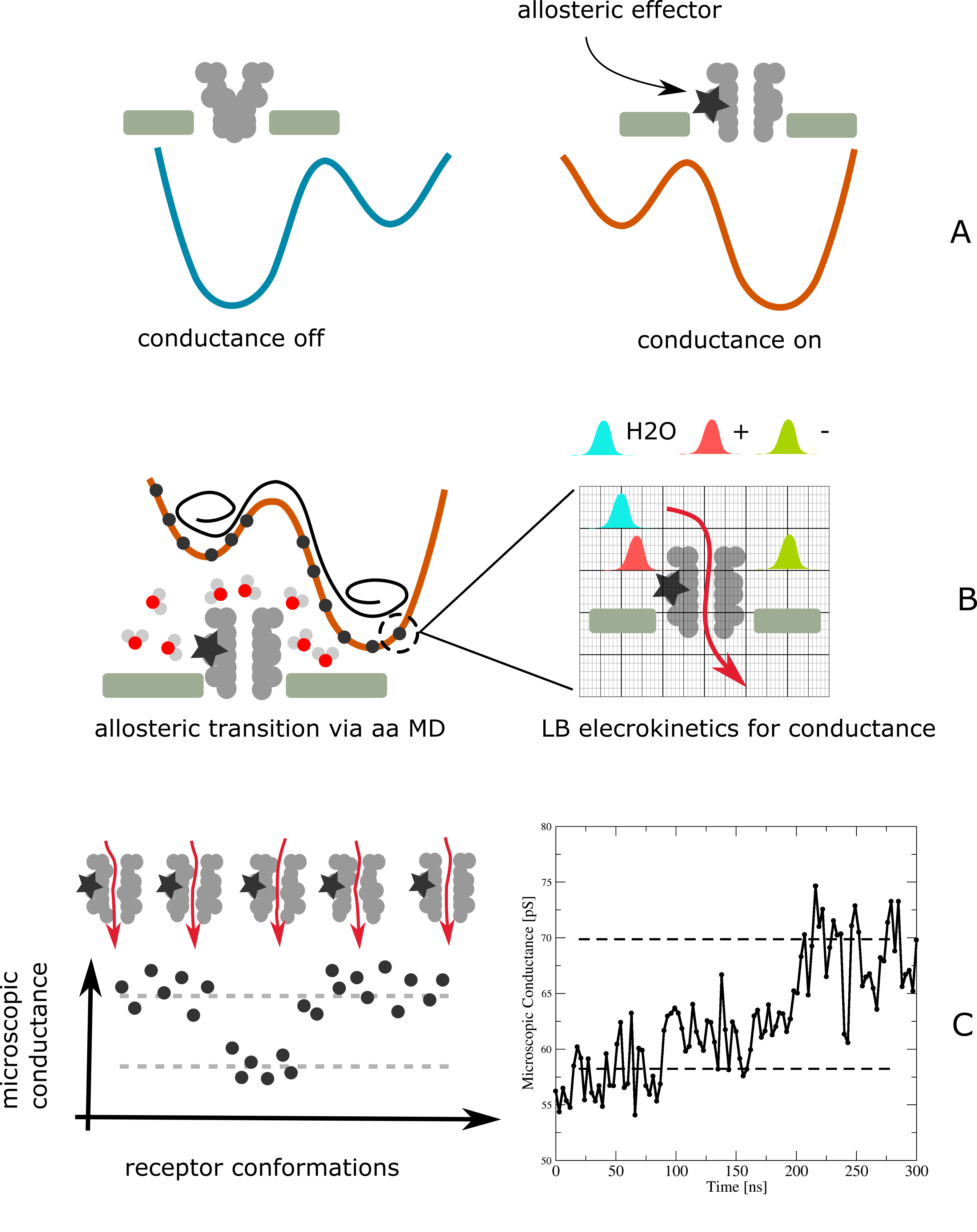
Multiscale methods offer a strategic advantage in simulating such receptors that lay at the basis of many basic neurological phenomena. The reasons is that neuroreceptors are large molecular assemblies with large stretches of membranes separating intra and extracellular regions and receptors (proteins) as gates. Binding small molecules (neurotransmitters) triggers spatially small but functionally very important structural changes (allosteric transitions) that open/close the gates to the passage of ions.
From the neurological viewpoint, it is key to quantify the allosteric response to the binding of nanomolar quantities of neurotransmitters. These can be natural or artificial, with important applications to medicinal chemistry. Once the allostery is measured and quantified, one can measure passage of ions in the receptor pore. To this purpose, electrokinetics raises a significant challenge, as it implies the presence of blocking elements (Magnesium for NMDA), since the process is an activated one. To achieve such ambitious program all-atoms simulations fall short of covering the wide stretch of spatial and temporal scales involved, ranging from picoseconds to minutes and with a number of atomic degrees of freedom in excess of millions. In essence, reproducing binding affinity (thermodynamics), allosteric transitions (large-scale, fine and coarse atomic motion) and electro-kinetics, is a task that involves too widely disparate scales to be liable to a single level of representation and simulation.
For these reasons, we have devised a new (three-level) multiscale method to study neuroreceptors starting from the reconstruction of the protein structure, as obtained from incomplete crystallographic data, to analyze the binding affinity of NMDA to its ligands, followed by the quantification of the allosteric motion, and finally to assess the modulation of the ionic passage.
Our approach begins by using homology modeling techniques to reconstruct the NMDA configuration in the phospholipidic environment, the simulation of the allosteric events by means of MD and finally by the study of the electrokinetic ion transport by means of a Lattice Boltzmann approach. The novelty of our approach relies on the use of LB electrokinetics to analyse the flow of ions in a probabilistic way. While other strategy can be powerful to study the passage of single or multiple charges in ion channels via the potential of mean force method roux2004theoretical or in a statistical sense, by solving the Poisson-Nernst-Planck equation kurnikova1999lattice, our approach takes into account the complete hydrodynamic modes of the saline solution flowing through the receptor matrix. In this way it is possible to observe direcly the effect of electrostatic forces and convection on the three-component solution made of neutral, cationic and anionic species. The approach thus allows selecting a large set of protein configurations so that the prediction of flux based on configuration allows direct comparative analysis with purely steric approaches smart1996hole. Finally, the proposed method can be enriched by combining the sampling of configurational space with adequate simulations of all atoms or coarse grained models, in order to follow the flow in different conditions.
The multiscale and multilevel approach is entirely novel in concept and application and demonstrates, for the first time to the best of our knowledge, that complex biological processes occurring in neuroscience can be tackled, thus providing a whole wealth of microscopic, dynamic and functional information that is unaccessible to current experimental techniques.
II Computational approach
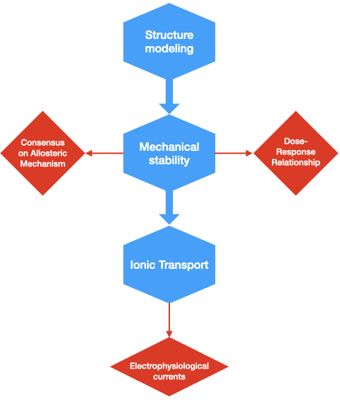
As shown in Fig 1, our computational approach involves a highly orchestrated network of computational methods and software tools, which can broadly classified within three basic levels:
-
1.
Structure modelling (Homology Methods);
-
2.
Mechanical stability (Molecular Dynamics and Molecular Mechanics);
-
3.
Ionic transport (Lattice Boltzmann).
The whole three-level procedure spans about six decades in time, from nanoseconds up to milliseconds, reaching up to timescales of physiological relevance. The ultimate outcome of clinical relevance is the dose response relationship, namely the current flowing across the neurotransmitter as a function of the dose of ligand.
To accomplish the three segments of the flowchart, we used three different softwares: Modeler eswar2006comparative for Homology Modeling and Gromacs abraham2015gromacs for All-Atoms MD. Most of simulations are based on the software Moebius from Lexma Technology, as it allows multiphysics simulations of multicomponent fluids and particles. Therefore, Moebius was then used for the Coarse-Grained MD and for the Lattice Boltzmann simulations. We utilized both the CPU and GPU architectures to accomplish the heavy-computing runs, as available on both the Gromacs and Moebius softwares.
II.1 Structure Modeling
The first level of the flowchart pertains to characterising the molecular structure of the neuroreceptor, consisting of the protein embedded in the cellular membrane. Homology modelling allows to construct the atomic-resolution model of NMDA GluN1A/GluN2B starting from its amino acid sequence and the available crystallographic three-dimensional structure (PDB id: 4PE5) karakas2014crystal. For crystallographic data from membrane proteins with a large amount of missing structures, such as for NMDA, is rather challenging for homology modeling.
Reconstructing the missing secondary structure is accomplished by resorting to homological techniques whereby the atomic-resolution model of the target protein from its amino acid sequence is constructed by comparing the experimental three-dimensional structure of a template homologous protein. The reconstruction relies on the identification of known secondary structures from homologous receptors acting as templates that resemble the structure of the query sequence and on an alignment that maps residues to residues in the template sequence. The identification of one or more known structures resembling the one of the query sequence and the alignment of residues in the query sequence to residues in the template sequence, benefits from the fact that NMDA structure is conserved amongst homologues.
II.2 Mechanical Stability and Allostery
The second level of the flowchart pertains to assessing the stability of the aforementioned molecular complex, including its interactions with the water solvent, and assessing the allosteric transition. Once ligands bind to the specific receptor sites, NMDA changes its shape, and altering the affinity for a ligand at a second site (e.g., either a receptor or a binding site); the ability of an effector molecule (ligand) to change the conformation and activity of a protein.
In allostery, the function of a receptor is modified by the interaction with its ligands, not only at the active site but also at a spatially distinct site of different specificity. In allostery, the interaction of the functional sites results in an altered affinity of ligand binding, thus depending on the dynamic interaction with the substrate. Ideally the conformational changes induced by the binding of the allosteric effector can be finely followed by brute force MD simulations at atomistic resolution. For instance, if the allosteric response path is of interest, starting from the substrate free equilibrated structure it is possible to introduce the effector and follow in time its conformational changes and fluctuations. This is the strategy used in our approach. In other situations, where both the APO and HOLO states are available, the thermal fluctuations in each state, and possible state interconversion can be simulated with brute force MD or by enhanced sampling techniques (e.g. parallel tempering, thermodynamic integration, metadynamics, etc.). However, the high resolution of the full atomistic approach is often limiting for very large systems. It is convenient therefore to use a simplified coarse-grained model. This is particularly challenging for membrane proteins that, for a proper treatment of allosteric process, an adequate level of controlled flexibility of the protein scaffold is required. In our approach we tested the capability of the OPEP CG force field chebaro2012; sterpone2014; sterpone.opep.2013 to reproduce the main fluctuations and conformational changes associated to the allosteric transition by comparing the results with atomistic modelling.
The AA protocols is as follows: we simulated both the APO (ligand free) and the HOLO (ligand bound) versions of NMDA. For the HOLO version, we positioned two Glutamate (GLU) and two Glycine (GLY) neurotransmitters molecules in the NMDA pockets of the relaxed model. This was obtained by docking them by means of the Chimera software chain-by-chain superimposition to achieve suboptimal overlap of the protein domains.
The OPEP force field has been designed to model peptide and protein folding and aggregation in solution. It is a multi-scale model that reserves an atomistic description to the backbone atoms and reduces the side chain amino acid to a unique bead. OPEP was designed, and progressively improved, for modelling soluble proteins chebaro2012; sterpone2014; sterpone.opep.2013 but its coupling with an explicit membrane environment is under way.
Within the scope of the present investigation, we adopted the available version with an adequate strategy to model the trans-membrane part of the protein. The test was performed on two models. In the first one the protein motion is fully flexible and its motion is controlled by the OPEP hamiltonian. Some restrains are applied to the trans-membrane part of the proteins so to mimic the spatial embedding of the membrane. The flexibility is key for monitoring the loops motions in the extra-membrane regions that control the allosteric response. The second model uses a more radical simplification, and is based on a floppy elastic network of the protein. On top of the elastic network, non-bonded sites interact via the OPEP non-bonded potential. To be noted that in several previous works the characterisation of protein allosteric paths are modelled by considering elastic networks rocks2017designing. In the CG approach for the allosteric effectors (GLN and GLY amino acids) instead we used the same OPEP model but their location in the binding site was restrained. It is worth noting that the OPEP force field was already successfully coupled to the hydrodynamic description based on LB sterpone2015, allowing the investigation of complex processes like amyloid aggregation chiricotto.jcp.2016; chiricotto.jpcl.2019, protein crowding sterpone2014; timr2020 and unfolding under shear flow sterpone.shear.2018; cattoen2018.
II.3 Ionic Currents
Finally, the third level pertains to simulating the ionic currents within the neuroreceptor, which is performed in order to quantify the conductivity of the neuroreceptor as a function of the applied voltage or salinity and for varying configurations of the receptor. The Lattice Boltzmann (LB) method provides a particularly useful approach to investigate the ionic response since it is rooted in the mesoscopic description of matter which perfectly maps the current representation of the receptor and its environment succibook.
LB is a very convenient computational approach since it is based on a over a Cartesian grid, that is, by using a uniform mesh based on cubic voxels. It is highly adaptable to reproduce the flow structure of single and multi-component system under the action of external or internal forces. In particular, LB has been shown in the past to be capable of reproducing the electro-kinetics of saline solutions in model devices with non-trivial geometries and locally charged surfaces MARCONI2011; melchionna2011electro.
The case of the NMDA receptor, however, presents a non trivial geometry within the pore region and highly localized forces at position stemming from charged or neutral atomic groups. In addition, the local electrostatic forces can be locally intense, often exhibiting rapid spatial modulations of the electrolytic densities due to the formation of disordered double layers. Such scenario poses several challenges to the computational scheme, particularly by endangering numerical stability as dictated by the Courant-Friedrich-Lewy stability condition . It is well-known that the presence of unit charges in simulation cannot be simulated by a direct LB approach raafatnia2014computing without proper treatment to enhance its stability.
To circument such limitations, we employed a multi-component Entropic Lattice Boltzmann Method, a powerful variant of the basic Lattice Boltzmann method based on a self-consistent tuning of the relaxation parameter so as to ensure compliance with local entropy growth (H theorem).
The LB approach to simulating the saline solution is based on tracking the evolution of each fluid component, with the index labelling the neutral aqueous medium (), and the positively () and negatively charged components (), via the discretized form of the density distribution function, named populations . Here, denotes the temporal coordinate and subscript labels a set of discrete speeds connecting the mesh points to its neighbors. The two ionic components are monovalent and characterised by charges density and velocity and, given the barycentric velocity , the relative velocity being denoted . The neuroreceptor and the membrane are described by the collection of particles at position being neutral or partially charged with charge valence provided by the force field.
In standard multicomponent LB the dynamics of each component then follows its own evolution equation:
| (1) |
where is a relaxation frequency related to the kinematic viscosity as , the lattice speed of sound, and the Maxwellian equilibrium given by MARCONI2011; MARCONI2011b,
| (2) |
and is a set of normalized weights. The force term is and the local force , being the sum of the self-consistent electrostatic forces, the inter-specie drag force characterized by the cross-diffusion coefficient , and the frictional force exerted by the receptor and membrane atoms on the fluid species, being proportional to the coefficient . In addition, is a function used to smear the particle charge on the LB grid, as used in Immersed Boundary method peskin2002immersed.
The electrostatic potential is the solution of the Poisson equation, in the aqueous medium of dielectric permittivity , being the unit electronic charge. In this study the LB solution is obtained on a cartesian grid of spacing nm, by employing the D3Q19 set of discrete speeds and associated weights, and by smearing particle charges over grid points. We chose a ionic cross-diffusivity of nm-2/ns and a frictional coefficient ns-1.
The complete multicomponent Entropic LB evolution is obtained by solving eq. 1 complemented by the minimization of the lattice H-function , which additionally provides a small local adjustment to the relaxation frequency to enforce stability. Under operating conditions we found that applying a filtering approach to populations to remove non-hydrodynamic modes kramer2019pseudoentropic significantly enhances numerical stability montessori2014regularized, providing the final conditions to simulate saline solutions in presence of unit charges.
III Results
In the following, we provide a summary of the main results of our analysis.
III.1 Homology Modeling
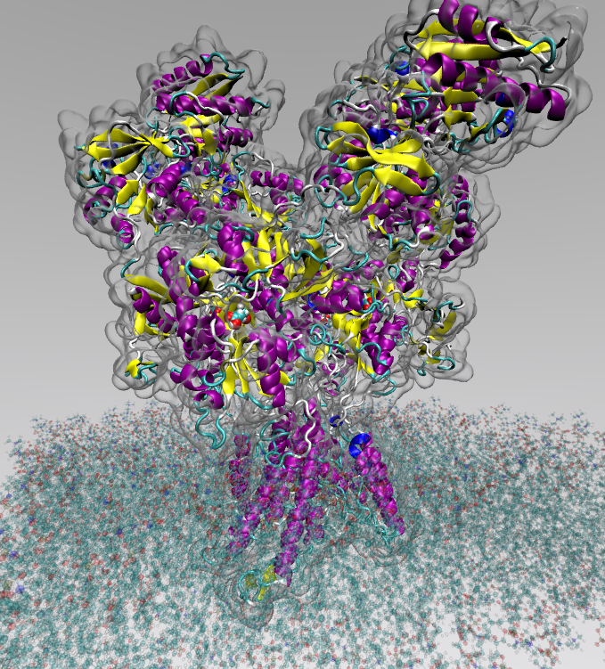
We first considered the tetrameric NMDA GluN1A/GluN2B structure from Rattus Norvegicus reconstructed by Homology Modeling at atomic-resolution model starting from the three-dimensional crystallographic structure karakas2014crystal.
The structure lacks several loops in the linker region between the ligand-binding domain and the missing loops were reconstructed chain-by-chain using the high precision DOPE-HR modeling protocol shen2006statistical, resulting in the most accurate available refinement method to obtain high quality structural stretches.
Subsequently, the receptor was relaxed using an energy minimization scheme first in vacuum by means of MD via a steepest descent algorithm to overcome/solve bad contacts and improve the overall quality of the structure quality.
Validation of both post-homology model and equilibrated structures was obtained by using MolProbity williams2018molprobity. Once relaxed, the receptor was embedded in a phospholipidic membrane and prepared for simulation by surrounding the entire system in water molecules at ambient temperature and as density corresponding to Kg/m3.
III.2 Molecular Dynamics and Molecular Mechanics simulations
Once the system was validated, we performed MD simulations at three increasing levels of coarse graining, namely i) All-atom resolution based on CHARMM36m force field, ii) Coarse-grained MD based on OPEP force field, iii) elastic network representation. We simulated both the APO (ligand free) and the HOLO (ligand bound) versions of NMDA. For the HOLO version, we positioned two Glutamate (GLU) and two Glycine (GLY) neurotransmitters molecules in the NMDA pockets of the relaxed model. This was obtained by docking them by means of the Chimera software chain-by-chain superimposition to achieve anoptimal overlap of the protein domains.
At first the all-atom resolution model was chosen by employing the CHARMM36m force field huang2013charmm36 to represent membrane proteins by taking into account bonding and non-bonding interactions. The former includes bonding, angular and torsional forces among peptidic groups while the latter include Van der Waals, electrostatic and hydrogen bond forces.
The atomistic simulations successfully model the allosteric effect, since upon the binding of the effector (GLN or GLY) we observe the opening of the channel associated to the specific reorganization of the extra-membrane region. This opening is observed in the timescale of ns.
A first-level coarse-grained model was utilized by employing the OPEP force field sterpone2018multi to represent amino acids fully explicitly for the peptidic backbone, inclusive of hydrogen atoms, while the lateral groups are represented by an effective particle. Such representation is particularly accurate for the backbone as it includes stretching, angular and torsional movements together with non-covalent and hydrogen bonds. The pseudo-particles for the lateral groups take into account the sterical and non-covalent interactions, together with explicit representations of the saline bridges.
The CG simulations were performed and contrasted against the atomistic one taken as reference. We observed a comparable opening of the channel. The result supports the use of this less time consuming model to generate a valid ensemble of configurations for further electrokinetic analysis. A detailed description of the trajectories and the conformational motions associated to the allosteric transition is reserved to a further work.
A further level of coarse graining was employed by using an elastic network representation. Here all intramolecular bonding and non-covalent forces were substituted with harmonic interactions that allow for a certain level of protein deformability. Intermolecular forces are still accounted for by means of the OPEP force field.
All three models showed to undergo the allosteric transition once the ligands have been positioned in the corresponding pockets.
The transition took place on the ns timescale and several attempts were observed before NMDA finally reached its stable configuration, which is supposed to be an open pore configuration. Importantely, the consensus of the three models to reproduce the transition lends a significant degree of confidence to the overall homology-based reconstruction and simulation models.
With the three representations of the neuroreceptor in place, we were able not only to generate a large number of configurations with the all-atom method, but also to harvest the relaxed structures by replacing the membrane and receptor by order two different levels of coarse graining, the OPEP and elastic network models.
III.3 Ionic passage
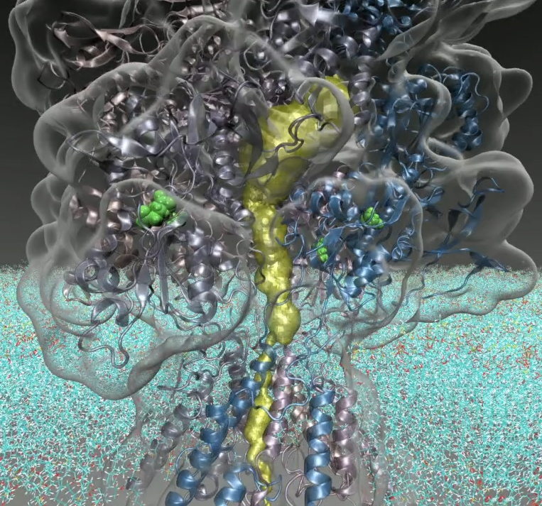
A purely steric analysis of the pore geometry is initially performed by finding the best route for a sphere with variable radius to squeeze through the channel. The method HOLE allows the analysis of the dimensions of the pore running through a structural model of an ion channel smart1996hole where the algorithm uses a Monte Carlo simulated annealing procedure. The method predicts conductance by using a simple empirically corrected ohmic model. However, ion permeation cannot be simply identified from the physical dimensions of the pore. For example, water within narrow hydrophobic pores can modulate permeation without even requiring steric occlusion of the pathway. Better methods have been proposed to account for hydrophobic gating, such as in the CHAP method klesse2019chap.
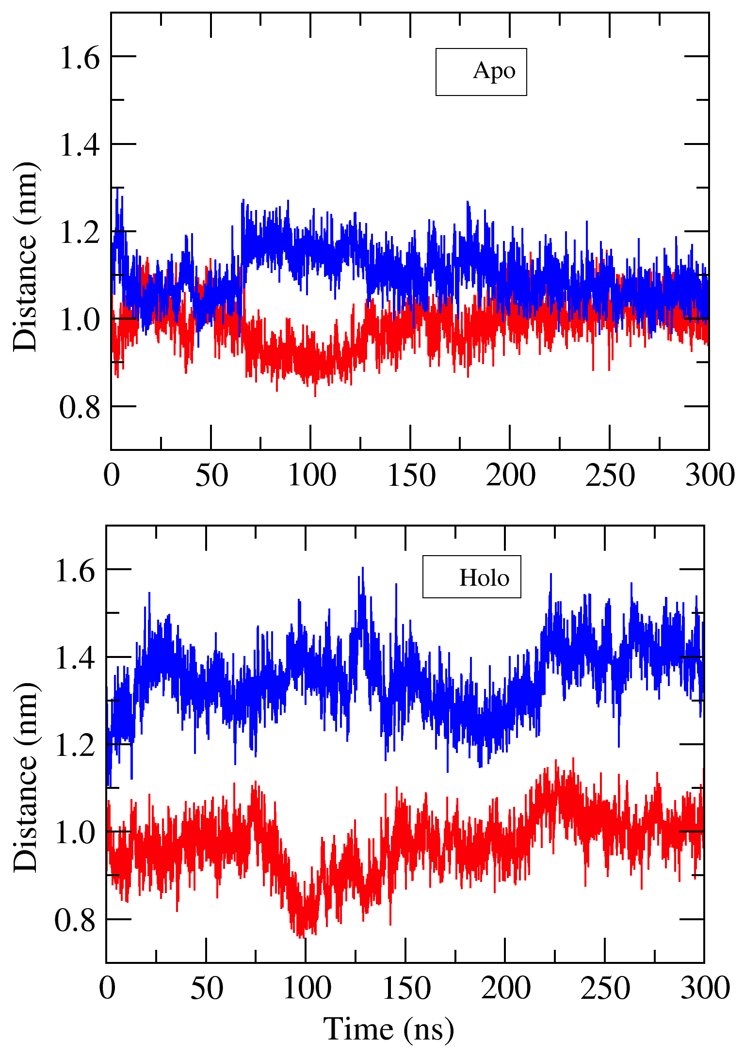
The effective passage of ions can take place in any of the small apertures and crevices present within the receptor matrix, that can be seen as an effective porous medium. Fig. 3 illustrates the steric passage within NMDA for a given receptor conformation. The available volume is rather funnel shaped and the receptor bottleneck is very narrow, with a lateral aperture being smaller than one Angstrom. By analysing the time evolution of the bottlenck distance, computed as the distance between the two GluN1A subunits considering the CoM of Val632 and Val2244, and between the GluN2B subunits Ile1426 and Ile3039, as reported in Fig. 4, we observe a differentiated behavior between the APO and HOLO states, suggesting that the allosteric transition has taken place. In accordance with the all-atom simulations, a similar behaviour has been observed analysing the bottleneck distances in the CG and Elastic Network simulations (data not shown); thus resulting in a coherent representation of the system allosteric movement under the influence of the ligands, for the three difference models. However, such narrow space renders a quantification of the total ionic resistance highly dependent on the multiple bottlenecks and conduction channels along the pathway.
The presence of small fluctuations and the presence of several charged groups along the pathway, renders the purely steric analysis qualitative and even hard to justify. Upon binding of the receptor with the ligands, the allosteric modification alters ionic resistance in several ways: the variations in the electrostatic environment along the pore extension, the fluctuating motion of the protein matrix in the channel, the presence of water that is advected and that alters the hydrophobic content of the pore, the sub-Angstrom modification of the pore bottlenecks. These conditions can result in modifications of the barrier crossing rates by orders of magnitude. Once again, the complex interplay of these conditions and their associated timescales rules out a computational approach based on a direct atomistic approach since it would require hundreds of nanoseconds of observation time for monitoring a single ion passage. Consequently, gathering sufficient statistics is out of reach. The proposed scheme instead provides an effective decoupling between the macromolecular motion and the ionic passage, thereby allowing to study the motion of the saline solution during the macromolecular evolution.
The simulation on ionic transport proceeds as follows. MD simulations deliver a time sequence of structural configurations of the neuroreceptor, . For a prescribed sequence of such configurations, a long-time (hundreds of nanoseconds) LB simulation is performed until the steady-state current, , supported by the given configuration at time is obtained. In passing, we note that this procedure also permits to accumulate significant statistics, due to the the fact that the structural changes of NMDA are pretty slow on the scale of the MD integration. The resulting current shows abrupt up and down changes in time, which we associate with opening (closing) of conductive channels within the receptor configuration. Needless to say, the structural dynamics of these channels is extremely rich, with abrupt morphological changes, such as sudden strictions which eventually quench an otherwise highly conductive channel and viceversa.
Functional response is quantified by considering both the APO and HOLO versions of NMDA and considering a single ns MD trajectory. Conformational analysis of the receptors and application of the HOLE method exhibit large fluctuations of the pore region by steric analysis, indicating that allosteric transition is effectively taking place (data not shown). Conductance was measured by extracting protein conformations, evenly spaced in time of the last ns of the all atom simulations of the equilibrated receptor for the putative close and open states.
Figure 5C shows the conductance (in picoSiemens) as a function of time. From this figure, several bursts of conductance are visible past the open-up event mark the genuine microscopic nature of conductance, driven by the underlyking fluctuations of the protein matrix. These fluctuations provide a neat signature of channel opening/closing events but with a clear trend towards stabilization of the open configuration. It is expected that at longer times, not covered by the present simulations, the open state will likely exhibit a consistent amount of short-lived closed configurations.
Summarizing, within our procedure, the neuroreceptor is treated as a slow time-changing molecular porous medium, and the geometrical flexibility of LB is leveraged to compute the “electrical permeability” of such molecular porous media “on-the-fly” succi1989three.
III.4 Summary
We have presented a new three-level multiscale method for the computational study of the NMDA neuroreceptor.
The computational framework combines three distinct representation levels:
i) Homology models for the characterisation of the protein structure, ii) various forms of Molecular Dynamics for the dynamical stability of the NMDA complex and finally iii) lattice Boltzmann simulations of ion transport across the neurotransmitter. The novelty of our approach relies on LB electrokinetics to analyse the mesoscopic flow of ions by taking into account the complete hydrodynamic modes of the saline solution as it flows inside the receptor matrix. It is thus possible to observe direcly the effect of electrostatic forces and convection on ionic currents at varying receptor configurational states. To this aim, the usage of mesoscopic representation of the protein via a coarse grained force field (such as OPEP used in this work) with the saline solution described by the Boltzmann picture provides equivalent levels of detail. In more general terms, the three-level multiscale method described here can be further extended to combine the evolution of the lagrangian representation of one subsystem, typically simulated via MD, with the eulerian representation of a complementary subsystem, simulated via LB, thereby taking into account the dynamical two-way exchange of forces.
The numerical results on NMDA show clear evidence of allosteric transitions stimulated by binding of Glutamate and Glycine ligands. Ionic transport across the NMDA complex show preliminary agreement with experimental data. Importantly, the provided scheme makes the study of the neuroreceptor functional response viable thanks to the high performances of the MD and LB components. In particular the MD component requires approx. 4 GPU hours per nanosecond, while the LB requires 1 CPU/hour per simulation of electrokinetics, that translates to 0.1 GPU/hour when running on a single GPU.
It is hoped and expected that the present three-level framework may pave the way to the computational study of a variety of fundamental multiscale biological processes. As presented in the text, such outcome compares very favourably with single-channel patch clamps, thus enabling future use of the present computational methodology to other types of neuroreceptors.
Acknowledgements
This paper is dedicated to Mike Klein, a pioneering and inspiring figure of molecular simulations for many decades.
SS wishes to acknowledge funding from the European Research Council under the Horizon 2020 Programme Grant Agreement n. 739964 (“COPMAT”).