Tissue hydraulics: physics of lumen formation and interaction
Abstract
Lumen formation plays an essential role in the morphogenesis of tissues during development. Here we review the physical principles that play a role in the growth and coarsening of lumens. Solute pumping by the cell, hydraulic flows driven by differences of osmotic and hydrostatic pressures, balance of forces between extracellular fluids and cell-generated cytoskeletal forces, and electro-osmotic effects have been implicated in determining the dynamics and steady-state of lumens. We use the framework of linear irreversible thermodynamics to discuss the relevant force, time and length scales involved in these processes. We focus on order of magnitude estimates of physical parameters controlling lumen formation and coarsening.
Lumen formation is ubiquitous in developmental biology Datta2011 ; Sigurbjornsdottir2014 ; Navis2015 ; blasky2015polarized ; navis2016pulling . The successful opening of fluid filled spaces within tissues is crucial for a zygote to grow into a topologically complex adult organism containing multiple cavities and networks of tubes. There are many prominent examples of lumen formation in development Navis2015 , and before turning to the physics of lumen formation and their interactions, we first discuss here some of these examples, illustrating the variety of roles lumen formation plays by contributing to morphogenesis, symmetry breaking, cell fate specification, or size control.
Lumen expansion driven by fluid flow, which is the essential process we discuss here, is one the fundamental mechanisms by which lumens can form Navis2015 . We review the physics of this process in more detail in section I. Fluid accumulation has been implicated or proposed to participate e.g. in zebrafish gut formation Bagnat2007 , brain ventricular expansion lowery2005initial ; zhang2010establishment , otic vesicle formation hoijman2015mitotic ; Swinburne2018 ; Mosaliganti2019 , formation of Kuppfer’s vesicle dasgupta2018cell or in the developing mouse salivary gland nedvetsky2014parasympathetic . During embryogenesis, fluid pumping by polarised cells in the epiblast, rather than cell apoptosis, acts as a lumen formation mechanism in the mouse pro-amniotic cavity Bedzhov2014 ; Kim2021-yy . The prospective pro-amniotic cavity forms in a similar way in the epiblast of human embryos, where a lumen opens in the centre of a rosette of polarised cells, and in the absence of apoptosis Shahbazi2016 .
Lumen coarsening. Larger lumens form in some cases through the coarsening of smaller lumens; we discuss this process in more detail in section II. In the zebrafish gut, multiple small lumens open via fluid accumulation driven by paracellular and transcellular ion transport Bagnat2007 ; Alvers2014 . These lumens subsequently fuse by tissue remodelling where cell-cell adhesions are lost in the tissue bridges separating neighbouring lumens. Fusion is a distinct process to lumen nucleation, as demonstrated by smoothened mutants that exhibit small lumens which fail to fuse Bagnat2007 ; Alvers2014 . Similarly in early lumen formation in zebrafish inner ear morphogenesis, two small, initially unconnected lumens appear to coalesce and fuse hoijman2015mitotic . A coarsening mechanism has also been reported during the formation of the blastocoel, the first lumen forming event in both mouse and human development Dumortier2019 ; Ryan2019 ; Zernicka-Goetz2005 ; Frankenberg2016 ; maitre2017mechanics . The successful formation of the blastocyst is crucial for the viability of the embryo Marikawa2012 ; Kim2021-yy , and occurs by the active pumping of fluid into the centre of the embryo by an outer layer of polarised cells. Initially, a large number of microlumens form in the intercellular space by hydraulic fracturing of cell-cell adhesions. Then, these combine into a single, large lumen (see Fig. 1a, where Dextran fluid labelling allows to visualise the transient fluid accumulation at cell-cell contacts Dumortier2019 ). Differences of cell contractility within the embryo are thought to provide directionality to this coarsening process and guide the final position of the blastocoel Dumortier2019 .

Interplay between lumen growth and epithelial mechanics. The interplay of luminal pressure and the rupture and healing dynamics of the surrounding epithelium has been proposed to act as a size control mechanism. In the mouse blastocyst, increased luminal pressure leads to increased tension in the trophectoderm, the epithelium lining the blastocoel. Above a critical tension, cell-cell adhesions cannot be maintained during mitosis, resulting in the temporary rupture of the blastocyst. This mechanism results in the cavity radius oscillating about some average value Chan2019 , following a mechanism which had also been proposed to explain tissue oscillations observed during Hydra regeneration futterer2003morphogenetic ; Kucken2008 . These oscillations have been modelled by considering a spherical elastic shell surrounding a pressurised lumen, which ruptures and forms a tear above a critical surface tension Ruiz-Herrero2017 . Following rupture, luminal fluid flows out of the tear, the tension decreases, and the tear heals at some lower tension. Denoting and the surface tensions at which the tear opens and closes, the 2D elastic modulus of the epithelium, and assuming , this process results in oscillations around a mean radius value for the lumen with the radius of the tensionless shell, and amplitude Ruiz-Herrero2017 .
Luminal pressure has also been studied, and directly measured, in the developing zebrafish inner ear Swinburne2018 ; Mosaliganti2019 . Here it has been proposed that hydrostatic pressure arising from stresses developing in the epithelium can act as a correcting size mechanism: indeed a decrease in size of the otic vesicle results in stress relaxation in the epithelium, lowering the hydrostatic pressure in the lumen and leading to faster growth Mosaliganti2019 .
On a smaller scale, lumen expansion has been associated during zebrafish sprouting angiogenesis with pressure-driven inverse membrane blebbing, local membrane protrusions resulting from membrane/cortex detachment gebala2016blood .
Lumen opening via electrostatic interactions. In the developing mouse aorta, a vascular lumen is initiated at cellular interfaces between endothelial cells strilic2009molecular . It has been suggested that repulsion of negatively charged tissue interfaces contribute to this lumen formation process Strilic2010 . Such repulsive interactions are thought to be due to charged “anti-adhesin” molecules at the cell surface, such as podocalyxin, which can be secreted into the lumen nucleation site, with electrostatic forces separating adhering cells. Podocalyxin is a transmembrane protein and a constituent of the glycocalix nielsen2008novel whose “anti-adhesive” effect has been documented e.g. through its effect in decreasing cell aggregation in MDCK cells and has been shown to be dependent on sialic acid Takeda2000 . Since electric charges are screened on short distances of nm in electrolyte solutions, physical arguments indicate that such electrostatic repulsive interactions occur if cells, or their glycocalyx layer, are in very close contact. We give in the box “Interaction of charged surfaces” order of magnitude estimates of the interaction of charged plates to illustrate this discussion. If such a close contact occurs, one may expect the arrangement of mucins on the cell surface, which evokes a polyelectrolyte brush kuo2018physical , to give rise to cellular interfaces interactions which correspond to the disjoining pressure of grafted polyelectrolyte on surfaces pincus1991colloid . Podocalyxin might also have a more indirect effect on cell adhesion, as it has also been shown to promote the formation of microvilli at the cell surface nielsen2007cd34 .
Lumen contribution to tissue patterning. As well as physically separating groups of cells, lumens contribute to patterning by the presence of signalling molecules in the luminal fluid, accumulating in microlumens durdu2014luminal . These microlumens can act as signalling hubs, as secreted diffusible molecules can easily reach cells enclosing the lumen, ensuring their coordinated response. During mouse blastocyst formation, the specification and segregation of the epiblast and primitive endoderm, with the latter being eventually in contact with the lumen, occurs concomitantly with lumen expansion and is impaired if lumen expansion does not proceed normally Ryan2019 .
Fluid exchange between cells and the extracellular medium has recently been shown to play a role in cell fate specification during oogenesis in C. elegans. Here, an hydraulic instability amplifies volume differences in germ cells, resulting in the establishment of a heterogeneous distribution of germ cell volumes. Smaller cells within this population tend to undergo apoptosis, thus providing an example of mechanically induced cell fate specification Chartier2021 .
Lumen formation in vitro. In vitro systems allow the mechanisms of lumen formation to be studied in isolation. An example of such an isolated lumen forming between two rat hepatocytes is shown in Fig. 1b. This process of lumen formation has been described theoretically with a model involving the balance of fluid pumping by the cells into the lumen with paracellular leakage out of it Dasgupta2018 . Hepatocytes have also been used to study the formation of anisotropic lumens which do not assume a spherical shape and can have a biased position along cellular interfaces Li2016 .
Madin-Darby Canine Kidney (MDCK) cells are a common in vitro system for studying lumen formation, as they readily self-organise into polarised epithelial spheres when grown in 3D McAteer1986 and can form with opposite polarities when grown in suspension (basal side facing the lumen) or in collagen gels wang1990steps . In cysts formed in collagen gels, the apical side is facing the lumen, and apicobasal polarity in MDCK cells is initiated by the sensing of collagen in the extra-cellular matrix (ECM) via 1-integrins Yu2005 . This signal induces reorganisation of the cytoskeleton, and the accumulation of apical polarity proteins, such as aPKC and Par3, at a region of the inward facing cell membrane called the apical membrane initiation site Yu2008 ; Bryant2010 ; Datta2011 ; Sigurbjornsdottir2014 . This is shown schematically in Fig. 1c. Having established an inward facing apical domain, the lumen begins to form by active pumping. As well as in spherical cysts, the mechanics and hydraulics of MDCK cells have long been studied in doming epithelial monolayers, which form local blisters by detachment from the substrate Cereijido1981-bz ; Tanner1983-fi ; Fernandez-Castelo1985-pi . Blisters are hydraulically connected through the lateral intercellular space (LIS), which is weakly compliant to hydrostatic pressure, or through flows occurring basally Timbs1996-gt . When the epithelium is subjected to a sudden increase of basal hydrostatic pressure Casares2015 , hydraulic fractures appear in the LIS which are reminiscent of the microlumens forming in the mouse embryo Dumortier2019 . Recently, epithelial blisters have been employed to demonstrate the ability of the epithelium to sustain large deformations at constant tension, a hallmark of superelasticity Latorre2018 .
Recent experiments have demonstrated the extent to which embryonic stem cells can self-organise Harrison2017 ; Sozen2018 ; Sozen2019 . Aggregates of a single stem cell lineage, epiblast cells, have been studied in vitro, with a particular emphasis on their ability to form lumens Bedzhov2014 (Fig. 1d). Similar to MDCK aggregates, an internal apical domain is established through a mechanism which is dependent on 1-integrin signaling Bedzhov2014 ; Mole2021 . In both mouse and human cells, successful lumen formation is coordinated by the transition from naive to primed pluripotent states. As cells exit naive pluripotency, an Oct4-governed transcriptional program results in the expression of podocalyxin, and ultimately lumen nucleation Shahbazi2017 .
In intestinal organoids, several studies have found a coupling between lumen dynamics and morphogensis. Lumen volume decrease driven by enterocytes, one of the intestinal organoid cell populations, contribute to bulging deformations of initially spherical organoids, possibly because luminal volume reduction results in increased compressive stresses in the epithelium Yang2020 . A recent work found that stem cell zones, regions of the organoid containing intestinal stem cells, could undergo fissions and deformation events which are dependent on phases of luminal expansion and collapse Tallapragada2021 . Merging of multiple intestinal organoids have also been shown to result in the formation of in vitro intestinal tubes which preserve a continuous lumen surrounded by a monolayer epithelium Sachs2017 .
Lumens in disease. Outside of developmental biology, the formation, maintenance or disruption of lumens play a role in e.g. polycystic kidney disease and cancer. In polycystic kidney disease, the renal epithelial tubules are abnormally enlarged, and ions pumps and transporters are mislocalized at the apical and basal interfaces of the epithelium marciano2017holey . Cancerous cells can disrupt normal tissue organisation in mammary glands by a process called luminal filling, associated with a loss of the luminal space muthuswamy2001erbb2 ; halaoui2017progressive . Conversely, several cancerous cell types, when cultured in vitro, spontaneously self-organise in a spheroid surrounding a single or multiple lumens, e.g. cancerous breast cells Alladin2020 or colorectal cancer cells Ashley2013a . Colorectal tumours also form multiple lumens surrounded by polarised epithelia in vivo; interestingly these spheroids can form with their apico-basal polarity oriented either towards or away from the central lumen Okuyama2016 (Fig. 1e-f).
Due to the fundamental physical mechanisms at play in lumen formation, these topics have attracted considerable attention within the biophysics community. In the next sections, we discuss some of the biophysical processes involved in lumen formation and interactions.
I Tissue pumping and the growth of single lumen
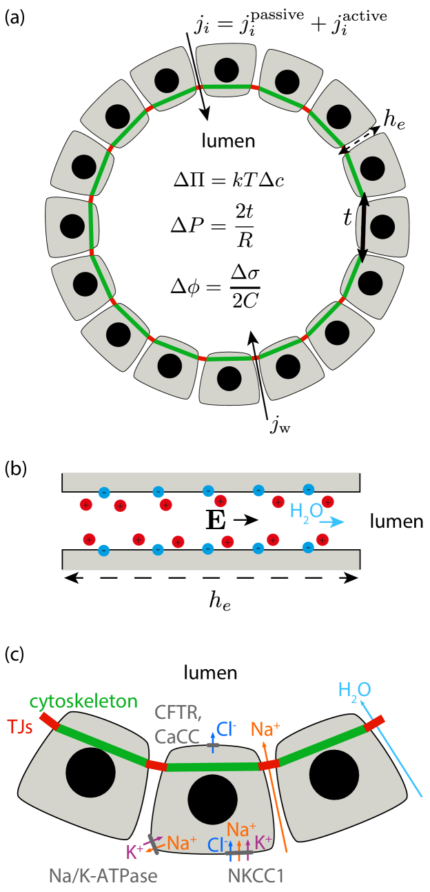
Irreversible thermodynamics of ion and water transport across an epithelium. To discuss the different mechanisms by which water can be transported across an epithelium, here we follow the framework of linear irreversible thermodynamics. In this framework, molecular fluxes are proportional to the chemical potential differences across the layer, with proportionality coefficients characterising the different couplings in the system kedem1963permeability ; de2013non . In the Box “Passive flux of water and ions across a layer” we give further details about the corresponding derivation, including the expression for the chemical potential of water and solutes. Assuming a dilute solution of ion types with concentrations and charges , the volumetric density flux of water towards the lumen is given by the expression
| (3) |
where is the difference of concentration across the layer, i.e. with the concentration in the lumen and the concentration outside, is the average concentration, is the osmotic pressure difference, the difference in electric potential, is the hydrostatic pressure difference, is the permeability of the layer to water and is a cross-coupling coefficient characterising the flow of water that is driven by differences of solute chemical potential across the epithelium (Fig. 2a). The flux is given in units of a permeation velocity. In the right-hand side of Eq. (3), the first two terms correspond to passive water flows due to differences in its chemical potential and to dissipative couplings with other molecules in the solution.
In the last term, we have also included a possible active contribution to water pumping. We are not aware of a known active mechanism of cellular water transport. We note however that apico-basal actomyosin cortical flows could in principle bring such an active contribution, due to friction forces between the paracellular fluid and the cortical cytoskeleton. Such friction forces have been proposed to allow for cellular swimming farutin2019crawling , and friction forces between the actomyosin cortex and the cell cytoplasm is thought to be at the origin of the cytoplasmic flow in the C.elegans embryo, with a velocity µm/s niwayama2011hydrodynamic . This large value compared to reported luminal growth velocities Mosaliganti2019 suggest that such flows could contribute to water pumping, if they exist with the right magnitude and direction.
The flux of solutes is given by
| (4) | ||||
where is the permeability of the layer to solute , and , are cross-coupling coefficients characterising the flow of solute that is driven by differences of chemical potential of water and other solutes, across the epithelium. Here has the usual units of a flux density, i.e. number of particles per unit area and time. As for the water flux, in the right-hand side of Eq. (4), the first three terms describe passive fluxes of solutes, while the last term describes active solute pumping by the epithelium. The coefficients form the matrix of phenomenological coefficients and encode all possible passive interactions between molecules across the layer. We now look at the interpretation of these parameters based on previous studies.
Transepithelial water flux and pressure difference. The epithelial permeability to water is characterised by and encodes the effective permeability to both paracellular flows across the LIS and transcellular flows occurring across the lipid membrane or aquaporins. For MDCK cells µm.s-1.Pa-1 Timbs1996-gt ; Latorre2018 . This number is comparable to typical values of lipid membranes water permeabilities µm.s-1.Pa-1 (corresponding to a membrane permeability coefficient µm.s-1 olbrich2000water ; this value depends on the aquaporin density in the membrane verkman2000structure ). Higher values have been reported for other epithelia ( times larger for the gallbladder and for the kidney proximal tubule fischbarg2010fluid ). During growth of the zebrafish inner ear, the water flux is of the order of µm/h Mosaliganti2019 ; corresponding to a driving pressure difference kPa with the values of reported above for MDCK cells, using Eq. (3) and neglecting cross-coupling coefficients. This relatively high pressure difference, compared to typical cytoskeletal pressures of Pa Salbreux2012 , is however consistent with physiological osmotic pressure differences: using Van’t Hoff law, a difference of concentration of mM across the epithelium yields an osmotic pressure difference kPa. Direct pressure measurements of the intraluminal pressure in the growing zebrafish otic vesicle, using a sensor coupled to a glass needle penetrating into the vesicle, indicate lower values for the excess pressure of the order of Pa Mosaliganti2019 ; similar values for are found in the blastocoel ( Pa) Dumortier2019 , in epithelial domes ( Pa) Latorre2018 , and in MDCK cysts (Pa narayanan2020osmotic which was found to decrease to Pa following inhibition of the CFTR channel). These observations suggest a mode of lumen growth which occurs in the regime .
We also note that the difference of pressure in Eq. (3) is not necessarily constant, and in particular dissipative processes associated to tissue deformation might also contribute to and further slow down, or entirely control, the speed of lumen expansion. Considering the epithelium as a purely fluid layer with two-dimensional viscosity , Laplace’s law leads to with the tension due to the epithelial deformation . For a spherical lumen, using Eq. (3) giving here , one would obtain . Thus, the relative role of tissue viscosity and fluid permeation in controlling the speed of lumen expansion is set by the dimensionless ratio . With a typical 3D tissue viscosity Pa.s marmottant2009role ; guevorkian2010aspiration , with µm the tissue thickness and µm, one obtains , indicating that water permeation can be the dominating dissipative process. More generally the state of the tissue can change as the lumen grows, for instance due to cellular deformations leading to elastic stresses Mosaliganti2019 , cell division and death, or cell volume change hoijman2015mitotic , leading to changes in tissue tension and, as a result of the law of Laplace, excess hydrostatic pressure within the lumen.
Ion permeability of the epithelium. The epithelial ion permeability, relating the ion flux density across the layer to a gradient in its chemical potential, is characterised by and, as well as , it effectively encodes the permeability for both paracellular and transcellular fluxes, including the effect of ion channels, pores, carriers and symporters. This permeability however excludes the effect of pumps: pumps are indeed actively transporting ions by consuming a source of chemical energy such as ATP; we will discuss them later. The paracellular ion permeability depends on the permeability of tight junctions, some of which can be cation/anion selective Krug2014-ef . For instance, tight MDCK monolayers, which are devoid of claudin-2 tight junction proteins that facilitate the permeation of small cations such as Na+ or K+, develop very large transepithelial potentials and very large electric resistance k\textOmegacm2. When transfected with claudin-2-cDNA, the resistance lowers to 150-500 \textOmegacm2 Furuse2001-jo . Measures of are scarce in the literature but an order of magnitude can be obtained from measurements of epithelial electric resistance Dasgupta2018 . Given that is an electric current, is an electric resistance times unit area, which for epithelial monolayers is typically in the range \textOmega.cm2 Sackin2013-fw . Using typical ion concentrations of mM butt2013physics and the electron charge, we can obtain an estimate for the permeability µm/s, in agreement with the permeability of Na+ in MDCK monolayers µm/s Fernandez-Castelo1985-pi .
If one imposes a difference in solute concentration across an epithelium surrounding a lumen of radius , both a water flux and a solute flux results from this difference. As a result of the solute flux, the solute concentration difference decays on a time scale . With the estimate for ion permeability above and µm, this gives equilibration occurring on a time between s and hours. Water fluxes driven by difference of osmotic pressures are sustained on longer time scales if active solute fluxes constantly compensate for this solute leakage, as we discuss below.
In leaky epithelia, with a low electric resistance, the difference of electric potential across the epithelium is small compared to tight epithelia ( mV) Reuss2008-nw ; Sackin2013-fw . Denoting the capacitance per unit area of the epithelium (with order of magnitude pF/µm2 Wegener1996-zc ), the transepithelial potential satisfies , where is the difference of charge density across the epithelium. Assuming that ions equilibrate quickly in the lumen, excess freely-diffusing charges in the lumen will be located in a thin layer covering the epithelium with a typical thickness given by the Debye-Hückel length scale (see Box “Interaction of charged surfaces”); giving rise to the charge difference . Assuming also that the solution in the basal side is electrically neutral, with and the volume and surface area of the lumen.
Ion and water interactions across the epithelium. The cross-coupling coefficients , and , which satisfy and due to the underlying Onsager reciprocal relations, stem from passive interactions between water and ions and different ions across the monolayer.
Cross-couplings between ion transport characterised by the coefficients can be attributed to cotransporters which transport several species simultaneously across the cell membrane. Symporters such as NKCC1 can use favorable differences in chemical potential of some of the transported species (e.g. Na+ in the case of NKCC1) to transport other molecules against their electrochemical potential (e.g. Cl- and K+ in the case of NKCC1 Barrett2000-gt ). The coefficients therefore effectively appear in models for ion transport across the monolayer which incorporate fluxes due to symporters, whose rate of transport is driven by concentration differences of transported ions benjamin1997quantitative ; Gin2007-oc .
The coefficients give rise to the osmotic Staverman’s reflection coefficients, staverman1951theory ; kedem1963permeability . For fully impermeable species which do not cross the epithelium, and the corresponding reflection coefficient . What is the possible origin of the cross coupling coefficients , , in epithelial monolayers? In Xenopus oocytes the Na+/glucose symporter has been shown to cotransport water Loo1996-tl which leads to a nonzero coefficient. Water can also in principle be dragged due to paracellular flows of solutes across paracellular junctions Ussing1989-hd . In the case of an uneven coupling between water with anions and cations, for instance because of symporters cotransporting water, or because some ionic species bind more strongly to the cell surface than others, the theory predicts an electro-osmotic coupling, i.e. a water flux driven by a transepithelial potential , in the absence of an osmotic pressure difference fischbarg2010fluid . In the corneal endothelium, a coupling between water flows and ionic current has been measured with a magnitude µm.cm2/(h µAmp) Sanchez2002-vh . Given that the electric resistance of the endothelium \textOmega.cm2, this leads to a measure for that can be used to obtain e.g. µm4/s.
For comparison, we can discuss the electro-osmotic flux that arises in the intercellular space as a response to an electric field, triggering the motion of the layer of freely diffusing ions located near the charged membrane sarkar2019field (Fig. 2b). We assume that an electric field with the tissue apico-basal height, oriented along the apico-basal axis, is present in the intercellular space. We also assume that cell membranes lining the intercellular space are charged, with the charge characterised by the cell membrane zeta-potential ; and that Na+ is providing the counterions. The electric field then gives rise to a plug flow in the intercellular space, whose velocity is given by the Helmholtz-Smoluchowski formula, , with the water viscosity butt2013physics . The corresponding flux of water across the epithelium is with the volumic fraction of intercellular space in the tissue. Identification with Eq. (3) then gives . With Pa.s, mV bondar2012monitoring , µm, nm and , one finds indeed a comparable value of µm4/s. With these numbers, a difference of electric potential of mV can elicit a water flow with velocity µm/s Mosaliganti2019 ; a sufficiently large number compared to reported flows in lumen formation of µm/h for this effect to play a role in practice. Whether or not these cross-coupling effects contribute to water pumping might depend on the type of epithelium fischbarg2010fluid .
Active solute pumping. The differences in ionic concentrations across the epithelium that drive passive water flows given by the mechanisms described in the previous paragraphs are set by the effect of ionic pumps, which generate an active flux density . In the steady state, the total flux . Although numerous pumping mechanisms in epithelial cells have been reported, see Reuss2008-nw ; Frizzell2012-kx for an extensive review, here we describe the main processes that have been proposed to lead to active transport of ions towards the lumen Frizzell2012-kx ; Navis2015 (Fig. 2c). By hydrolysing ATP, the Na+/K+-ATPase pump transports three Na+ from the cytoplasm to the interstitial fluid and two K+ in the opposite direction per cycle with a pumping rate of the order of s-1 Reuss2008-nw . The activity of the Na+/K+-ATPase pump, as well as its basolateral distribution following the establishment of the cell apico-basal polarity, has been reported to be required for lumen formation, e.g. inhibition of the Na+/K+-ATPase pump by ouabain blocks lumen formation Bagnat2007 . The Na+/K+-ATPase pump generates an electrochemical driving force for the entry of Na+ into the cell, which through symporters such as NKCC1 leads to the import of other ions such as Cl- Barrett2000-gt . Chloride is then exported to the lumen through channels such as CaCC or CFTR. Inhibition/activation of these channels leads to decreased/increased lumen size li2004relationship ; Bagnat2007 . Altogether, this leads to ionic flux of Cl- across the monolayer which is indirectly actively driven. In turn, this generates an electrochemical gradient across the epithelium which draws a passive paracellular flow of Na+, i.e. across the intercellular space, into the lumen. As another possible active mechanism of osmolyte transport to the lumen, exocytic vesicles have been observed concomitantly with lumen initiation in several systems Bryant2010 ; Ryan2019 .
A simple model for the growth of a spherical lumen. We now discuss how the previous ingredients can be used to derive a simple picture for the growth of a spherical lumen. For simplicity, we do not take into account here the different passive cross-coupling terms between water flux and ion chemical potential difference, conversely cross-couplings between ion fluxes and water chemical potential difference, and active water transport. For a spherical lumen of radius , conservation of lumen volume and number of molecules and Eqs. (3) and (4) give for the rate of change of the radius and lumen concentration ,
| (10) | ||||
where , and the difference of hydraulic pressure satisfies Laplace’s law with the surface tension in the monolayer. The surface tension of the monolayer might depend on and through the constitutive law of the monolayer. In Eq. (10) we have made the approximation that the volume change in the lumen is entirely due to the water flux across the epithelium. For simplicity, we can consider to be constant. Assuming a density of µm-2 Na+/K+-ATPase pumps Ewart1995-hy with a pumping rate of s-1 Reuss2008-nw , µm-2 s-1, whose range is consistent with pumping rates measured across MDCK monolayers Cereijido1981-bz ; Simmons1981-ok ; Latorre2018 , as well as net Na+ fluxes measured across the rabbit corneal endothelium lim1982analysis . Similar equations have been used in recent studies Gin2010-wf ; Ruiz-Herrero2017 ; Mosaliganti2019 ; Chan2019 , although the transepithelial potential is not always explicitely considered.
To clarify the discussion, we consider a simple case of a pump-leak mechanism where an anion with charge and concentration is pumped towards the lumen with an active flux density , while a corresponding cation with charge with concentration diffuses through the epithelium passively. The external concentrations are also taken to be fixed, ; and the permeability of the epithelium to both ions taken to be equal, . At low epithelial capacitance and for constant lumen radius , the steady-state concentration differences are and the transepithelial potential ( here as we assume small concentration differences). In this expression, is a characteristic electric potential which is about mV, an order of magnitude comparable to measured transepithelial potentials. With the ranges discussed above µm and µm.s-1, this gives ranging from mM to mM - values towards the end of this range are consistent with physiological concentration of ions of mM butt2013physics .
The equilibrium radius for the lumen is then given by the balance of hydrostatic and osmotic pressure (), giving . Depending on the constitutive law for the monolayer tension and dependency of the active flux density on the lumen size, one expects the corresponding equilibrium lumen size given by the radius to be stable or unstable: e.g. if the epithelial tension is independent of strain and the active flux density is constant, the equilibrium state is unstable as an increase of the radius leads to a decrease in the hydrostatic pressure and further flow towards the lumen; this point is further discussed in the next section. Elastic stresses in the epithelium would generally tend to stabilise the lumen Mosaliganti2019 . Indeed, considering an elastic epithelium with surface tension given by with and the steady-state epithelial tension and radius, a perturbation of the radius and the epithelial area elastic modulus, the pressure change in the lumen due to a change in epithelium radius is given by , which, for , appears as a stabilisation term for lumen growth in Eq. (10). Physically, an increase in lumen radius leads to a positive tension in the epithelium due to elastic stretching, increasing the excess hydrostatic pressure in the lumen and favouring fluid expulsion.
The steady-state relations obtained above indicate that lumens do not need to be perfectly sealed to grow: the balance between the active pumping density flux and leakage with rate can set up a difference of concentration gradient allowing for lumen expansion. Indeed, pumping of ions within a forming cavity leads to a positive osmotic pressure which favours the growth of the cavity and counteracts its tendency to close due to the cortical surface tension Dasgupta2018 . Leakage of ions is opposing the growth of the cavity, due to the reduction of the osmotic pressure associated to ion loss. A dynamic balance between these effects implies that for any value of the leakage magnitude, a threshold rate of pumping can compensate and ensure that the cavity is growing Dasgupta2018 .
In a dynamically growing lumen, the luminal concentrations are also changing by dilution, due to incoming water fluxes. Keeping explicit forms for the flux densities and , the change of concentration in the lumen can indeed be written:
| (11) |
such that a constant luminal concentration can be maintained during lumen expansion if the water and solute fluxes balance according to Mosaliganti2019 . If the role of the hydrostatic pressure difference can be neglected compared to the osmotic pressure difference and the influx of solute is constant, the model then predicts the relaxation to a constant solute concentration in the lumen, and convergence to a linear radius increase .
Flexoelectricity. In this discussion we have incorporated a simple description of electric effects. A recent theoretical work has studied a possible role in lumen formation for flexoelectricity, the establishment of an electric current in the tissue that is sensitive to tissue curvature Duclut2019a . Flexoelectricity can lead to an effective negative surface tension of the inner interface of the tissue facing the lumen, generally favoring growth and allowing for lumen nucleation, even when the osmotic pressure difference is unfavorable to lumen expansion.
II Lumen interactions: Physics of hydraulic flows in tissues
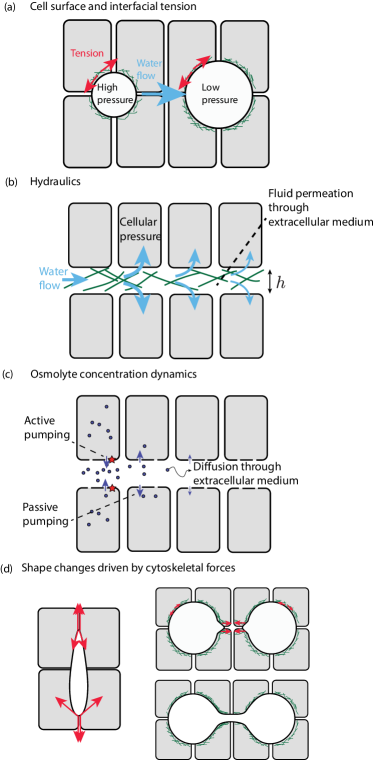
Lumen size instabilities. We now discuss interactions of several lumens in biological tissues. As discussed in the introduction, large lumen formation can occur through intermediate steps involving the growth and fusion of multiple smaller micro-lumens. In recent years several key points on the physics of lumen communication have been made. If lumens are enclosed by cellular interfaces under tension due to the action of the acto-myosin cytoskeleton Salbreux2012 , this results in an excess hydrostatic pressure in the lumen given by the law of Laplace, , with the epithelial surface tension and the radius of curvature. Such configuration is fundamentally unstable if the tension is constant (Fig. 3a): indeed a reduction of the radius of curvature leads to a higher hydrostatic pressure difference , which favours expulsion of the fluid out of the cavity and further reduction of the radius . If the osmotic pressure difference between the lumen and the extracellular space, , is constant, a critical radius determines whether the lumen collapses (for ) or expands indefinitely (for ). By contrast, if the total osmolyte number is fixed inside the lumen, osmotic pressure provides a stabilising effect as constriction of the lumen leads to an increase in osmolyte concentration, and an increase in the lumen osmotic pressure difference , triggering a water flow which restores the lumen initial size.
Because of this fundamental instability, when several spherical cavities subjected to equal surface tension are brought into contact, a flow arises directed from smaller cavities towards larger cavities (Fig. 3a). In binary mixtures that phase separate, a similar physical instability gives rise to Ostwald ripening. In this dynamic process, small droplets of one of the mixture component progressively coarsen through a diffusive flux which favours the growth of bigger droplets at the expense of smaller droplets yao1993theory . As droplets coarsen, the interfacial energy of the mixture is reduced, such that the end result, in a finite system, is a single droplet rich in one component, surrounded by a medium rich in the second component. Similarly, one would expect such a process of lumen coarsening in a tissue to eventually result in a single lumen. This is indeed the dynamics that has been experimentally observed in mouse blastocysts Dumortier2019 . In line with the picture of water flowing from microlumens of high pressure to microlumens of lower pressures, the position of the emerging single lumen can be biased experimentally by mixing cells with higher and lower surface tension, through knock out of Myh9 Dumortier2019 ; as expected the cells surrounding the winning lumen are more likely to have a lower tension. A characteristic scaling law of growth controls Ostwald ripening, such that the mean droplet radius increases with time as for diffusion-limited growth marqusee1983kinetics . It is unclear if a similar scaling relation can also be determined in the process of lumen fusion and coarsening; notably the physics of matter exchange might differ between classical Ostwald ripening and the coarsening of biological lumens Verge-Serandour2021 .
Extracellular water flows. How does water flow in response to gradients of hydrostatic pressure? It seems reasonable to assume that water flows in the extracellular space of a tissue according to Darcy’s law, such that a gradient of hydrostatic pressure in the extracellular fluid, , drives a flow of the extracellular fluid with average velocity , according to the relation Ranft2012 :
| (12) |
where is the coarse-grained cell velocity in the tissue, and is a coefficient of hydraulic resistance. The Darcy equation above describes flows in porous media; here the coefficient of hydraulic resistance is associated to flow of the interstitial fluid through the extracellular matrix or through intercellular spaces in the tissue. If the extracellular fluid is flowing with viscosity in extracellular channels with volumetric density , and each cellular channel is filled with a filamentous mesh with filaments separated by a distance , the hydraulic resistance (de1979scaling , we take into account here a numerical prefactor here corresponding to Poiseuille flow in a tube of radius ). The typical time scale for pressure equilibration between lumens is then ; with a characteristic distance between lumens. With the viscosity of water Pa.s, pN.µm-1Salbreux2012 , nm yurchenco1987basement , and µm, this gives s using . This timescale is roughly in line with the duration of lumen coarsening observed in the formation of the mouse blastocoel Dumortier2019 . We note however that the estimate for this timescale is highly sensitive to the actual value of the extracellular mesh size or gap size between cells, and that non-filamentous extracellular structures, such as linker proteins between cells, could also limit extracellular flows. In the limit where the extracellular space is devoid of extracellular matrix and flows occur in intercellular channels of size separating cells with size , and . In the Xenopus embryonic ectoderm, interstitial gaps at tricellular junctions have been measured to have a characteristic size µm with a characteristic cell size µm Barua2017 ; resulting in faster equilibration time scale s. In addition, other dissipative processes, for instance associated to cellular deformations and rearrangements, might also limit the deformation of lumens in morphogenetic processes.
This analysis of flow between lumens is modified if one takes into account that water can also flow across the cell membrane (Dasgupta2018 , Fig. 3b). The competing processes of flow through the intercellular space and through the cell membrane give rise to a characteristic screening length,
| (13) |
with the width of the extracellular space, and the membrane permeability to water. This length scale arises by combining Darcy’s law, Eq. (12), with an equation for the conservation of matter, , which takes into account losses of fluid through the cell membrane, here assuming cylindrical channels with diameter and a cellular pressure . If one considers a situation with fixed cellular pressure, above this length-scale, extracellular flows are screened by absorption of the fluid into the surrounding cells. What is the order of magnitude of this length scale? With µm and µm.s-1.Pa-1 (discussed in section I), one finds mm. This large length scale ( cell lengths, taking a typical cell diameter of µm) indicates that hydraulic communication between lumens through the extracellular space can be relevant to their dynamics. As noted above, the permeation constant however varies strongly with the extracellular matrix mesh size , and the length scale for the dimension of the intercellular space is also likely to vary significantly, so that the precise value of the length scale could vary between experimental systems. Also, this discussion assumes a constant cellular pressure and therefore does not take into account the possible role of transcellular flows.
Extracellular osmolyte diffusion. What is the dynamics of osmolytes (Fig. 3c)? Osmolytes passively flow in the extracellular space and a diffusion flux should occur to balance concentration in different microlumens. Here as well such diffusive fluxes are screened by exchange with the intracellular space, through pumping of osmolytes across the cell membranes. The corresponding screening length scale can be written Dasgupta2018
| (14) |
with the diffusion constant of osmolytes in the extracellular medium, and the membrane osmolyte permeability. A typical diffusion constant for ions in a tissue is µm2.s-1 suenson1974diffusion . Taking again µm and µm/s, one finds µm. This length scale arises from a competition between diffusion in the extracellular space and reabsorption in the cell. In a thick epithelium, this length scale could limit paracellular ion exchange across the epithelium, as ions would diffuse into the cells before crossing the epithelium. This length scale also determines, in principle, whether neighbouring microlumens share solutes that are pumped towards the lumens.
The length-scale involved in Eq. (14) arises from a similar competition of processes to the diffusion-degradation model involved in morphogen gradient formation and spread, determining a patterning length scale with the morphogen diffusion constant and an effective degradation rate Kicheva2007 . In Eq. (14), osmolytes are assumed to flow passively through the membrane instead of binding to cell membrane receptors and being internalised. In both cases, this length-scale determine the range of cell-cell communication occuring through diffusible molecules. The presence of the factor in Eq. (14) indicates that the length scale increases with the size of the intercellular space. Interestingly microlumens forming in the zebrafish lateral line primordium have been called “luminal hubs”, as cells sharing a common lumen exhibits a coordinated behaviour as a result of FGF concentrating specifically in the shared lumen durdu2014luminal .
Nonspherical lumens. The shape of forming lumens is not necessarily spherical (Fig. 3d): indeed due to the Young-Dupré force balance equation at the junctions between several cells, the interface surfaces can in principle deform away from a spherical shape to establish force balance at the cellular junctions. In liver cells cultured in vitro, early lumens open preferentially towards the tissue interface with the free medium, away from the extracellular matrix Li2016 . This asymmetric lumen growth process has been suggested to arise from differences in junctional tensions across cellular junctions. In the zebrafish otic vesicle, the lumen grows anisotropically hoijman2015mitotic . Such anisotropic shape is initiated early before the lumen starts to expand through the formation of an anisotropic apical surface, and is maintained through differential behaviour of the cells facing the lumens, notably through inhomogeneous epithelial thinning hoijman2015mitotic . In MDCK cysts, the lumen solidity, a geometric measure of shape regularity that compares a given shape to its convex hull, changes with its size, with smaller lumens exhibiting more irregular shapes vasquez2020physical . In general, one expects that lumen with volume close to the cell size are more irregular, due to the role of single cell mechanical effects playing a more visible role at this scale.
Differences in interfacial tension could also in principle drive lumen fusion, bringing lumens into contact for them to undergo coalescence by contact (Fig. 3d). During zebrafish gut development, the intestinal tube forms in several stages, starting with the initiation of multiple small lumens within a solid rod of endodermal cells, followed by lumen fusion and resolution Alvers2014 . This fusion process could occur through de-adhesion of cellular interfaces in the bridge connecting adjacent lumens, of through contraction of the cellular interfaces participating to the bridge Alvers2014 . More generally, coalescence by direct contact (Fig. 3d) or “ripening” based on fluid flows between lumens (Fig. 3a) are two processes that could be responsible for lumen fusion, separately or in combination.
III Conclusion
The physics of lumen formation involves multiple length scales and a complex interplay of hydraulic flows, cell mechanics, and electric and electro-osmotic effects. This topic has attracted significant interest in recent years, as it is becoming evident that the dynamics and mechanics of lumen formation plays a key role in morphogenetic processes during development. Stem cells aggregates have the ability to spontaneous form a lumen, indicating that lumen formation is a fundamental self-organising ability of biological tissues. How this self-organisation occurs is a key question at the interface of physics and biology. The interplay of lumen formation with cellular forces and pumping, but also with cell polarity, morphogen gradient and cell signalling, and electric forces and currents leads to a large range of situations and rich physical behaviours.
Acknowledgement
GS thanks Jacques Prost for discussions and helpful feedback on the manuscript. ATS, MKW, and GS acknowledge support from the Francis Crick Institute, which receives its core funding from Cancer Research UK (FC001317), the UK Medical Research Council (FC001317), and the Wellcome Trust (FC001317).
Competing interests
The authors have no competing interests to declare.
References
- (1) A. Datta, D. M. Bryant, and K. E. Mostov, “Molecular regulation of lumen morphogenesis,” Current Biology, vol. 21, no. 3, pp. R126–R136, 2011.
- (2) S. Sigurbjörnsdóttir, R. Mathew, and M. Leptin, “Molecular mechanisms of de novo lumen formation,” Nature Reviews Molecular Cell Biology, vol. 15, no. 10, pp. 665–676, 2014.
- (3) A. Navis and M. Bagnat, “Developing pressures: fluid forces driving morphogenesis,” Current Opinion in Genetics and Development, vol. 32, pp. 24–30, 2015.
- (4) A. J. Blasky, A. Mangan, and R. Prekeris, “Polarized protein transport and lumen formation during epithelial tissue morphogenesis,” Annual review of cell and developmental biology, vol. 31, pp. 575–591, 2015.
- (5) A. Navis and C. M. Nelson, “Pulling together: Tissue-generated forces that drive lumen morphogenesis,” in Seminars in cell & developmental biology, vol. 55, pp. 139–147, Elsevier, 2016.
- (6) M. Bagnat, I. D. Cheung, K. E. Mostov, and D. Y. Stainier, “Genetic control of single lumen formation in the zebrafish gut,” Nature Cell Biology, vol. 9, no. 8, pp. 954–960, 2007.
- (7) L. A. Lowery and H. Sive, “Initial formation of zebrafish brain ventricles occurs independently of circulation and requires the nagie oko and snakehead/atp1a1a. 1 gene products,” Development, vol. 132, no. 9, pp. 2057–2067, 2005.
- (8) J. Zhang, J. Piontek, H. Wolburg, C. Piehl, M. Liss, C. Otten, A. Christ, T. E. Willnow, I. E. Blasig, and S. Abdelilah-Seyfried, “Establishment of a neuroepithelial barrier by claudin5a is essential for zebrafish brain ventricular lumen expansion,” Proceedings of the National Academy of Sciences, vol. 107, no. 4, pp. 1425–1430, 2010.
- (9) E. Hoijman, D. Rubbini, J. Colombelli, and B. Alsina, “Mitotic cell rounding and epithelial thinning regulate lumen growth and shape,” Nature communications, vol. 6, no. 1, pp. 1–13, 2015.
- (10) I. A. Swinburne, K. R. Mosaliganti, S. Upadhyayula, T. L. Liu, D. G. Hildebrand, T. Y. Tsai, A. Chen, E. Al-Obeidi, A. K. Fass, S. Malhotra, F. Engert, J. W. Lichtman, T. Kirchhausen, E. Betzig, and S. G. Megason, “Lamellar projections in the endolymphatic sac act as a relief valve to regulate inner ear pressure,” eLife, vol. 7, pp. 1–34, 2018.
- (11) K. R. Mosaliganti, I. A. Swinburne, C. U. Chan, N. D. Obholzer, A. A. Green, S. Tanksale, L. Mahadevan, and S. G. Megason, “Size control of the inner ear via hydraulic feedback,” eLife, vol. 8, pp. 1–30, 2019.
- (12) A. Dasgupta, M. Merkel, M. J. Clark, A. E. Jacob, J. E. Dawson, M. L. Manning, and J. D. Amack, “Cell volume changes contribute to epithelial morphogenesis in zebrafish kupffer’s vesicle,” Elife, vol. 7, p. e30963, 2018.
- (13) P. I. Nedvetsky, E. Emmerson, J. K. Finley, A. Ettinger, N. Cruz-Pacheco, J. Prochazka, C. L. Haddox, E. Northrup, C. Hodges, K. E. Mostov, et al., “Parasympathetic innervation regulates tubulogenesis in the developing salivary gland,” Developmental cell, vol. 30, no. 4, pp. 449–462, 2014.
- (14) I. Bedzhov and M. Zernicka-Goetz, “Self-organizing properties of mouse pluripotent cells initiate morphogenesis upon implantation,” Cell, vol. 156, no. 5, pp. 1032–1044, 2014.
- (15) Y. S. Kim, R. Fan, L. Kremer, N. Kuempel-Rink, K. Mildner, D. Zeuschner, L. Hekking, M. Stehling, and I. Bedzhov, “Deciphering epiblast lumenogenesis reveals proamniotic cavity control of embryo growth and patterning,” Science Advances, vol. 7, Mar. 2021.
- (16) M. N. Shahbazi, A. Jedrusik, S. Vuoristo, G. Recher, A. Hupalowska, V. Bolton, N. M. E. Fogarty, A. Campbell, L. G. Devito, D. Ilic, Y. Khalaf, K. K. Niakan, S. Fishel, and M. Zernicka-Goetz, “Self-organization of the human embryo in the absence of maternal tissues.,” Nature Cell Biology, vol. 18, no. 6, pp. 700–8, 2016.
- (17) A. L. Alvers, S. Ryan, P. J. Scherz, J. Huisken, and M. Bagnat, “Single continuous lumen formation in the zebrafish gut is mediated by smoothened-dependent tissue remodeling,” Development (Cambridge), vol. 141, no. 5, pp. 1110–1119, 2014.
- (18) J. G. Dumortier, M. Le Verge-Serandour, A. F. Tortorelli, A. Mielke, L. De Plater, H. Turlier, and J. L. Maître, “Hydraulic fracturing and active coarsening position the lumen of the mouse blastocyst,” Science, vol. 365, no. 6452, pp. 465–468, 2019.
- (19) A. Q. Ryan, C. J. Chan, F. Graner, and T. Hiiragi, “Lumen expansion facilitates epiblast-primitive endoderm fate specification in the mouse blastocyst formation,” Developmental Cell, p. 575282, 2019.
- (20) M. Zernicka-Goetz, “Cleavage pattern and emerging asymmetry of the mouse embryo,” Nature Reviews Molecular Cell Biology, vol. 6, no. 12, pp. 919–928, 2005.
- (21) S. R. Frankenberg, F. R. de Barros, J. Rossant, and M. B. Renfree, “The mammalian blastocyst,” Wiley Interdisciplinary Reviews: Developmental Biology, vol. 5, no. 2, pp. 210–232, 2016.
- (22) J.-L. Maître, “Mechanics of blastocyst morphogenesis,” Biology of the Cell, vol. 109, no. 9, pp. 323–338, 2017.
- (23) Y. Marikawa and V. B. Alarcon, “Creation of trophectoderm, the first epithelium, in mouse preimplantation development,” Results and Problems in Cell Differentiation, vol. 55, pp. 165–184, 2012.
- (24) S. Dasgupta, K. Gupta, Y. Zhang, V. Viasnoff, and J. Prost, “Physics of lumen growth,” Proceedings of the National Academy of Sciences of the United States of America, vol. 115, no. 21, pp. E4751–E4757, 2018.
- (25) A. Manninen, “Epithelial polarity - Generating and integrating signals from the ECM with integrins,” Experimental Cell Research, vol. 334, no. 2, pp. 337–349, 2015.
- (26) A. Z. Wang, G. K. Ojakian, and W. J. Nelson, “Steps in the morphogenesis of a polarized epithelium. I. Uncoupling the roles of cell-cell and cell-substratum contact in establishing plasma membrane polarity in multicellular epithelial (MDCK) cysts,” Journal of Cell Science, vol. 95, no. 1, pp. 137–151, 1990.
- (27) H. Okuyama, J. Kondo, Y. Sato, H. Endo, A. Nakajima, J. M. Piulats, Y. Tomita, T. Fujiwara, Y. Itoh, A. Mizoguchi, M. Ohue, and M. Inoue, “Dynamic change of polarity in primary cultured spheroids of human colorectal adenocarcinoma and its role in metastasis,” American Journal of Pathology, vol. 186, no. 4, pp. 899–911, 2016.
- (28) C. J. Chan, M. Costanzo, T. Ruiz-Herrero, G. Mönke, R. J. Petrie, M. Bergert, A. Diz-Muñoz, L. Mahadevan, and T. Hiiragi, “Hydraulic control of mammalian embryo size and cell fate,” Nature, vol. 571, no. 7763, pp. 112–116, 2019.
- (29) C. Fütterer, C. Colombo, F. Jülicher, and A. Ott, “Morphogenetic oscillations during symmetry breaking of regenerating hydra vulgaris cells,” EPL (Europhysics Letters), vol. 64, no. 1, p. 137, 2003.
- (30) M. Kücken, J. Soriano, P. A. Pullarkat, A. Ott, and E. M. Nicola, “An osmoregulatory basis for shape oscillations in regenerating hydra,” Biophysical Journal, vol. 95, pp. 978–985, 2008.
- (31) T. Ruiz-Herrero, K. Alessandri, B. V. Gurchenkov, P. Nassoy, and L. Mahadevan, “Organ size control via hydraulically gated oscillations,” Development (Cambridge), vol. 144, no. 23, pp. 4422–4427, 2017.
- (32) V. Gebala, R. Collins, I. Geudens, L.-K. Phng, and H. Gerhardt, “Blood flow drives lumen formation by inverse membrane blebbing during angiogenesis in vivo,” Nature Cell Biology, vol. 18, no. 4, pp. 443–450, 2016.
- (33) H.-J. Butt, K. Graf, and M. Kappl, Physics and Chemistry of Interfaces. John Wiley & Sons, 2013.
- (34) T. D. Perez and W. J. Nelson, “Cadherin Adhesion: Mechanisms and Molecular Interactions,” Handbook of Experimental Pharmacology, vol. 5, no. 165, pp. 3–21, 2004.
- (35) P. Panorchan, M. S. Thompson, K. J. Davis, Y. Tseng, K. Konstantopoulos, and D. Wirtz, “Single-molecule analysis of cadherin-mediated cell-cell adhesion,” Journal of Cell Science, vol. 119, no. 1, pp. 66–74, 2006.
- (36) P. Katsamba, K. Carroll, G. Ahlsen, F. Bahna, J. Vendome, S. Posy, M. Rajebhosale, S. Price, T. M. Jessell, A. Ben-Shaul, L. Shapiro, and B. H. Honig, “Linking molecular affinity and cellular specificity in cadherin-mediated adhesion,” Proceedings of the National Academy of Sciences of the United States of America, vol. 106, no. 28, pp. 11594–11599, 2009.
- (37) “The podocalyxin charge was estimated using the amino acid sequence from https://www.uniprot.org/uniprot/o00592, and the protein charge calculator at https://pepcalc.com/protein-calculator.php..”
- (38) D. Kerjaschki, A. T. Vernillo, and M. G. Farquhar, “Reduced sialylation of podocalyxin - The major sialoprotein of the rat kidney glomerulus - in aminonucleoside nephrosis,” American Journal of Pathology, vol. 118, no. 3, pp. 343–349, 1985.
- (39) A. Varki, R. D. Cummings, J. D. Esko, P. Stanley, G. W. Hart, M. Aebi, A. G. Darvill, T. Kinoshita, N. H. Packer, J. H. Prestegard, R. L. Schnaar, and P. H. Seeberger, “Essentials of Glycobiology, Chapter 14,” in Essentials of Glycobiology, Cold Spring Harbor Laboratory Press, 2nd ed., 2009.
- (40) B. Strilić, T. Kučera, J. Eglinger, M. R. Hughes, K. M. McNagny, S. Tsukita, E. Dejana, N. Ferrara, and E. Lammert, “The molecular basis of vascular lumen formation in the developing mouse aorta,” Developmental cell, vol. 17, no. 4, pp. 505–515, 2009.
- (41) B. Strilić, J. Eglinger, M. Krieg, M. Zeeb, J. Axnick, P. Babál, D. J. Müller, and E. Lammert, “Electrostatic cell-surface repulsion initiates lumen formation in developing blood vessels,” Current Biology, vol. 20, no. 22, pp. 2003–2009, 2010.
- (42) J. S. Nielsen and K. M. McNagny, “Novel functions of the cd34 family,” Journal of cell science, vol. 121, no. 22, pp. 3683–3692, 2008.
- (43) T. Takeda, W. Y. Go, R. A. Orlando, and M. G. Farquhar, “Expression of podocalyxin inhibits cell-cell adhesion and modifies junctional properties in Madin-Darby canine kidney cells,” Molecular Biology of the Cell, vol. 11, no. 9, pp. 3219–3232, 2000.
- (44) J. C.-H. Kuo, J. G. Gandhi, R. N. Zia, and M. J. Paszek, “Physical biology of the cancer cell glycocalyx,” Nature Physics, vol. 14, no. 7, pp. 658–669, 2018.
- (45) P. Pincus, “Colloid stabilization with grafted polyelectrolytes,” Macromolecules, vol. 24, no. 10, pp. 2912–2919, 1991.
- (46) J. S. Nielsen, M. L. Graves, S. Chelliah, A. W. Vogl, C. D. Roskelley, and K. M. McNagny, “The cd34-related molecule podocalyxin is a potent inducer of microvillus formation,” PloS one, vol. 2, no. 2, p. e237, 2007.
- (47) S. Durdu, M. Iskar, C. Revenu, N. Schieber, A. Kunze, P. Bork, Y. Schwab, and D. Gilmour, “Luminal signalling links cell communication to tissue architecture during organogenesis,” Nature, vol. 515, no. 7525, pp. 120–124, 2014.
- (48) N. T. Chartier, A. Mukherjee, J. Pfanzelter, S. Furthauer, B. T. Larson, A. W. Fritsch, A. Rana, M. Kreysing, F. Julicher, and S. W. Grill, “A hydraulic instability drives the cell death decision in the nematode germline,” Nature Physics, 2021.
- (49) Q. Li, Y. Zhang, P. Pluchon, J. Robens, K. Herr, M. Mercade, J.-P. Thiery, H. Yu, and V. Viasnoff, “Extracellular matrix scaffolding guides lumen elongation by inducing anisotropic intercellular mechanical tension,” Nature Cell Biology, vol. 18, no. 3, pp. 311–318, 2016.
- (50) J. A. McAteer, A. P. Evan, E. E. Vance, and K. D. Gardner, “MDCK cysts: An in vitro model of epithelial cyst formation and growth,” Journal of Tissue Culture Methods, vol. 10, no. 4, pp. 245–248, 1986.
- (51) W. Yu, A. Datta, P. Leroy, L. E. O’Brien, G. Mak, T. S. Jou, K. S. Matlin, K. E. Mostov, and M. M. Zegers, “1-integrin orients epithelial polarity via Rac1 and laminin,” Molecular Biology of the Cell, vol. 16, no. 2, pp. 433–445, 2005.
- (52) W. Yu, A. M. Shewan, P. Brakeman, D. J. Eastburn, A. Datta, D. M. Bryant, Q. W. Fan, W. A. Weiss, M. M. Zegers, and K. E. Mostov, “Involvement of RhoA, ROCK I and myosin II in inverted orientation of epithelial polarity,” EMBO Reports, vol. 9, no. 9, pp. 923–929, 2008.
- (53) D. M. Bryant, A. Datta, A. E. Rodríguez-Fraticelli, J. Peränen, F. Martín-Belmonte, and K. E. Mostov, “A molecular network for de novo generation of the apical surface and lumen,” Nature Cell Biology, vol. 12, no. 11, pp. 1035–1045, 2010.
- (54) M. Cereijido, J. Ehrenfeld, S. Fernàndez-Castelo, and I. Meza, “Fluxes, junctions, and blisters in cultured monolayers of epithelioid cells (MDCK),” Annals of the New York Academy Sciences, vol. 372, pp. 422–441, 1981.
- (55) C. Tanner, D. A. Frambach, and D. S. Misfeldt, “Transepithelial transport in cell culture. A theoretical and experimental analysis of the biophysical properties of domes,” Biophys. J., vol. 43, pp. 183–190, Aug. 1983.
- (56) S. Fernández-Castelo, J. J. Bolívar, R. López-Vancell, G. Beaty, and M. Cereijido, “Ion Transport in MDCK Cells,” in Tissue Culture of Epithelial Cells (M. Taub, ed.), pp. 37–50, Boston, MA: Springer US, 1985.
- (57) M. M. Timbs and K. R. Spring, “Hydraulic properties of MDCK cell epithelium,” Journal of Membrane Biology, vol. 153, pp. 1–11, Sept. 1996.
- (58) L. Casares, R. Vincent, D. Zalvidea, N. Campillo, D. Navajas, M. Arroyo, and X. Trepat, “Hydraulic fracture during epithelial stretching,” Nature Materials, vol. 14, no. 3, pp. 343–351, 2015.
- (59) E. Latorre, S. Kale, L. Casares, M. Gómez-González, M. Uroz, L. Valon, R. V. Nair, E. Garreta, N. Montserrat, A. del Campo, B. Ladoux, M. Arroyo, and X. Trepat, “Active superelasticity in three-dimensional epithelia of controlled shape,” Nature, vol. 563, no. 7730, pp. 203–208, 2018.
- (60) S. E. Harrison, B. Sozen, N. Christodoulou, C. Kyprianou, and M. Zernicka-Goetz, “Assembly of embryonic and extraembryonic stem cells to mimic embryogenesis in vitro,” Science, vol. 356, no. 6334, p. eaal1810, 2017.
- (61) B. Sozen, G. Amadei, A. Cox, R. Wang, E. Na, S. Czukiewska, L. Chappell, T. Voet, G. Michel, N. Jing, D. M. Glover, and M. Zernicka-goetz, “Self-assembly of embryonic and two extra-embryonic stem cell types into gastrulating embryo-like structures,” Nature Cell Biology, vol. 20, no. August, 2018.
- (62) B. Sozen, A. L. Cox, J. De Jonghe, M. Bao, F. Hollfelder, D. M. Glover, and M. Zernicka-Goetz, “Self-Organization of Mouse Stem Cells into an Extended Potential Blastoid,” Developmental Cell, vol. 51, no. 6, pp. 698–712.e8, 2019.
- (63) M. Mole, A. Weberling, S. Fishel, M. Zernicka-goetz, A. Weberling, and R. Fa, “Integrin b1 coordinates survival and morphogenesis of the embryonic lineage upon implantation and pluripotency transition,” Cell Reports, vol. 34, 2021.
- (64) M. N. Shahbazi, A. Scialdone, N. Skorupska, A. Weberling, G. Recher, M. Zhu, A. Jedrusik, L. G. Devito, L. Noli, I. C. MacAulay, C. Buecker, Y. Khalaf, D. Ilic, T. Voet, J. C. Marioni, and M. Zernicka-Goetz, “Pluripotent state transitions coordinate morphogenesis in mouse and human embryos,” Nature, vol. 552, no. 7684, pp. 239–243, 2017.
- (65) Q. Yang, S.-L. Xue, C. J. Chan, M. Rempfler, D. Vischi, F. M. Gutierrez, T. Hiiragi, E. Hannezo, and P. Liberali, “Cell fate coordinates mechano-osmotic forces in intestinal crypt morphogenesis,” bioRxiv, 2020.
- (66) N. P. Tallapragada, H. M. Cambra, T. K. Wald, S. Jalbert, D. M. Abraham, O. D. Klein, and A. M. Klein, “Inflation-collapse dynamics drive patterning and morphogenesis in intestinal organoids,” Cell Stem Cell, vol. 28, pp. 1–17, 2021.
- (67) N. Sachs, Y. Tsukamoto, P. Kujala, P. J. Peters, and H. Clevers, “Intestinal epithelial organoids fuse to form self-organizing tubes in floating collagen gels,” Development, vol. 144, pp. 1107–1112, 2017.
- (68) D. K. Marciano, “A holey pursuit: lumen formation in the developing kidney,” Pediatric Nephrology, vol. 32, no. 1, pp. 7–20, 2017.
- (69) S. K. Muthuswamy, D. Li, S. Lelievre, M. J. Bissell, and J. S. Brugge, “Erbb2, but not erbb1, reinitiates proliferation and induces luminal repopulation in epithelial acini,” Nature Cell Biology, vol. 3, no. 9, pp. 785–792, 2001.
- (70) R. Halaoui, C. Rejon, S. J. Chatterjee, J. Szymborski, S. Meterissian, W. J. Muller, A. Omeroglu, and L. McCaffrey, “Progressive polarity loss and luminal collapse disrupt tissue organization in carcinoma,” Genes & Development, vol. 31, no. 15, pp. 1573–1587, 2017.
- (71) A. Alladin, L. Chaible, L. G. del Valle, R. Sabine, M. Loeschinger, M. Wachsmuth, J. K. Hériché, C. Tischer, and M. Jechlinger, “Tracking cells in epithelial acini by light sheet microscopy reveals proximity effects in breast cancer initiation,” eLife, vol. 9, pp. 1–20, 2020.
- (72) N. Ashley, T. M. Yeung, and W. F. Bodmer, “Stem cell differentiation and lumen formation in colorectal cancer cell lines and primary tumors,” Cancer Research, vol. 73, no. 18, pp. 5798–5809, 2013.
- (73) O. Kedem and A. Katchalsky, “Permeability of composite membranes. part 1.—electric current, volume flow and flow of solute through membranes,” Transactions of the Faraday Society, vol. 59, pp. 1918–1930, 1963.
- (74) S. R. De Groot and P. Mazur, Non-equilibrium thermodynamics. Courier Corporation, 2013.
- (75) A. Farutin, J. Étienne, C. Misbah, and P. Recho, “Crawling in a fluid,” Physical Review Letters, vol. 123, no. 11, p. 118101, 2019.
- (76) R. Niwayama, K. Shinohara, and A. Kimura, “Hydrodynamic property of the cytoplasm is sufficient to mediate cytoplasmic streaming in the caenorhabiditis elegans embryo,” Proceedings of the National Academy of Sciences, vol. 108, no. 29, pp. 11900–11905, 2011.
- (77) K. Olbrich, W. Rawicz, D. Needham, and E. Evans, “Water permeability and mechanical strength of polyunsaturated lipid bilayers,” Biophysical Journal, vol. 79, no. 1, pp. 321–327, 2000.
- (78) A. Verkman and A. K. Mitra, “Structure and function of aquaporin water channels,” American Journal of Physiology-Renal Physiology, vol. 278, no. 1, pp. F13–F28, 2000.
- (79) J. Fischbarg, “Fluid transport across leaky epithelia: central role of the tight junction and supporting role of aquaporins,” Physiological Reviews, vol. 90, no. 4, pp. 1271–1290, 2010.
- (80) G. Salbreux, G. Charras, and E. Paluch, “Actin cortex mechanics and cellular morphogenesis,” Trends in Cell Biology, vol. 22, no. 10, pp. 536–545, 2012.
- (81) V. Narayanan, L. E. Schappell, C. R. Mayer, A. A. Duke, T. J. Armiger, P. T. Arsenovic, A. Mohan, K. N. Dahl, J. P. Gleghorn, and D. E. Conway, “Osmotic gradients in epithelial acini increase mechanical tension across e-cadherin, drive morphogenesis, and maintain homeostasis,” Current Biology, vol. 30, no. 4, pp. 624–633, 2020.
- (82) P. Marmottant, A. Mgharbel, J. Käfer, B. Audren, J.-P. Rieu, J.-C. Vial, B. Van Der Sanden, A. F. Marée, F. Graner, and H. Delanoë-Ayari, “The role of fluctuations and stress on the effective viscosity of cell aggregates,” Proceedings of the National Academy of Sciences, vol. 106, no. 41, pp. 17271–17275, 2009.
- (83) K. Guevorkian, M.-J. Colbert, M. Durth, S. Dufour, and F. Brochard-Wyart, “Aspiration of biological viscoelastic drops,” Physical Review Letters, vol. 104, no. 21, p. 218101, 2010.
- (84) S. M. Krug, J. D. Schulzke, and M. Fromm, “Tight junction, selective permeability, and related diseases,” Semin. Cell Dev. Biol., vol. 36, pp. 166–176, Dec. 2014.
- (85) M. Furuse, K. Furuse, H. Sasaki, and S. Tsukita, “Conversion of zonulae occludentes from tight to leaky strand type by introducing claudin-2 into Madin-Darby canine kidney I cells,” J. Cell Biol., vol. 153, pp. 263–272, Apr. 2001.
- (86) H. Sackin and L. G. Palmer, “Chapter 7 - Electrophysiological Analysis of Transepithelial Transport,” in Seldin and Giebisch’s The Kidney (Fifth Edition) (R. J. Alpern, O. W. Moe, and M. Caplan, eds.), pp. 177–216, Academic Press, Jan. 2013.
- (87) L. Reuss, “Chapter 2 - Mechanisms of Ion Transport Across Cell Membranes and Epithelia,” in Seldin and Giebisch’s The Kidney (Fourth Edition) (R. J. Alpern and S. C. Hebert, eds.), pp. 35–56, San Diego: Academic Press, Jan. 2008.
- (88) J. Wegener, M. Sieber, and H. J. Galla, “Impedance analysis of epithelial and endothelial cell monolayers cultured on gold surfaces,” Journal of Biochemical and Biophysical Methods, vol. 32, pp. 151–170, July 1996.
- (89) K. E. Barrett and S. J. Keely, “Chloride secretion by the intestinal epithelium: molecular basis and regulatory aspects,” Annual Reviews of Physiology, vol. 62, pp. 535–572, 2000.
- (90) B. Benjamin and E. Johnson, “A quantitative description of the na-k-2cl cotransporter and its conformity to experimental data,” American Journal of Physiology-Renal Physiology, vol. 273, no. 3, pp. F473–F482, 1997.
- (91) E. Gin, E. J. Crampin, D. A. Brown, T. J. Shuttleworth, D. I. Yule, and J. Sneyd, “A mathematical model of fluid secretion from a parotid acinar cell,” Journal of Theoretical Biology, vol. 248, pp. 64–80, Sept. 2007.
- (92) A. Staverman, “The theory of measurement of osmotic pressure,” Recueil des Travaux Chimiques des Pays-Bas, vol. 70, no. 4, pp. 344–352, 1951.
- (93) D. D. Loo, T. Zeuthen, G. Chandy, and E. M. Wright, “Cotransport of water by the Na+/glucose cotransporter,” Proceedings of the National Academy of Sciences of the United States of America, vol. 93, pp. 13367–13370, Nov. 1996.
- (94) H. H. Ussing and K. Eskesen, “Mechanism of isotonic water transport in glands,” Acta Physiologica Scandinavica, vol. 136, pp. 443–454, July 1989.
- (95) J. M. Sánchez, Y. Li, A. Rubashkin, P. Iserovich, Q. Wen, J. W. Ruberti, R. W. Smith, D. Rittenband, K. Kuang, F. P. J. Diecke, and J. Fischbarg, “Evidence for a Central Role for Electro-Osmosis in Fluid Transport by Corneal Endothelium,” Journal of Membrane Biology, vol. 187, pp. 37–50, May 2002.
- (96) N. Sarkar, J. Prost, and F. Jülicher, “Field induced cell proliferation and death in a model epithelium,” New Journal of Physics, vol. 21, no. 4, p. 043035, 2019.
- (97) O. V. Bondar, D. Saifullina, I. Shakhmaeva, I. Mavlyutova, and T. Abdullin, “Monitoring of the zeta potential of human cells upon reduction in their viability and interaction with polymers,” Acta Naturae, vol. 4, no. 1 (12), 2012.
- (98) R. A. Frizzell and J. W. Hanrahan, “Physiology of epithelial chloride and fluid secretion,” Cold Spring Harbor Perspectives in Medicine, vol. 2, p. a009563, June 2012.
- (99) H. Li, I. A. Findlay, and D. N. Sheppard, “The relationship between cell proliferation, cl- secretion, and renal cyst growth: a study using cftr inhibitors,” Kidney International, vol. 66, no. 5, pp. 1926–1938, 2004.
- (100) H. S. Ewart and A. Klip, “Hormonal regulation of the Na(+)-K(+)-ATPase: mechanisms underlying rapid and sustained changes in pump activity,” American Journal of Physiology, vol. 269, pp. C295–311, Aug. 1995.
- (101) N. L. Simmons, “Ion transport in ’tight’ epithelial monolayers of MDCK cells,” Journal of Membrane Biology, vol. 59, pp. 105–114, Apr. 1981.
- (102) J. J. Lim and H. H. Ussing, “Analysis of presteady-state na+ fluxes across the rabbit corneal endothelium,” The Journal of Membrane Biology, vol. 65, no. 3, pp. 197–204, 1982.
- (103) E. Gin, E. M. Tanaka, and L. Brusch, “A model for cyst lumen expansion and size regulation via fluid secretion,” Journal of Theoretical Biology, vol. 264, pp. 1077–1088, June 2010.
- (104) C. Duclut, N. Sarkar, J. Prost, and F. Jülicher, “Fluid pumping and active flexoelectricity can promote lumen nucleation in cell assemblies,” Proceedings of the National Academy of Sciences of the United States of America, vol. 116, no. 39, pp. 19264–19273, 2019.
- (105) J. H. Yao, K. Elder, H. Guo, and M. Grant, “Theory and simulation of ostwald ripening,” Physical Review B, vol. 47, no. 21, p. 14110, 1993.
- (106) J. Marqusee and J. Ross, “Kinetics of phase transitions: Theory of ostwald ripening,” The Journal of Chemical Physics, vol. 79, no. 1, pp. 373–378, 1983.
- (107) M. Verge-Serandour and H. Turlier, “A hydro-osmotic coarsening theory of biological cavity formation,” bioRxiv, 2021.
- (108) J. Ranft, J. Prost, F. Jülicher, and J.-F. Joanny, “Tissue dynamics with permeation,” European Physical Journal E, vol. 35, no. 46, 2012.
- (109) P.-G. De Gennes and P.-G. Gennes, Scaling concepts in polymer physics. Cornell university press, 1979.
- (110) P. D. Yurchenco and G. C. Ruben, “Basement membrane structure in situ: evidence for lateral associations in the type iv collagen network.,” The Journal of Cell Biology, vol. 105, no. 6, pp. 2559–2568, 1987.
- (111) D. Barua, S. E. Parent, and R. Winklbauer, “Mechanics of fluid-filled interstitial gaps. ii. gap characteristics in xenopus embryonic ectoderm,” Biophysical Journal, vol. 113, pp. 923–936, 2017.
- (112) M. Suenson, D. Richmond, and J. Bassingthwaighte, “Diffusion of sucrose, sodium, and water in ventricular myocardium,” American Journal of Physiology-Legacy Content, vol. 227, no. 5, pp. 1116–1123, 1974.
- (113) A. Kicheva, P. Pantazis, T. Bollenbach, Y. Kalaidzidis, T. Bittig, F. Jülicher, and M. González-Gaitán, “Kinetics of morphogen gradient formation,” Science, vol. 315, no. 5811, pp. 521–525, 2007.
- (114) C. G. Vasquez, V. T. Vachharajani, C. Garzon-Coral, and A. R. Dunn, “Physical basis of lumen shape and stability in a simple epithelium,” BioRxiv, p. 746792, 2020.