Polarization-Resolved Extreme Ultraviolet Second Harmonic Generation from LiNbO3
Abstract
Second harmonic generation (SHG) spectroscopy ubiquitously enables the investigation of surface chemistry, interfacial chemistry as well as symmetry properties in solids. Polarization-resolved SHG spectroscopy in the visible to infrared regime is regularly used to investigate electronic and magnetic orders through their angular anisotropies within the crystal structure. However, the increasing complexity of novel materials and emerging phenomena hamper the interpretation of experiments solely based on the investigation of hybridized valence states. Here, polarization-resolved SHG in the extreme ultraviolet (XUV-SHG) is demonstrated for the first time, enabling element-resolved angular anisotropy investigations. In non-centrosymmetric LiNbO3, elemental contributions by lithium and niobium are clearly distinguished by energy dependent XUV-SHG measurements. This element-resolved and symmetry-sensitive experiment suggests that the displacement of Li ions in LiNbO3, which is known to lead to ferroelectricity, is accompanied by distortions to the Nb ion environment that breaks the inversion symmetry of the NbO6 octahedron as well. Our simulations show that the measured second harmonic spectrum is consistent with Li ion displacements from the centrosymmetric position by 0.5 while the Nb-O bonds are elongated/contracted by displacements of the O atoms by 0.1 . In addition, the polarization-resolved measurement of XUV-SHG shows excellent agreement with numerical predictions based on dipole-induced SHG commonly used in the optical wavelengths. This constitutes the first verification of the dipole-based SHG model in the XUV regime. The findings of this work pave the way for future angle and time-resolved XUV-SHG studies with elemental specificity in condensed matter systems.
Nonlinear spectroscopies have become an indispensable tool to characterize material properties and dynamics. Second-order optical nonlinearities proportional to the second-order susceptibility, , are especially relevant due to their distinct selection rules beyond the angular momentum selection rule. Factors such as the bulk symmetry of the crystal and the presence of an inversion center determines whether second-order susceptibilities are nonzeroBoyd (2008). Owing to these properties, second-order nonlinear spectroscopies, such as second harmonic generation (SHG) with optical and infrared wavelengths, is widely used as an interfacial and surface probe of electronic properties of solid state materials Heinz et al. (1985).
Due to the nonlinear process, a high electric field strength is needed to generate experimentally detectable SHG signals. In this regard, advances in pulsed laser sources with a high peak field strength greatly accelerated the adoption of this technique. In the optical regime, besides direct measurement of SHG, the angular anisotropy of SHG is often used to characterize electronic and magnetic orders in solids, offering an ultra-sensitive probe of crystalline symmetry Torchinsky and Hsieh (2017). For example, SHG angular anisotropy has been used to characterize the symmetries of ferroic materials Denev et al. (2008); Padmanabhan et al. (2018), multipolar order Jin et al. (2020), and chiral structures Fiebig et al. (2005); Fichera et al. (2020). In systems with strong electron correlations – from unconventional superconductors to quantum spin liquid – the sensitivity afforded by SHG has also revealed important phases that elude previous investigations Harter et al. (2017); Zhao et al. (2017); Laurita et al. (2019). Free-electron lasers (FELs) present a similar opportunity to extend the capabilities of SHG into the extreme ultraviolet (XUV) and soft X-ray regime Lam et al. (2018). Recently, nonlinear X-ray and XUV spectroscopies have been the subjects of both theoretical Minerbi and Shwartz (2019) and experimental Glover et al. (2012); Szlachetko et al. (2016); Beye et al. (2019); Bohinc et al. (2019); Shwartz et al. (2014) works as a result of the available high intensity light sources. Among these techniques, XUV-SHG is particularly attractive as the core-level specificity of XUV radiation and the unique selection rules of SHG can be united. Further, short pulse durations pushing to the attosecond regime that are available at the current and upcoming generation of FELs is expected to enable XUV-SHG studies with exceptional time resolution Duris et al. (2020). Experimentally, distinct elemental edges can be probed using XUV-SHG Lam et al. (2018); Yamamoto et al. (2018). Following these proof of principle works, XUV-SHG was also shown to probe material properties with high sensitivity to the chemical environment around the select elements Berger et al. (2020) and was demonstrated as a spectroscopic tool capable of probing buried interfaces Schwartz et al. (2020).
In contrast to optical SHG that measures an average response across the elements forming the valence orbitals, XUV-SHG probes the element-specific core level states, allowing the separation of elemental contributions in the measured anisotropies. The elemental specificity of XUV-SHG is particularly attractive for materials where emergent behavior is rooted in single ion displacements in the unit cell. For instance, in ferroelectric materials, a spontaneous polarization forms as a result of unit cell distortions that break the inversion symmetry. In particular for the ferroelectric material studied in this article, LiNbO3, the spontaneous polarization establishes as a result of Li displacement relative to the Nb-O octahedron in the unit cell.
Here, polarization-resolved XUV-SHG is demonstrated for the first time and applied to study the nature of symmetry-breaking ion displacement in ferroelectric LiNbO3. Spectroscopy measurements covering the Li and Nb edges were conducted in concert with polarization-resolved studies at an FEL. Resonant features relating to the two elements are observed and assigned using ab initio density functional perturbation theory (DFPT). The angular anisotropy of a selected resonance is resolved which is well reproduced by the theory of nonlinear polarization based on the DFPT calculation.
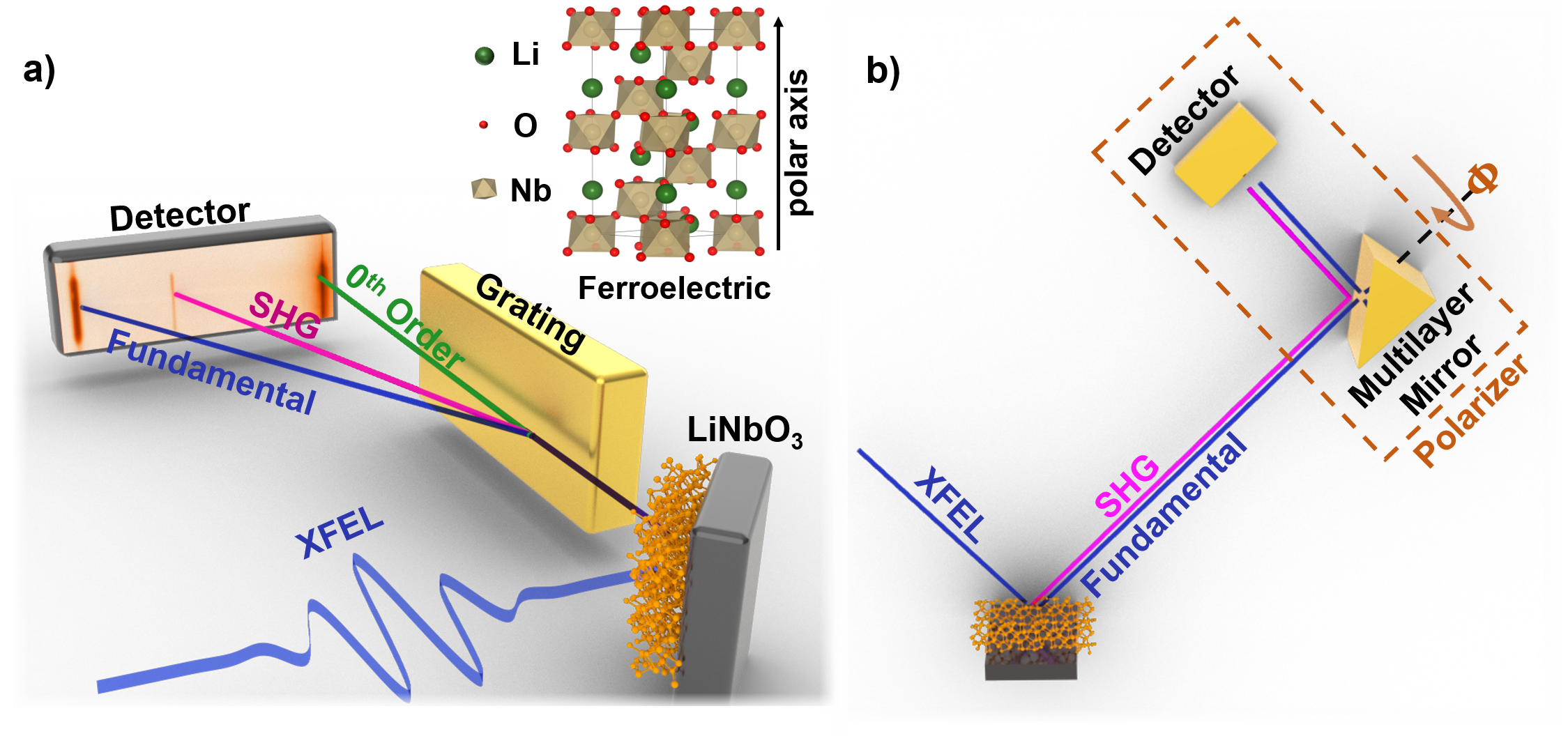
The experiments were performed at the BL1 of SACLA in Japan Owada et al. (2018). A 30-fs -polarized FEL pulse was tuned to energies between 28 eV and 33 eV with 0.5 eV steps and incident on an -cut LiNbO3 crystal (Fig. S5) at 45∘ with respect to the sample surface. The incoming photon energies are referred as the fundamental in the remaining text. The second harmonic response of the sample was analyzed in two separate experiments.
In the first experiment, as illustrated in Fig. 1(a), the second harmonic and the reflected fundamental were dispersed by a grating (1200 groove/mm, 30-002, Shimadzu) and captured using a microchannel plate detector (MCP) (Rectangular, Hamamatsu Photonics) coated with CsI. The images of the detector were captured with a camera (IPX-VGA120-LMCN, Imperx Inc.). This measurement was used to retrieve the second order susceptibility spectrum, . We emphasize that both the fundamental and the second harmonic light were simultaneously recorded on the detector, allowing a comparison of the shot-to-shot variation in the photon flux and energy. The photon energy jitter was approximately 0.2% of the fundamental frequency and was leveraged to increase the spectral resolution (see Supplementary Material Section S1). Shot-to-shot fluctuations in the photon flux of the fundamental was used to extract the second-order susceptibility [Fig. S2(b)].
In the second experiment [Fig. 1(b)], the polarization of the second harmonic was investigated with an XUV polarizer. Specifically, the second harmonic emitted from the sample was reflected off a multilayer mirror at Brewster’s angle, such that only the -polarized portion of the incident light was reflected. The reflected light was then detected by an MCP. The multilayer mirror and the MCP were rotated azimuthally around the beam axis [ in Fig. 1(b)] between to in 3∘ steps with respect to the laboratory frame. As the multilayer mirror was rotated, the portion of light -polarized with respect to the mirror surface with the new orientation is refracted into the mirror substrate and absorbed, while the -polarized component is reflected. The multilayer mirror was coated such that the second harmonic is preferentially reflected over the fundamental, making the separation of the two possible Attwood (1999). Nonetheless, a small fraction of fundamental was able to reach the MCP due to non-perfect extinction on the multilayer mirror. Inspecting Fig. S3, one can estimate that the fundamental and second harmonic intensities are approximately the same order of magnitude. As a result of the residual fundamental at the detector, the polarization of the fundamental and the second harmonic can be resolved simultaneously.
A detailed analysis procedure for the spectral data is presented in the Section S1 of the Supplementary Material. Briefly, each FEL shot imprints a 2D image on the MCP detector. Each image contains peaks associated with the incident fundamental and the emitted second harmonic at their respective frequencies. The quality of each shot was determined by fitting a Gaussian to the fundamental peak and assessing the quality of the fit. Approximately 5% of all shots at each energy were discarded on the basis of R2 values less than . The remaining shots were binned with respect to the fundamental energy and intensity. The second order susceptibility at each energy was extracted using the quadratic relationship between the fundamental intensity and its second harmonic.
The detector in the polarization-resolved experiment measures both the reflected fundamental and the emitted second harmonic as a function of angle as shown in Fig. 1(b). A detailed procedure for the data analysis steps for the polarization experiment is presented in the Supplementary Material Section S2. Briefly, the fundamental and the second harmonic response were separated by a linear background subtraction. At lower incident fundamental intensities, the measured voltage was linear with respect to the intensity of the incident fundamental while at higher incident fundamental intensities a quadratic relationship is observed.
These experiments were corroborated by theoretical calculations. The linear response of LiNbO3 was simulated with first-principles density functional theory Hohenberg and Kohn (1964) using the excitingGulans et al. (2014) full potential all electron augmented linearized planewave package. Both centrosymmetric and noncentrosymmetric structures of LiNbO3 were investigated. The Brillouin zone was sampled with a -point centered -point grid within the local density approximation Perdew and Zunger (1981). The Li core 1 and Nb semi-core 4 and 4 electrons were included in the self-consistent field calculation loop to extract their respective linear responses. The excited states of the system were accessed through time-dependent density functional theory (TDDFT) simulations using the random phase approximation (RPA) kernel implemented within excitingVast et al. (2002); Sagmeister and Ambrosch-Draxl (2009) with a -point chosen to be the same as that of the aforementioned k-point grid. Plots of imaginary part of dielectric function are shown in Fig. S12. No characteristic differences to the linear dielectric response were found between the non-polar and polar phases. The experimental and theoretical results show good agreement except the overestimation of the first peak at 36 eV as shown in Fig. S12, which is a common trait in the level of theory used here. To properly sample the peak, many-body effects would need to be included, but is beyond the scope of the present work and not necessary for the current level of analysis. The level of theory employed in the linear response calculation was kept at the same level as the second harmonic response calculation for consistency and ease of comparison. The second harmonic response formalism by SharmaSharma and Ambrosch-Draxl (2004) implemented within excitingGulans et al. (2014) was used to calculate the second order susceptibility of LiNbO3. Here, lifetime broadening was also employed to account for the high oscillatory behavior of high-energy states as detailed in the work of LamLam et al. (2018). A total of 120 empty states were included to account for excitation up to double of the incoming photon frequency. Molecular dynamics simulations were performed to correctly reflect the finite temperature of the system (see SI Section S4 for details).
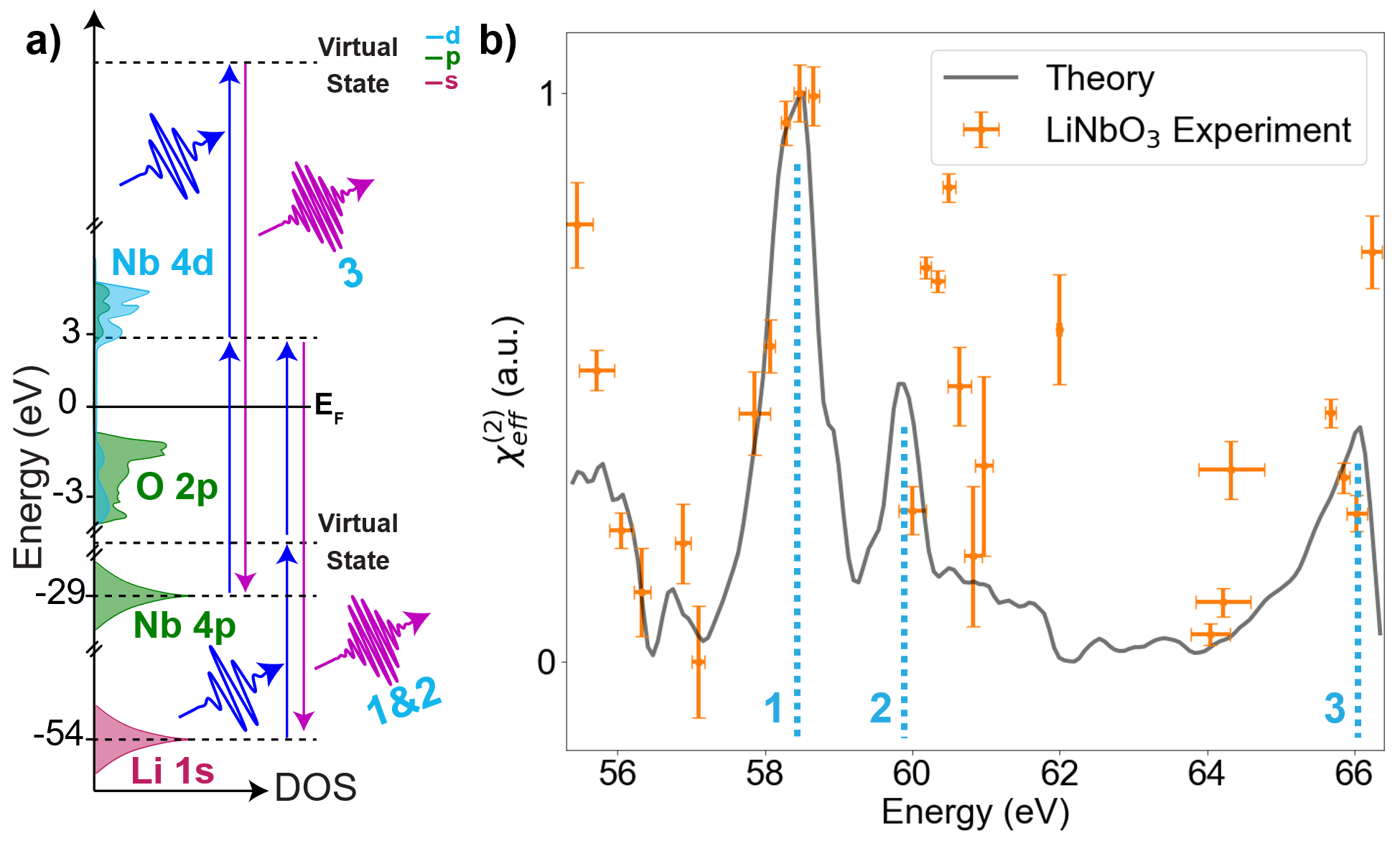
Considering the electronic density of states, the core level transitions that can be accessed by XUV-SHG is schematically shown in Fig. 2(a). Half resonant transitions from Li 1 core states to the conduction band states with majority Nb 4 character, along with resonant transitions originating from Nb 4 core-like states fall within the range of the fundamental energies studied. Though the availability of density of states is vital for observing XUV-SHG, it is not the only factor that governs the transition probability. Experimentally, the measured second order susceptibility spectrum, , is a direct measurement of the allowed transitions within the selection rules of XUV-SHG. In Fig. 2(b) the measured is overlaid with the theoretically calculated spectrum for LiNbO3. The theoretically calculated spectrum is derived as a weighted sum of the individual tensor elements of the C3v point group under consideration of the experimental geometry (see SI Section S3 for details). The two half-resonant features around 58 eV correspond to transitions from Li 1 to conduction band states of majority Nb 4 character. These features report on the ferroelectric displacement in the unit cell involving the Li ions. Our TDDFT calculations (SI Section S4) are consistent with an inversion symmetry breaking displacement of Li ions by .5 . The resonant feature at 58 eV is similar to the feature observed in a previous XUV-SHG study on LiOsO3Berger et al. (2020). The previous study reported an enhancement of the amplitude with inversion symmetry breaking in the unit cell by Li displacements. In fact, the behavior of the two Li containing compounds with ferroelectric displacements, LiNbO3 and LiOsO3 is very similar at this energy range. The key difference in the spectra is highlighted in Fig. 2(b) as transition #3. This highlighted feature in Fig. 2(b) stems from half-resonant transitions from the Nb edge, pointing towards inversion symmetry breaking not only around the Li ion but also the Nb ion. Molecular dynamics simulations of the Nb ion environment in the presence of the ferroelectric displacement show that the amplitude of at eV is consistent with a modulation of the Nb-O bonds by 0.1. The molecular dynamics simulations also show that antiferrodistortive rotations of the NbO6 does not substantially contribute to the amplitude of the spectrum shown in Fig. 2(b). This result is consistent with a picture of ferroelectric displacement of the Li ion that is stabilized by distortions to the NbO6 octahedra that break the inversion symmetry around the Nb ion in the unit cell Toyoura et al. (2015); Barker and Loudon (1967); Inbar and Cohen (1996, 1997). No evidence of such inversion symmetry breaking around the Os ion in LiOsO3 was observed Berger et al. (2020). This contrast between the two cases demonstrates the strength of the unique selection rules of XUV-SHG and is a direct visualization of element specificity of the method.
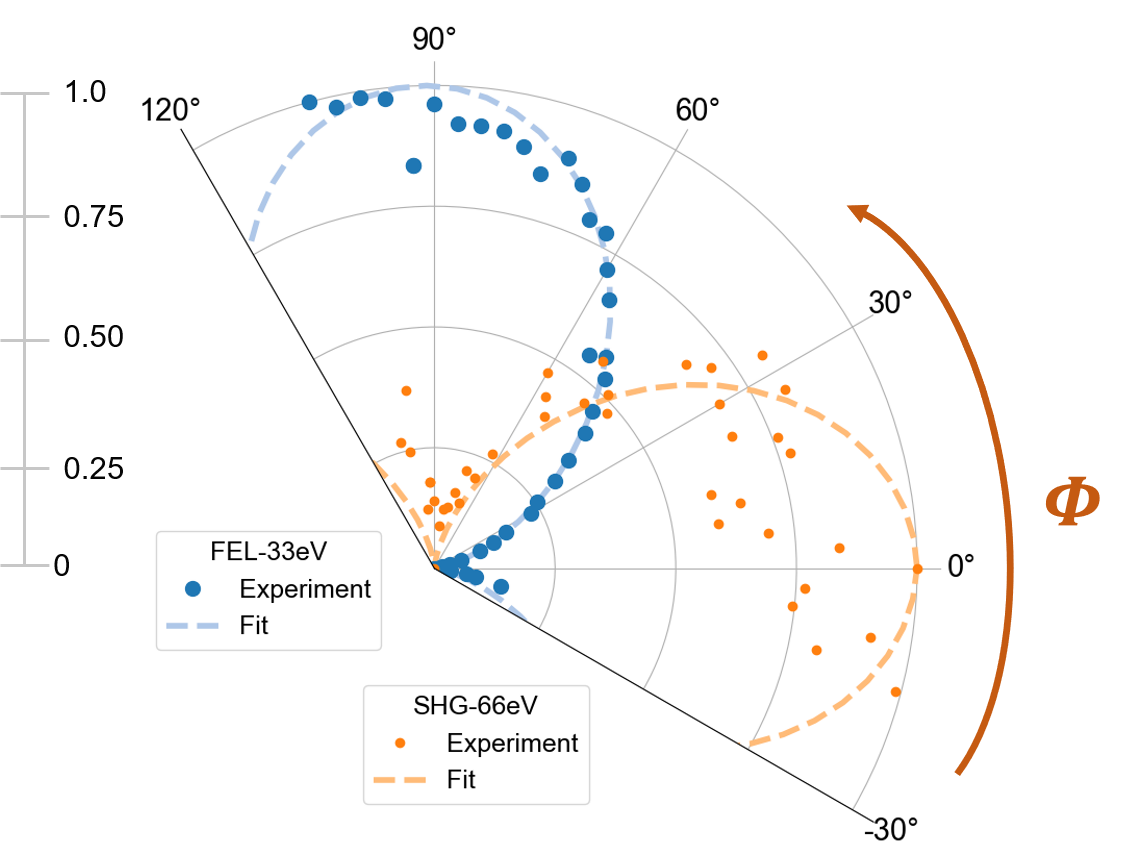
To further demonstrate the capabilities of energy-resolved SHG, we conducted polarization-resolved measurements at a single energy. Along with the fundamental ( eV) that initiates SHG, the polarization of SHG at eV is resolved and shown as a polar plot in Fig. 3. The expected -polarization of the fundamental is recovered while the second harmonic is measured to be -polarized. Using the well established arguments for the crystal symmetry and a detailed tensor analysis, the polarization of the second harmonic can be calculated and decomposed into four distinct channels characterized by the polarization of the incident fundamental and the detection channel of the polarization. All four cases are shown in Fig. S8 as a function of in plane rotation of -cut and -cut LiNbO3. The calculated polar plots for the 4 distinct channels of polarization ( in out, in out, in out, in out) show distinct patterns demonstrating the symmetry of the crystal. Note that, as expected, the 3-fold symmetry of the bulk crystal is reproduced by the polar plots for the -cut of the crystal. For -cut LiNbO3, when it is incident with a -polarized fundamental, the majority of the emitted second harmonic is calculated to be -polarized [Fig. S7(b) & (c)] at eV, in excellent agreement with the measured polarization of the second harmonic. The emitted -polarized SHG constitutes the emission of the orthogonal polarization to the polarization of the fundamental. The emission of the orthogonal polarization can be attributed to the energy dependent tensor as the emission of -polarized SHG is indeed symmetry allowed as shown in Fig. S7(b) & (c) for a different XUV energy. The observed -polarized SHG, thus cannot be solely explained by symmetry considerations and highlights the nonlinear nature of the light-matter interaction in this regime.
The findings presented here demonstrate the feasibility of an XUV-SHG study with angular resolution and the possibility to extend SHG rotational anisotropy studies into XUV wavelengths. The long established theory of nonlinear polarization up to the dipole contributions is shown to be adequate to explain the measured polarization for a bulk noncentrosymmetric material in the extreme ultraviolet. Our spectral findings suggest that inversion symmetry breaking in the ferroelectric phase of LiNbO3 may not be limited to Li ion displacements. Contributions to the spectrum from Nb ions suggest inversion symmetry breaking around the Nb ions is important as well. We envision that the demonstrated principles can be fully extended to time-resolved studies that leverage the excellent time resolution afforded by the short XUV pulses, paving the way towards attosecond nonlinear spectroscopies on surfaces or buried interfaces with element specificity.
I ACKNOWLEDGEMENTS
M. Z., C. S. and A. A. acknowledge support by the Max Planck Society (Max Planck Research Group). M. Z. acknowledges support by the Federal Ministry of Education and Research (BMBF) under “Make our Planet Great Again – German Research Initiative” (Grant No. 57427209 “QUESTforENERGY”) implemented by DAAD. J. F. acknowledges Department of Energy grant number DE SC-0012375. W.D. acknowledges support from the Joint Center for Artificial Photosynthesis, a DOE Energy Innovation Hub, supported through the Office of Science of the U.S. Department of Energy, under Award No. DE-SC0004993. A.Z. acknowledges support from the Miller Institute for Basic Research in Science. Measurements were performed at BL1 of SACLA with the approval of the Japan Synchrotron Radiation Research Institute (JASRI) (Proposal No. 2019B8066). This work was supported by the SACLA Basic Development Program 2018-2020. The authors would like to acknowledge the supporting members of the SACLA facility. Additional measurements were performed at beamline 6.3.2 of the Advanced Light Source, a U.S. DOE Office of Science User Facility under contract no. DE-AC02-05CH11231. This research used resources of the National Energy Research Scientific Computing Center, a DOE Office of Science User Facility supported by the Office of Science of the U.S. Department of Energy under Contract No. DE-AC02-05CH11231. This work also used the Extreme Science and Engineering Discovery Environment (XSEDE), which is supported by National Science Foundation grant number ACI-1548562. C. W. acknowledges support by the National Science Foundation REU Program grant number 1852537. M. Z. acknowledges funding by the W. M. Keck Foundation, funding from the UC Office of the President within the Multicampus Research Programs and Initiatives (M21PL3263), and funding from Laboratory Directed Research and Development Program at Berkeley Lab (107573). We acknowledge Shukai Yu for providing the sample for this study. C. U. is grateful for discussions with Yue Sun.
References
- Boyd (2008) R. W. Boyd, Nonlinear Optics, 3rd ed. (Academic Press, 2008).
- Heinz et al. (1985) T. F. Heinz, M. M. T. Loy, and W. A. Thompson, “Study of Si(111) Surfaces by Optical Second-Harmonic Generation: Reconstruction and Surface Phase Transformation,” Phys. Rev. Lett. 54, 63–66 (1985).
- Torchinsky and Hsieh (2017) D. H. Torchinsky and D. Hsieh, Rotational Anisotropy Nonlinear Harmonic Generation (Springer, 2017).
- Denev et al. (2008) S. Denev, A. Kumar, M. D. Biegalski, H. W. Jang, C. M. Folkman, A. Vasudevarao, Y. Han, I. M. Reaney, S. Trolier-McKinstry, C. B. Eom, D. G. Schlom, and V. Gopalan, “Magnetic Color Symmetry of Lattice Rotations in a Diamagnetic Material,” Phys. Rev. Lett. 100, 257601 (2008).
- Padmanabhan et al. (2018) H. Padmanabhan, Y. Park, D. Puggioni, Y. Yuan, Y. Cao, L. Gasparov, Y. Shi, J. Chakhalian, J. M. Rondinelli, and V. Gopalan, “Linear and nonlinear optical probe of the ferroelectric-like phase transition in a polar metal, LiOsO3,” Appl. Phys. Lett. 113, 122906 (2018).
- Jin et al. (2020) W. Jin, E. Drueke, S. Li, A. Admasu, R. Owen, M. Day, K. Sun, S. Cheong, and L. Zhao, “Observation of a ferro-rotational order coupled with second-order nonlinear optical fields,” Nat. Phys. 16, 42–46 (2020).
- Fiebig et al. (2005) M. Fiebig, V. V. Pavlov, and R. V. Pisarev, “Second-harmonic generation as a tool for studying electronic and magnetic structures of crystals: review,” J. Opt. Soc. Am. B 22, 96 (2005).
- Fichera et al. (2020) B. T. Fichera, A. Kogar, L. Ye, B. Gökce, A. Zong, J. G. Checkelsky, and N. Gedik, “Second harmonic generation as a probe of broken mirror symmetry,” Phys. Rev. B 101, 241106 (2020).
- Harter et al. (2017) J. W. Harter, Z. Y. Zhao, J.-Q. Yan, D. G. Mandrus, and D. Hsieh, “A parity-breaking electronic nematic phase transition in the spin-orbit coupled metal Cd2Re2O7,” Science 356, 295–299 (2017).
- Zhao et al. (2017) L. Zhao, C. A. Belvin, R. Liang, D. A. Bonn, W. N. Hardy, N. P. Armitage, and D. Hsieh, “A global inversion-symmetry-broken phase inside the pseudogap region of YBa2Cu3Oy,” Nat. Phys. 13, 250–254 (2017).
- Laurita et al. (2019) N. J. Laurita, A. Ron, J. W. Han, A. Scheie, J. P. Sheckelton, R. W. Smaha, W. He, J. J. Wen, J. S. Lee, Y. S. Lee, M. R. Norman, and D. Hsieh, “Evidence for a parity broken monoclinic ground state in the kagomé antiferromagnet herbertsmithite,” (2019), arXiv:1910.13606 .
- Lam et al. (2018) R. K. Lam, S. L. Raj, T. A. Pascal, C. D. Pemmaraju, L. Foglia, A. Simoncig, N. Fabris, P. Miotti, C. J. Hull, A. M. Rizzuto, et al., “Soft X-Ray Second Harmonic Generation as an Interfacial Probe,” Phys. Rev. Lett. 120, 023901 (2018).
- Minerbi and Shwartz (2019) E. Minerbi and S. Shwartz, “Difference frequency generation of ultraviolet from x-ray pulses in opaque materials,” J. Opt. Soc. Am. B 36, 624 (2019).
- Glover et al. (2012) T. E. Glover, D. M. Fritz, M. Cammarata, T. K. Allison, S. Coh, J. M. Feldkamp, H. Lemke, D. Zhu, Y. Feng, R. N. Coffee, M. Fuchs, S. Ghimire, J. Chen, S. Shwartz, D. A. Reis, S. E. Harris, and J. B. Hastings, “X-ray and optical wave mixing,” Nature 488, 603–608 (2012).
- Szlachetko et al. (2016) J. Szlachetko, J. Hoszowska, J. Dousse, M. Nachtegaal, W. Błachucki, Y. Kayser, J. Sà, M. Messerschmidt, S. Boutet, G. J. Williams, et al., “Establishing nonlinearity thresholds with ultraintense X-ray pulses,” Sci. Rep. 6, 33292 (2016).
- Beye et al. (2019) M. Beye, R. Y. Engel, J. O. Schunck, S. Dziarzhytski, G. Brenner, and P. S. Miedema, “Non-linear soft x-ray methods on solids with MUSIX—the multi-dimensional spectroscopy and inelastic x-ray scattering endstation,” J. Condens. Matter Phys. 31, 014003 (2019).
- Bohinc et al. (2019) R. Bohinc, G. Pamfilidis, J. Rehault, P. Radi, C. Milne, J. Szlachetko, F. Bencivenga, F. Capotondi, R. Cucini, L. Foglia, et al., “Nonlinear XUV-optical transient grating spectroscopy at the Si L –edge,” Appl. Phys. Lett. 114, 181101 (2019).
- Shwartz et al. (2014) S. Shwartz, M. Fuchs, J. B. Hastings, Y. Inubushi, T. Ishikawa, T. Katayama, D. A. Reis, T. Sato, K. Tono, M. Yabashi, et al., “X-Ray Second Harmonic Generation,” Phys. Rev. Lett. 112, 163901 (2014).
- Duris et al. (2020) J. Duris, S. Li, T. Driver, E. G. Champenois, J. P. MacArthur, A. A. Lutman, Z. Zhang, P. Rosenberger, J. W. Aldrich, R. Coffee, G. Coslovich, et al., “Tunable isolated attosecond X-ray pulses with gigawatt peak power from a free-electron laser,” Nat. Photon. 14, 30–36 (2020).
- Yamamoto et al. (2018) S. Yamamoto, T. Omi, H. Akai, Y. Kubota, Y. Takahashi, Y. Suzuki, Y. Hirata, K. Yamamoto, R. Yukawa, K. Horiba, et al., “Element Selectivity in Second-Harmonic Generation of GaFeO3 by a Soft-X-Ray Free-Electron Laser,” Phys. Rev. Lett. 120, 223902 (2018).
- Berger et al. (2020) E. Berger, S. Jamnuch, C. Uzundal, C. Woodahl, H. Padmanabhan, P. Manset, Y. Hirata, I. Matsuda, V. Goplan, Y. Kubota, et al., “Direct observation of symmetry-breaking in a ‘ferroelectric’ polar metal,” (2020), arXiv:2010.03134 .
- Schwartz et al. (2020) C. P. Schwartz, S. L. Raj, S. Jamnuch, C. J. Hull, P. Miotti, K. Lam, D. Nordlund, C. B. Uzundal, C. D. Pemmaraju, L. Foglia, et al., “Ångström-resolved Interfacial Structure in Organic-Inorganic Junctions,” (2020), arXiv:2005.01905 .
- Owada et al. (2018) S. Owada, K. Togawa, T. Inagaki, T. Hara, T. Tanaka, Y. Joti, T. Koyama, K. Nakajima, H. Ohashi, Y. Senba, et al., “A soft X-ray free-electron laser beamline at SACLA: the light source, photon beamline and experimental station,” J. Synchrotron Rad. 25, 282–288 (2018).
- Attwood (1999) D. Attwood, Soft X-Rays and Extreme Ultraviolet Radiation: Principles and Applications (Cambridge University Press, 1999).
- Hohenberg and Kohn (1964) P. Hohenberg and W. Kohn, “Inhomogeneous Electron Gas,” Phys. Rev. 136, B864–B871 (1964).
- Gulans et al. (2014) A. Gulans, S. Kontur, C. Meisenbichler, D. Nabok, P. Pavone, S. Rigamonti, S. Sagmeister, U. Werner, and C. Draxl, “exciting: a full-potential all-electron package implementing density-functional theory and many-body perturbation theory,” J. Condens. Matter Phys. 26, 363202 (2014).
- Perdew and Zunger (1981) J. P. Perdew and A. Zunger, “Self-interaction correction to density-functional approximations for many-electron systems,” Phys. Rev. B 23, 5048–5079 (1981).
- Vast et al. (2002) N. Vast, L. Reining, V. Olevano, P. Schattschneider, and B. Jouffrey, “Local Field Effects in the Electron Energy Loss Spectra of Rutile TiO2,” Phys. Rev. Lett. 88, 037601 (2002).
- Sagmeister and Ambrosch-Draxl (2009) S. Sagmeister and C. Ambrosch-Draxl, “Time-dependent density functional theory versus Bethe–Salpeter equation: an all-electron study,” Phys. Chem. Chem. Phys. 11, 4451 (2009).
- Sharma and Ambrosch-Draxl (2004) S. Sharma and C. Ambrosch-Draxl, “Second-Harmonic Optical Response from First Principles,” Phys. Scr. T109, 128 (2004).
- Toyoura et al. (2015) K. Toyoura, M. Ohta, A. Nakamura, and K. Matsunaga, “First-principles study on phase transition and ferroelectricity in lithium niobate and tantalate,” J. Appl. Phys. 118, 064103 (2015).
- Barker and Loudon (1967) A. S. Barker and R. Loudon, “Dielectric Properties and Optical Phonons in LiNbO3,” Phys. Rev. 158, 433–445 (1967).
- Inbar and Cohen (1996) I. Inbar and R. E. Cohen, “Comparison of the electronic structures and energetics of ferroelectric LiNbO3 and LiTaO3,” Phys. Rev. B 53, 1193–1204 (1996).
- Inbar and Cohen (1997) I. Inbar and R. E. Cohen, “Origin of ferroelectricity in LiNbO3 and LiTaO3,” Ferroelectrics 194, 83–95 (1997).