Photoinduced Transient States of Antiferromagnetic Orderings in \ceLa1/3Sr2/3FeO3 and \ceSrFeO_3 Thin Films Observed through Time-resolved Resonant Soft X-ray Scattering
Abstract
The relationship between the magnetic interaction and photoinduced dynamics in antiferromagnetic perovskites is investigated in this study. In \ceLa1/3Sr2/3FeO3 thin films, commensurate spin ordering is accompanied by charge disproportionation, whereas \ceSrFeO_3 thin films show incommensurate helical antiferromagnetic spin ordering due to increased ferromagnetic coupling compared to \ceLa1/3Sr2/3FeO3. To understand the photoinduced spin dynamics in these materials, we investigate the spin ordering through time-resolved resonant soft X-ray scattering. In \ceLa1/3Sr2/3FeO3, ultrafast quenching of the magnetic ordering within 130 fs through a nonthermal process is observed, triggered by charge transfer between the Fe atoms. We compare this to the photoinduced dynamics of the helical magnetic ordering of \ceSrFeO_3. We find that the change in the magnetic coupling through optically induced charge transfer can offer an even more efficient channel for spin-order manipulation.
I Introduction
In the last century, the challenges in material science have mainly included the observation and manipulation of charges in materials, for application in semiconductor electronics. In the past decades, the utilization of spins, i.e., spintronics, has attracted considerable interest. The faster and energy-efficient manipulation of spins is one of the main issues in spintronics. Optical control of spins with ultrashort laser pulses is an important research topic for the ultrafast control of magnetization Kirilyuk et al. (2010). The first reported ultrafast control involved photoinduced demagnetization in ferromagnetic Ni within 1 ps Beaurepaire et al. (1996). Subsequently, the photoinduced charge and spin dynamics have been studied extensively through diffraction and spectroscopy in ferro-, antiferro-, and ferrimagnetic materials Stamm et al. (2007); Stanciu et al. (2007); Koopmans et al. (2010); Radu et al. (2011); Johnson et al. (2012); Lee et al. (2012); Caviglia et al. (2013); Beaud et al. (2014); Först et al. (2014); Forst et al. (2015); Tsuyama et al. (2016).
Antiferromagnets are expected to be the key material for the photocontrol of magnetic orderings. In the case of ferromagnetic materials, the magnetic moments need to be transferred from the spin to the other degrees-of-freedom, such as the lattice, when the magnetic moments are changed. However, due to the quenched total magnetic moments of antiferromagnets, angular momentum transfer between the spin and other systems is not necessary in antiferromagnets, enabling ultrafast magnetization changes. This was clarified by comparing the ferro- and antiferromagnetic phases of Dy Thielemann-Kühn et al. (2017).
Several perovskite oxides with antiferromagnetic orderings have been discovered, whose properties can be controlled through elemental substitution and doping because of the strong interactions between the electrons, lattice, and spin degree-of- freedom Imada et al. (1998). Taking advantage of this feature, it is expected that perovskite oxide materials suitable for the fast optical control of magnetism can be discovered.
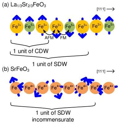
The equilibrium magnetic properties of \ceLa_1-Sr_FeO3 perovskites can be controlled based on the La/Sr composition. \ceLa1/3Sr2/3FeO3 thin films exhibit a charge disproportionation (CD) of 3Fe3.67+ Fe5+ + 2Fe3+ accompanying antiferromagnetic ordering below 190 K Wadati et al. (2005); Sichel-Tissot et al. (2013). The high valence state of \ceFe^5+ is realized as , where denotes an oxygen 2p hole Abbate et al. (1992). This charge disproportionation phase results in a charge density wave (CDW) with threefold periodicity as that of the crystal lattice, and a spin density wave (SDW) of sixfold periodicity along the 111 directions, as revealed by a neutron diffraction study (see Fig. 1 (a))Imada et al. (1998). \ceSrFeO_3 includes the \ceFe^4+ ion and shows a helimagnetic phase with incommensurate periodicity, which occurs because of the competition between the nearest neighbor ferromagnetic and the next-nearest neighbor antiferromagnetic interaction of the conducting 3d electrons, as indicated by neutron scattering results Takeda et al. (1972). was reported to be 134 K Takeda et al. (1972) (bulk), K Chakraverty et al. (2013), K Rogge et al. (2019) (thin films). A multiple helimagnetic phase in \ceSrFeO_3 was argued Chakraverty et al. (2013); Ishiwata et al. (2011), which could possibly host skyrmion crystals Ishiwata et al. (2020).
Resonant soft X-ray scattering (RSXS) Fink et al. (2013) is a powerful tool for revealing the ordered structures in solids, such as the magnetic, charge, and orbital ordering Schüßler-Langeheine et al. (2001); Zhou et al. (2011); Partzsch et al. (2011); Wadati et al. (2012); Fink et al. (2013); Matsuda et al. (2015); Yamamoto et al. (2018). RSXS can be performed with the core level absorption process, and the interaction between X-rays and a specific element can be enhanced. Hence, RSXS can be applied to thin films with small sample volumes. X-ray polarization is sensitive to the orbitals of the valence electrons and RSXS can detect orbital and spin ordering, which are coupled through spin-orbital interaction. The magnetic ordering of irons in \ceLa1/3Sr2/3FeO3 Okamoto et al. (2010); Yamamoto et al. (2018) and \ceSrFeO_3 Chakraverty et al. (2013) have been observed through RSXS at the Fe L2,3 absorption edge (2p 3d, eV).
In this study, we report the photoinduced magnetic dynamics of \ceLa1/3Sr2/3FeO3 and \ceSrFeO_3 thin films determined through time-resolved RSXS measurements. We ascertain the ultrafast melting of the \ceLa1/3Sr2/3FeO3 magnetic ordering through charge transfer between Fe ions, which can be attributed to the strong coupling between the charge and spin in the system. Based on the comparison between \ceLa1/3Sr2/3FeO3 and \ceSrFeO_3, the effect of the electronic properties on the dynamics of the photoinduced quenching of the magnetic orderings is examined.
II Experimental
La1/3Sr2/3FeO3 thin films with 40-nm thicknesses were grown epitaxially on \ceSrTiO3 (111) substrates using the pulsed laser deposition method. The details can be found in Ref. Minohara et al. (2016). By oxidizing \ceSrFeO_2.5, \ceSrFeO_3 thin films with nm thicknesses were fabricated on \ceSrTiO3(100) substrates using the pulsed laser deposition method. \ceSrFeO_3 was obtained by annealing SrFeO2.5 in an ozone atmosphere at 300 for 6 h with the sample exposed to UV light in a UV/O3 DRY CLEANER UV-1 (SAMCO Inc).
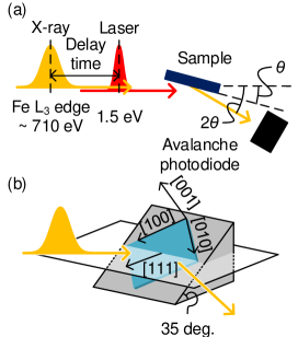
In order to elucidate the photoinduced dynamics of the antiferromagnetic ordering in \ceLa1/3Sr2/3FeO3 and \ceSrFeO_3, we performed time-resolved RSXS measurements using the pump-probe method at the slicing facility UE56/1-ZPM Helmholtz-Zentrum, Berlin Holldack et al. (2014) The experimental setup is shown in Fig. 2. A Ti:Sapphire laser (, 1.5 eV) was employed for photoexcitation, with a pulse duration of 50 fs. The laser and X-ray pulse frequencies were 3 and 6 kHz, respectively, and signals with and without pump laser excitation were obtained alternately. The signals without excitation were used for normalizing the pumped signals. The scattered X-rays were detected using an avalanche photo diode. \ceLa1/3Sr2/3FeO3 was investigated using 100-fs X-ray pulses generated through the laser slicing technique Holldack et al. (2014); the total time resolution was . \ceSrFeO_3 was mounted on a wedge-shaped jig at an angle of 55 deg. in order to orient the [111] direction in the scattering plane, and the temperature was set to 35 K. We set the X-ray photon energy and polarization to the Fe L3 edge ( eV) and horizontal, respectively, for obtaining the magnetic signals. In the \ceSrFeO_3 case, the time resolution was approximately 50 ps, corresponding to the pulse width of the synchrotron X-ray radiation. Static \ceLa1/3Sr2/3FeO3 measurements were performed with a diffractometer at the Soft X-ray beamline BL-16A, Photon Factory, KEK, Japan Nakao et al. (2014). The experimental geometry and temperature were equivalent to the time-resolved measurement, and a silicon drift detector was used.
III Results and Discussion
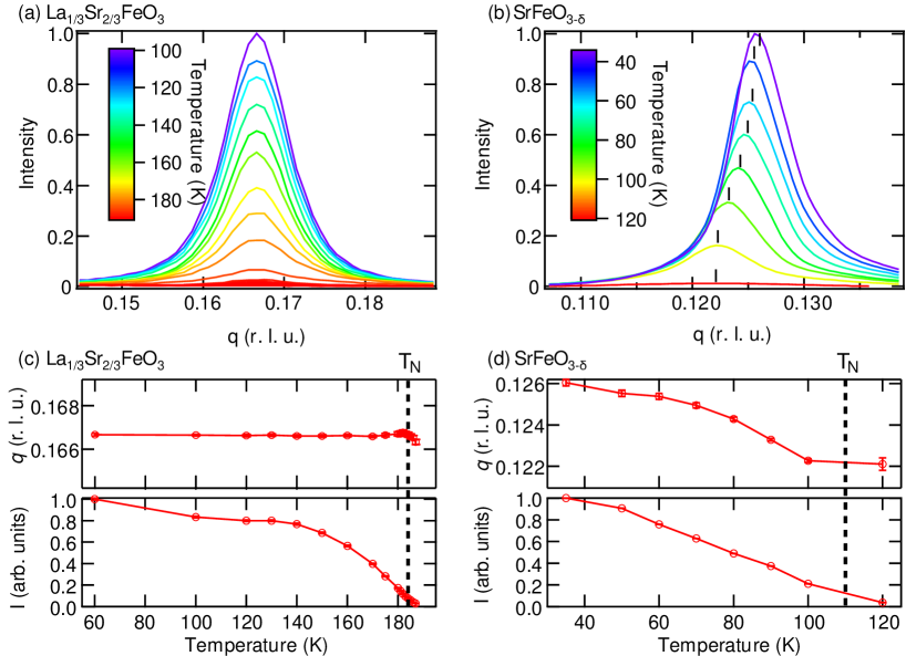
Figure 3 depicts the antiferromagnetic ordering peaks of (a) \ceLa1/3Sr2/3FeO3 and (b) \ceSrFeO_3. The diffraction peaks were scanned along the [111] direction. The temperature dependence of and the peak intensities of \ceLa1/3Sr2/3FeO3 and \ceSrFeO_3 are displayed in Figs. 3(c,d) respectively. For the \ceLa1/3Sr2/3FeO3 thin films, the peak position is fixed at in the entire temperature range below , as shown in Fig 3(a), and is estimated to be 182 K. The \ceSrFeO_3 diffraction peak appears below , and the peak position shifts according to the temperature. is also in the same range as those of previous reports Chakraverty et al. (2013); Rogge et al. (2019). depends on the oxygen vacancy , and decreases from 134 K for to 80 K for MacChesney et al. (1965), which implies a small for our sample. of the \ceSrFeO_3 helimagnetic ordering was reported to be 0.128-0.112 (\ceSrFeO_2.87) Reehuis et al. (2012); Takeda et al. (1972), 0.13 (\ceSrFeO_3) or 0.125 (\ceSr_0.99Co_0.01FeO3) Chakraverty et al. (2013); our observed is in a similar range. In the heating and cooling cycle, thermal hysteresis was observed for the peak intensity and peak positions. According to previous resistivity measurement reports Chakraverty et al. (2013); Rogge et al. (2019), thermal hysteresis was observed at , and our observed thermal hysteresis reflects this phenomenon.
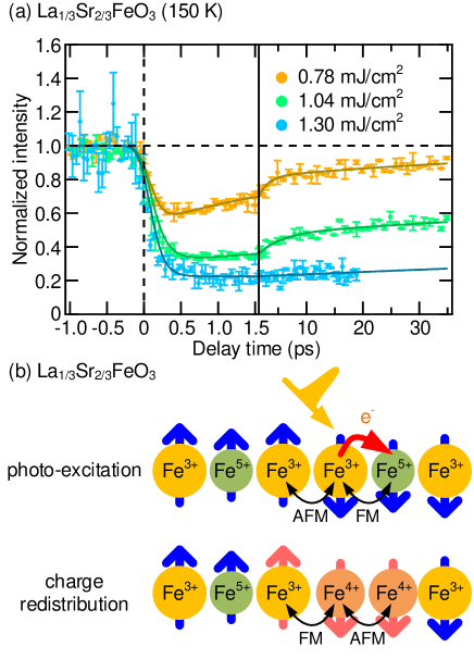
We first discuss the \ceLa1/3Sr2/3FeO3 dynamics. The time evolution of the magnetic peak intensity after laser pumping, revealed in the laser slicing mode, is shown in Fig. 4(a). Rapid reduction in the diffraction intensity is observed within a time resolution of 130 fs. The decreased diffraction intensity recovers 30 ps after laser excitation in the lower fluence regime; however, the recovery is slower at higher fluence. In order to discuss the results quantitatively, we fitted them using the function depicted by Eq. (1) for the decay and recovery processes. The fitting function is as follows:
| (1) |
where , , and are the corresponding time constants, denotes the convolution, and is the Heaviside step function. To consider time resolution, the fitting function was convoluted by a Gaussian with a full-width-at-half-maximum of . The parameters extracted from the experimental delay scans are summarized in Table 1. The time scale was estimated to be ps.
For the nonthermal \ceLa1/3Sr2/3FeO3 process with an ultrashort time scale of , we consider that the ultrafast magnetization dynamics occurs because of the ultrafast photo-melting of the charge ordering, which destroys the magnetic ordering due to the strong coupling between the charge and spin. Demagnetization due to the photo-melting of the charge ordering has also been observed, for example, in NdNiO3 thin films Caviglia et al. (2013). Figure 4(b) illustrates the origins of the ultrafast melting of the magnetic ordering. Due to the strong onsite Hund’s rule coupling, excitation with linearly polarized light at 1.5 eV can result only in transitions between the ferromagnetically coupled sites Ishikawa et al. (1998). Optical conductivity measurement has demonstrated that the optical gap of 0.15 eV has a strong interatomic transition characteristic Ishikawa et al. (1998). Therefore, the excited electron would be transferred only in the ferromagnetically coupled Fe3+ - Fe5+ - Fe3+ sites, and the observation in this study suggests that the excitation induces charge transfer in a unit of the charge-ordering site of Fe3+ - Fe5+ - Fe3+ into Fe3+ - Fe4+ - Fe4+, as shown in Fig. 4(b). It has been suggested that this charge transfer is a metastable state accompanying Fe-O bond-length change, in a previous report on the photoinduced transient states of \ceLa1/3Sr2/3FeO3 Zhu et al. (2018). Transfer in the charge ordering induces changes in the magnetic interactions. Fe3+ - Fe4+ ions have ferromagnetic interactions, whereas the Fe4+ - Fe4+ sites have antiferromagnetic ones, as shown in Fig. 4(b).
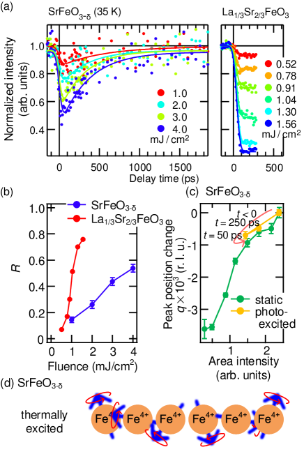
Furthermore, time-resolved RSXS measurements were performed for 35 and 80 K \ceSrFeO_3 thin films as shown in Fig. 5(a) with a time resolution of ; the experimental results for \ceLa1/3Sr2/3FeO3 with a similar time resolution are also shown in the right panel for comparison. Due to , the fast decay process cannot be resolved in Fig. 5(a), and we focus on the changes in the magnetic ordering peak intensity. We fitted using the following single exponential function
| (2) |
which is the limit of Eq. (1) as , , and .
| Fluence [mJ/cm2] | [ps] | [ps] | |
|---|---|---|---|
| 0.78 | 0.51 | 1.7 | 60 |
| 1.04 | 0.72 | 3.5 | 210 |
| 1.30 | 0.78 | - | 550 |
The degrees of quenching , defined by Eq. (2), of the \ceLa1/3Sr2/3FeO3 and \ceSrFeO_3 magnetic ordering are plotted as a function of the laser fluence in Fig. 5(b). For \ceLa1/3Sr2/3FeO3, the quenching of the peak intensity reaches at a fluence of approximately ; however, the \ceSrFeO_3 diffraction intensity decreases by less than 60 % of the initial intensity below a fluence of . This suggests that the antiferromagnetic ordering of \ceLa1/3Sr2/3FeO3 is quenched with less excitation energy compared to \ceSrFeO_3. The peak positions of the photoinduced transient states and the heating process are plotted as a function of the peak area intensity in Fig. 5(c). Both curves are similar, indicating that the observed photoinduced quenching of the \ceSrFeO_3 helimagnetic ordering is triggered by the same mechanism as the temperature-induced change.
SrFeO_3 is a metallic material and 1.5 eV laser excitation corresponds to the interband transition Fujioka et al. (2012). The photoinduced quenching of the helimagnetic ordering in \ceSrFeO_3 can be interpreted as the result of the increase in the spin and electron temperatures, as depicted in Fig. 5(d). On the other hand, \ceLa1/3Sr2/3FeO3 is insulating and its magnetic orderings disappear because of the charge transfer between Fe ions induced locally by photo excitation, as discussed above. The photoinduced dynamics of the antiferromagnetic orderings reflect the versatile electronic feature of perovskite oxides.
IV Conclusion
In this study, we examined the photoinduced dynamics of the antiferromagnetic orderings in two perovskites, \ceLa1/3Sr2/3FeO3 and \ceSrFeO_3, through time-resolved RSXS. The magnetic ordering in \ceLa1/3Sr2/3FeO3 thin films was quenched within 130 fs. This ultrafast dynamics can be explained based on the photoinduced charge transfer between Fe ions, which induces a change in the strong magnetic interactions. The process is nonthermal. The spin moment can be cancelled with the nearest neighbors in antiferromagnetic \ceLa1/3Sr2/3FeO3 through this ultrafast process, leading to ultrafast photoinduced change in the magnetic ordering. Compared to helimagnetic \ceSrFeO_3, the magnetic ordering of \ceLa1/3Sr2/3FeO3 was destroyed at lower fluence. Spin manipulation in helical systems has been found to be more energy efficient than in ferromagnets. An even more energy efficient process can be realized in \ceLa1/3Sr2/3FeO3. Ultrafast changes in the magnetic ordering of antiferromagnetic perovskite thin films were discovered, and the photoinduced dynamics was shown to be controlled by tuning the electronic feature through perovskite doping. The photoinduced dynamics of the spin order in antiferromagnetic perovskite thin films is important for integrating functional oxides into spintronic device architectures.
Acknowledgements.
We thank Yasutuki Hirata for the productive discussions and HZB for the allocation of the synchrotron radiation beamtime, Karsten Holldack and Rolf Mitzner for experimental support. This work was performed under the approval of the Photon Factory Program Advisory Committee (Proposal Nos. 2016PF-BL-19B, 2015G556, 2015S2-007, 2013G058, 2013G661). This work was partially supported by the Japan Society for the Promotion of Science (JSPS) KAKENHI Grant Nos. 19H05824, 19H01816, 19K23430, and 17K14334, and the MEXT Quantum Leap Flagship Program (MEXT Q-LEAP) Grant No. JPMXS0118068681. K. Y. acknowledges the support from the ALPS program of the University of Tokyo.References
- Kirilyuk et al. (2010) A. Kirilyuk, A. V. Kimel, and T. Rasing, Rev. Mod. Phys. 82, 2731 (2010).
- Beaurepaire et al. (1996) E. Beaurepaire, J.-C. Merle, A. Daunois, and J.-Y. Bigot, Phys. Rev. Lett. 76, 4250 (1996).
- Stamm et al. (2007) C. Stamm, T. Kachel, N. Pontius, R. Mitzner, T. Quast, K. Holldack, S. Khan, C. Lupulescu, E. F. Aziz, M. Wietstruk, H. A. Dürr, and W. Eberhardt, Nat. Mater. 6, 740 (2007).
- Stanciu et al. (2007) C. D. Stanciu, F. Hansteen, A. V. Kimel, A. Kirilyuk, A. Tsukamoto, A. Itoh, and T. Rasing, Phys. Rev. Lett. 99, 047601 (2007).
- Koopmans et al. (2010) B. Koopmans, G. Malinowski, F. Dalla Longa, D. Steiauf, M. Fähnle, T. Roth, M. Cinchetti, and M. Aeschlimann, Nat. Mater. 9, 259 (2010).
- Radu et al. (2011) I. Radu, K. Vahaplar, C. Stamm, T. Kachel, N. Pontius, H. A. Dürr, T. A. Ostler, J. Barker, R. F. L. Evans, R. W. Chantrell, A. Tsukamoto, A. Itoh, A. Kirilyuk, T. Rasing, and A. V. Kimel, Nature 472, 205 (2011).
- Johnson et al. (2012) S. L. Johnson, R. A. de Souza, U. Staub, P. Beaud, E. Möhr-Vorobeva, G. Ingold, A. Caviezel, V. Scagnoli, W. F. Schlotter, J. J. Turner, O. Krupin, W.-S. Lee, Y.-D. Chuang, L. Patthey, R. G. Moore, D. Lu, M. Yi, P. S. Kirchmann, M. Trigo, P. Denes, D. Doering, Z. Hussain, Z.-X. Shen, D. Prabhakaran, and A. T. Boothroyd, Phys. Rev. Lett. 108, 037203 (2012).
- Lee et al. (2012) W. Lee, Y. Chuang, R. Moore, Y. Zhu, L. Patthey, M. Trigo, D. Lu, P. Kirchmann, O. Krupin, M. Yi, M. Langner, N. Huse, J. Robinson, Y. Chen, S. Zhou, G. Coslovich, B. Huber, D. Reis, R. Kaindl, R. Schoenlein, D. Doering, P. Denes, W. Schlotter, J. Turner, S. Johnson, M. Först, T. Sasagawa, Y. Kung, A. Sorini, A. Kemper, B. Moritz, T. Devereaux, D.-H. Lee, Z. Shen, and Z. Hussain, Nat. Commun. 3, 838 (2012).
- Caviglia et al. (2013) A. D. Caviglia, M. Först, R. Scherwitzl, V. Khanna, H. Bromberger, R. Mankowsky, R. Singla, Y.-D. Chuang, W. S. Lee, O. Krupin, W. F. Schlotter, J. J. Turner, G. L. Dakovski, M. P. Minitti, J. Robinson, V. Scagnoli, S. B. Wilkins, S. A. Cavill, M. Gibert, S. Gariglio, P. Zubko, J.-M. Triscone, J. P. Hill, S. S. Dhesi, and A. Cavalleri, Phys. Rev. B 88, 220401 (2013).
- Beaud et al. (2014) P. Beaud, A. Caviezel, S. O. Mariager, L. Rettig, G. Ingold, C. Dornes, S.-W. Huang, J. A. Johnson, M. Radovic, T. Huber, T. Kubacka, A. Ferrer, H. T. Lemke, M. Chollet, D. Zhu, J. M. Glownia, M. Sikorski, A. Robert, H. Wadati, M. Nakamura, M. Kawasaki, Y. Tokura, S. L. Johnson, and U. Staub, Nat. Mater. 13, 923 (2014).
- Först et al. (2014) M. Först, R. I. Tobey, H. Bromberger, S. B. Wilkins, V. Khanna, A. D. Caviglia, Y.-D. Chuang, W. S. Lee, W. F. Schlotter, J. J. Turner, M. P. Minitti, O. Krupin, Z. J. Xu, J. S. Wen, G. D. Gu, S. S. Dhesi, A. Cavalleri, and J. P. Hill, Phys. Rev. Lett. 112, 157002 (2014).
- Forst et al. (2015) M. Forst, A. D. Caviglia, R. Scherwitzl, R. Mankowsky, P. Zubko, V. Khanna, H. Bromberger, S. B. Wilkins, Y. D. Chuang, W. S. Lee, W. F. Schlotter, J. J. Turner, G. L. Dakovski, M. P. Minitti, J. Robinson, S. R. Clark, D. Jaksch, J. M. Triscone, J. P. Hill, S. S. Dhesi, and A. Cavalleri, Nat. Mater. 14, 883 (2015).
- Tsuyama et al. (2016) T. Tsuyama, S. Chakraverty, S. Macke, N. Pontius, C. Schüßler-Langeheine, H. Y. Hwang, Y. Tokura, and H. Wadati, Phys. Rev. Lett. 116, 256402 (2016).
- Thielemann-Kühn et al. (2017) N. Thielemann-Kühn, D. Schick, N. Pontius, C. Trabant, R. Mitzner, K. Holldack, H. Zabel, A. Föhlisch, and C. Schüßler-Langeheine, Phys. Rev. Lett. 119, 197202 (2017).
- Imada et al. (1998) M. Imada, A. Fujimori, and Y. Tokura, Rev. Mod. Phys. 70, 1039 (1998).
- Wadati et al. (2005) H. Wadati, D. Kobayashi, H. Kumigashira, K. Okazaki, T. Mizokawa, A. Fujimori, K. Horiba, M. Oshima, N. Hamada, M. Lippmaa, M. Kawasaki, and H. Koinuma, Phys. Rev. B 71, 035108 (2005).
- Sichel-Tissot et al. (2013) R. J. Sichel-Tissot, R. C. Devlin, P. J. Ryan, J.-w. Kim, and S. J. May, Appl. Phys. Lett. 103, 212905 (2013).
- Abbate et al. (1992) M. Abbate, F. M. F. de Groot, J. C. Fuggle, A. Fujimori, O. Strebel, F. Lopez, M. Domke, G. Kaindl, G. A. Sawatzky, M. Takano, Y. Takeda, H. Eisaki, and S. Uchida, Phys. Rev. B 46, 4511 (1992).
- Takeda et al. (1972) T. Takeda, Y. Yamaguchi, and H. Watanabe, J. Phys. Soc. Jpn. 33, 967 (1972).
- Chakraverty et al. (2013) S. Chakraverty, T. Matsuda, H. Wadati, J. Okamoto, Y. Yamasaki, H. Nakao, Y. Murakami, S. Ishiwata, M. Kawasaki, Y. Taguchi, Y. Tokura, and H. Y. Hwang, Phys. Rev. B 88, 220405 (2013).
- Rogge et al. (2019) P. C. Rogge, R. J. Green, R. Sutarto, and S. J. May, Phys. Rev. Mater. 3, 084404 (2019).
- Ishiwata et al. (2011) S. Ishiwata, M. Tokunaga, Y. Kaneko, D. Okuyama, Y. Tokunaga, S. Wakimoto, K. Kakurai, T. Arima, Y. Taguchi, and Y. Tokura, Phys. Rev. B 84, 054427 (2011).
- Ishiwata et al. (2020) S. Ishiwata, T. Nakajima, J.-H. Kim, D. S. Inosov, N. Kanazawa, J. S. White, J. L. Gavilano, R. Georgii, K. M. Seemann, G. Brandl, P. Manuel, D. D. Khalyavin, S. Seki, Y. Tokunaga, M. Kinoshita, Y. W. Long, Y. Kaneko, Y. Taguchi, T. Arima, B. Keimer, and Y. Tokura, Phys. Rev. B 101, 134406 (2020).
- Fink et al. (2013) J. Fink, E. Schierle, E. Weschke, and J. Geck, Reports Prog. Phys. 76, 056502 (2013).
- Schüßler-Langeheine et al. (2001) C. Schüßler-Langeheine, E. Weschke, A. Grigoriev, H. Ott, R. Meier, D. Vyalikh, C. Mazumdar, C. Sutter, D. Abernathy, G. Grübel, and G. Kaindl, J. Electron Spectros. Relat. Phenomena 114-116, 953 (2001).
- Zhou et al. (2011) S. Y. Zhou, Y. Zhu, M. C. Langner, Y.-D. Chuang, P. Yu, W. L. Yang, A. G. Cruz Gonzalez, N. Tahir, M. Rini, Y.-H. Chu, R. Ramesh, D.-H. Lee, Y. Tomioka, Y. Tokura, Z. Hussain, and R. W. Schoenlein, Phys. Rev. Lett. 106, 186404 (2011).
- Partzsch et al. (2011) S. Partzsch, S. B. Wilkins, J. P. Hill, E. Schierle, E. Weschke, D. Souptel, B. Büchner, and J. Geck, Phys. Rev. Lett. 107, 057201 (2011).
- Wadati et al. (2012) H. Wadati, J. Okamoto, M. Garganourakis, V. Scagnoli, U. Staub, Y. Yamasaki, H. Nakao, Y. Murakami, M. Mochizuki, M. Nakamura, M. Kawasaki, and Y. Tokura, Phys. Rev. Lett. 108, 047203 (2012).
- Matsuda et al. (2015) T. Matsuda, S. Partzsch, T. Tsuyama, E. Schierle, E. Weschke, J. Geck, T. Saito, S. Ishiwata, Y. Tokura, and H. Wadati, Phys. Rev. Lett. 114, 236403 (2015).
- Yamamoto et al. (2018) K. Yamamoto, Y. Hirata, M. Horio, Y. Yokoyama, K. Takubo, M. Minohara, H. Kumigashira, Y. Yamasaki, H. Nakao, Y. Murakami, A. Fujimori, and H. Wadati, Phys. Rev. B 97, 075134 (2018).
- Okamoto et al. (2010) J. Okamoto, D. J. Huang, K. S. Chao, S. W. Huang, C.-H. Hsu, A. Fujimori, A. Masuno, T. Terashima, M. Takano, and C. T. Chen, Phys. Rev. B 82, 132402 (2010).
- Minohara et al. (2016) M. Minohara, M. Kitamura, H. Wadati, H. Nakao, R. Kumai, Y. Murakami, and H. Kumigashira, J. Appl. Phys. 120, 025303 (2016).
- Holldack et al. (2014) K. Holldack, J. Bahrdt, A. Balzer, U. Bovensiepen, M. Brzhezinskaya, A. Erko, A. Eschenlohr, R. Follath, A. Firsov, W. Frentrup, L. Le Guyader, T. Kachel, P. Kuske, R. Mitzner, R. Müller, N. Pontius, T. Quast, I. Radu, J.-S. Schmidt, C. Schüßler-Langeheine, M. Sperling, C. Stamm, C. Trabant, and A. Föhlisch, J. Synchrotron Radiat. 21, 1090 (2014).
- Nakao et al. (2014) H. Nakao, Y. Yamasaki, J. Okamoto, T. Sudayama, Y. Takahashi, K. Kobayashi, R. Kumai, and Y. Murakami, J. Phys. Conf. Ser. 502, 012015 (2014).
- MacChesney et al. (1965) J. B. MacChesney, R. C. Sherwood, and J. F. Potter, J. Chem. Phys. 43, 1907 (1965).
- Reehuis et al. (2012) M. Reehuis, C. Ulrich, A. Maljuk, C. Niedermayer, B. Ouladdiaf, A. Hoser, T. Hofmann, and B. Keimer, Phys. Rev. B 85, 184109 (2012).
- Ishikawa et al. (1998) T. Ishikawa, S. K. Park, T. Katsufuji, T. Arima, and Y. Tokura, Phys. Rev. B 58, R13326 (1998).
- Zhu et al. (2018) Y. Zhu, J. Hoffman, C. E. Rowland, H. Park, D. A. Walko, J. W. Freeland, P. J. Ryan, R. D. Schaller, A. Bhattacharya, and H. Wen, Nat. Commun. 9, 1799 (2018).
- Fujioka et al. (2012) J. Fujioka, S. Ishiwata, Y. Kaneko, Y. Taguchi, and Y. Tokura, Phys. Rev. B 85, 155141 (2012).