Geometric effects induce anomalous size-dependent active transport in structured environments
Abstract
Variations of transport efficiency in structured environments between distinct individuals in actively self-propelled systems is both hard to study and poorly understood. Here, we study the transport of a non-tumbling Escherichia coli strain, an active-matter archetype with intrinsic size variation but fairly uniform speed, through a periodic pillar array. We show that long-term transport switches from a trapping dominated state for shorter cells to a much more dispersive state for longer cells above a critical bacterial size set by the pillar array geometry. Using a combination of experiments and modeling, we show that this anomalous size-dependence arises from an enhancement of the escape rate from trapping for longer cells caused by nearby pillars. Our results show that geometric effects can lead to size being a sensitive tuning knob for transport in structured environments, with implications in general for active matter systems and, in particular, for the morphological adaptation of bacteria to structured habitats, spatial structuring of communities and for anti-biofouling materials design.
Structural features of environments have been recently shown to have a significant impact on the motility phases of active matter systems [1, 2, 3, 4, 5, 6, 7, 8, 9, 10, 11, 12]. However, much less is known about how the interplay between variations in individual particle geometry and environmental structure affects macroscopic transport. For motile bacterial systems, in particular, such effects [13, 14, 15, 16, 17] can have implications for tunable transport in structured habitats. These implications arise because bacteria come in a variety of shapes and sizes across species [18, 19, 20] and even within a single strain [21, 22, 23]. It has been suggested that such widely distributed shapes and sizes are a consequence of adaptation to a diversity of features in their environments ranging from mechanical properties to nutrient availability [18]. In particular, the optimization of transport or dispersal is known to provide a strong selective pressure for bacterial morphology evolution [18, 24, 25, 26]. For bacteria that live in structured or porous environments such as soil or tissue [27, 28], proximity to a surface involves a whole host of physical interactions. These include hydrodynamic [29, 30, 31, 32], electrostatic [33], and steric [34] forces as well as flow induced effects [15, 35, 14], which could affect transport in a geometry dependent manner [36, 16, 37]. Here we study the possibility that the interplay between minor, intrinsic variations in the geometry of swimmers and structural features of the environment could lead to significant transport effects at the macroscopic scale.
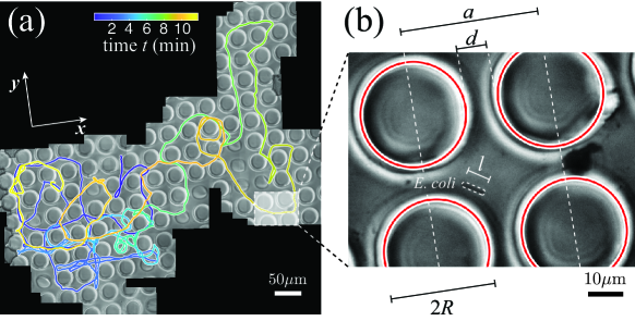
One of the main challenges for such a study is that, in a standard microscope setting, a freely moving individual cell can only be observed, with adequate resolution of individual geometry, over a length scale similar to that of the cell size ( m). Meaningful statistics for its long-range transport over the millimeter scale is therefore hard to obtain. Here, we resolved this issue by following individual bacteria via a tracking microscope, where the microscope stage is adjusted in real-time to recenter the cell of interest in the field of view [25]. To provide a structured environment, we fabricated a rectangular microfluidic channel, mm wide and 30 m deep, embedded with square arrays of micropillars using a standard soft photolithography method (see Methods). A typical pillar array with pillar radii m and lattice size m (with the closest gap thus m) is shown in Fig. 1(a). To focus on purely geometrical effects on transport that are applicable to generic active matter systems, we used a smooth swimming Escherichia coli strain, HCB437, which avoids any potential active response by the bacteria switching between run and tumble phases [38]. Additionally, this strain shows a natural length variation from m between individuals allowing us to examine the effects of microscopic geometry on macroscopic transport. We visualized the transport of these bacteria through the pillar array at a high ( or ) magnification over millimeters by reconstructing trajectories. This was done by stitching together single image frames during the course of tracking (see Supplementary Material) as shown in Fig. 1(b). Even though the tracked bacterium navigates the pillar array over a long distance, its detailed movement and orientation can still be resolved by visiting every single frame with a submicron resolution (Fig. 1(b)).
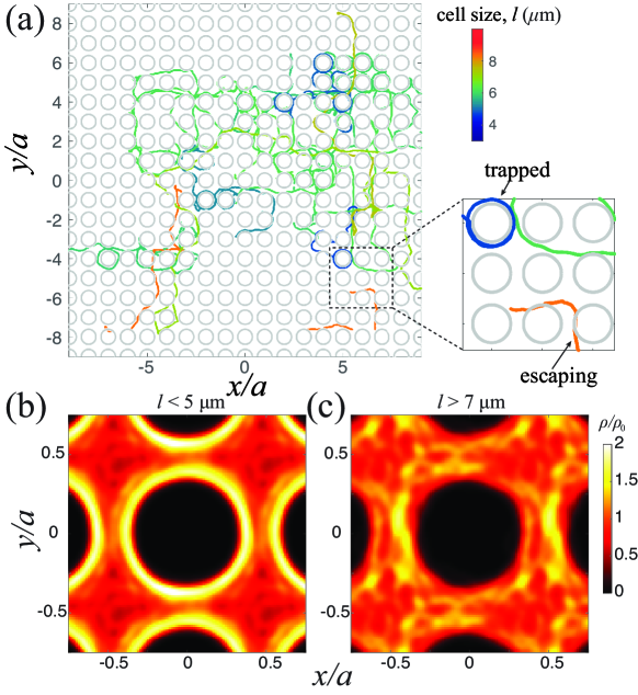
We first examined in detail the trajectories of several bacteria with different sizes. Independent of size, bacteria are constrained to move within the open spaces between pillars and the presence of noise leads to an overall diffusive trajectory at long times. However, we noticed two qualitatively different modes of motility depending on size. Short cells ( m), on the one hand, frequent the pillar surfaces and move mostly in circular patterns, due to effective hydrodynamic trapping [32, 30, 36] by the pillar array (Fig. 2(a), also see Movie S1). Distinct from a plane-wall-induced circulation [29], this circulation around the pillar is bidirectional, regardless of the chirality embedded in the flagellar filaments (Supplementary Material, Fig. S7). Longer cells, on the other hand, appear to escape from such traps, resulting in more persistent movement along the directions of two orthogonal lattice vectors (here, - and -axes) (Fig. 2(a), also see Movie S2). Thus, longer cells, despite feeling an increased confinement, showed an anomalous increase in their net transport.
To further quantify these distinct effects of the pillar array on bacteria with different sizes, we computed the probability distribution of the centers of bacteria in space within a single unit cell. As shown in Fig. 2(b) and (c), shorter bacteria are concentrated near the pillar surface (consistent with a hydrodynamic attraction [14]), while longer cells spend more time in the channels, confirming a size-dependent trapping effect. It is worth noting that tracking the front end of a cell provides essentially the same distribution near the pillar surface (Supplementary Material, Fig. S5), suggesting a negligible role of cell length in volume exclusion.
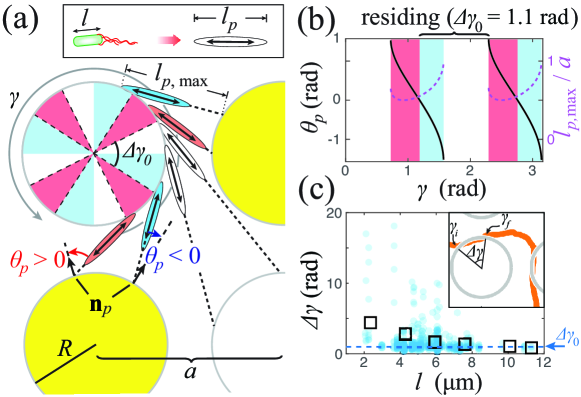
To understand the mechanism governing this effect, we considered the geometric constraints on a swimming bacterium due to neighboring pillars. For simplicity, the bacterium is regarded as a rod-shaped pusher of length , which is the effective hydrodynamic size set by the flow profile of the entire swimmer including the cell body (of length ) and the flagella (Fig. 3a, inset). Such a pusher tends to circulate around a single pillar in its natural state (without any neighboring pillars), due to the known hydrodynamic attraction between a solid surface and a generic pusher swimmer, including both bacteria and synthetic microswimmers [39, 37]. In the presence of the neighboring pillars, the allowable pusher sizes are restricted, with the maximum length determined by the geometry (Fig. 3a). In addition to the length constraint, a pusher is also subjected to hydrodynamic interactions from the neighboring pillars. Here, we consider a pusher circulating in the counter-clockwise direction and assume the pusher’s orientation is always tangential to the pillar surface that it is circulating around. Depending on the orientation of the pusher relative to the pillar lattice (denoted by angle ), the pusher may experience a torque from the nearest-neighbor pillar that either promotes or inhibits its circulation around the pillar. Such distinct effects are determined by the orientation angle of the pusher relative to the surface normal of the nearest-neighbor pillar (Fig. 3a). Here, we consider only the normal component of the hydrodynamic force from the pillar, associated with the anisotropic drag coefficients in the presence of a nearby wall [40, 41]. For , the normal force from the nearest-neighbor pillar provides a torque that tends to tip the pusher toward the center of the pillar it is circulating around, leading to an effective attraction to the pillar surface. For , the torque due to the nearest-neighbor pillar tends to tip the pusher further away from the center causing the pusher to escape (likely along the tangent to the pillar surface). Considering the tangential components of the forces does not alter the directions of these torques, as long as the drag coefficient normal to the pillar surface dominates. It should be noted that we only treated the above pusher in a resistive-force-type manner to signify the geometric roles played by the nearest-neighbor pillar. More qualitative and quantitative insights of the hydrodynamic interactions requires resolving the flow field associated with a full pusher model including no-slip boundaries with pillar geometries [30].
This nearest-neighbor effect leads to a series of alternating attractive and repulsive zones along the perimeter of the pillar (with ), determined by the sign of . Figure 3b shows the calculated and (see Supplementary Material) for a counter-clockwise circulating pusher and a pillar lattice that is consistent with the experimental setting (). The attractive () and repulsive () zones are shaded in red and blue, respectively. Zones that lack any constraints from the nearest-neighbor pillar (in white) are also considered attractive, due to the natural circulating state of pushers (in the absence of the neighboring pillars). This result shows four continuous attractive zones (blue plus white) along the perimeter of the pillar, with each spanning an arc angle rad, centered near , , , and , respectively.
To facilitate a more quantitative comparison between the experiment and the theoretical picture, we measured a residency arc angle , which is the difference between the two angles where the bacterium enters () and escapes () the vicinity of the pillar surface (Fig. 3c, inset). The bacteria continuously circulates the pillar over an arc subtending this angle without leaving the surface. The experimental residency arc angles , plotted against cell lengths , are shown in Fig. 3c. At short cell lengths ( m), can span over larger angles (), corresponding to the presence of multi-turn circulations. As increases, becomes restricted only to small angles (e.g., for m). Such a restriction in the distribution of is responsible for the decrease in its mean with increasing . For sufficiently long ( m), the mean falls beneath the size of the computed attractive zone ( rad), consistent with a highly constrained pusher that is unable to bypass any repulsive zones (as required for circulations beyond a single attractive zone). We also note that there is no such size dependence of for sufficiently large gaps among pillars (Supplementary Material, Fig. S6), which further validates our geometric arguments.
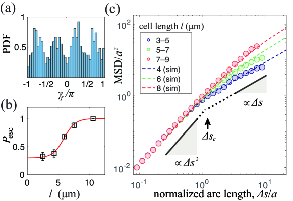
To further validate our model, we investigated the statistics of the angles, , at which bacteria escaped the pillar surface. The probability density function (PDF) of shows peaks near the diagonal directions of the pillar lattice (Fig. 4a), consistent with the predicted locations of the repulsive zones (Fig. 3b). This highly anisotropic distribution of also justifies the use of a single escaping probability (within all repulsive zones only) for characterizing the size-dependent geometric effects. Such an escaping probability is consistent with the stochastic nature of the competition between both the hydrodynamic attraction from the orbited pillar and the “repulsive” contribution from the nearest-neighboring one, associated with the fluctuating orientation and location of a microswimmer. If we consider that bacteria only escape within repulsive zones, can be obtained as , where and correspond respectively to the number of escaping events and the number of repulsive zones that bacteria cross during their travel along the pillars’ perimeters. Noting that the angular separation between the centers of two adjacent repulsive zones is and neglecting the detailed escaping locations, the number is given by , where corresponds to the index of an escaping event and denotes the integer part of a number. The escaping probability is thus the inverse of an ensemble average (denoted by ), i.e., . We computed this probability from experiments for different groups of cell body lengths . As shown in Fig. 4b, there is a transition from a low escaping probability () for relatively shorter cells ( m) to a high escaping probability () for relatively longer ones ( m). A hyperbolic-tangent fit of the experimental data yields a function (solid curve in Fig. 4b) with the critical cell body length m demarcating the distinct escaping behaviors.
To show how this geometric effect manifests itself in the long-time transport of bacteria, we simulated bacterial trajectories by a kinematic model (using the above ): a bacterium that reaches a pillar surface stays on the surface and continues circulating the pillar if it is within an attractive zone or it escapes with a probability if it enters a repulsive zone (in red in Fig. 3b). Here, we assumed that a bacterium moves at a constant speed , which is a fairly good approximation for the non-tumbling mutant (Supplementary Material, Fig. S8). After escaping along the direction tangential to the pillar’s circumference, the bacterium moves at constant speed until it reaches an attractive or repulsive zone of the next pillar along its trajectory. A white noise in swimming direction was extracted from experimental trajectories (Supplementary Material, Fig. S2) and introduced to the model swimmer when it is away from the pillar surface. We then computed the corresponding mean squared-displacement (MSD) for comparison with the experimental data (Fig. 4c). To secure optimal convergence, the MSD values were binned by path lengths and sampled over all trajectories within the same size group. For all experimental MSD (dots in Fig. 4c), cell body lengths ( - m) were grouped every m to secure a sufficient number () of long trajectories ( m) within each size category. All data points with less than 10 sampled trajectories were excluded. Each simulated MSD of the corresponding (dashed line in Fig. 4c) was computed over 100 model swimmers with each of them traveling in total path length from a random starting position. As shown in Fig. 4c, the simulated MSD reproduces well the characteristics of bacterial transport. In both experimental and simulated results, each MSD- curve contains a ballistic regime for small and a diffusive regime for large . The size of the ballistic regime can be characterized by a ballistic length (Fig. 4c), set by the crossover point between two distinct scaling regimes for transport (with MSD exponents and ). Interestingly, the ballistic length for relatively shorter cells, reminiscent of the effective reorientation time scale for point-like particles diffusing in obstacle networks [42, 43]. While converging at short , the long-time MSD- curves are noticeably higher for longer cells, corresponding to longer ballistic lengths at longer cell lengths and thus suggesting a finite-length effect. Again, longer cells, despite feeling an increased confinement, display an anomalous increase in their MSD due to the geometric effects of the neighboring pillars. These quantitative agreements between the experiment and theory further confirms the dominant role played by geometry.
Our study illustrates the role of individual morphology in the transport of active particles in general and bacteria, in particular, through periodic structured media. In living systems, this surprising enhancement of transport for longer cells, combined with a maximal cell size set by interstitial spaces of the lattice, suggests an optimal cell size potentially determined by the geometry of a porous environment [44, 18, 24]. Similar geometry-sensitive effects in transport of wild-type strains may already be present, but not explicitly identified, in other studies of bacterial transport [17]. Generalizing our geometric effects to such 3D environments will enable targeted design of environmental geometry for desired size-dependent transport and collective motion [13, 45, 46]. For a non-periodic lattice, it is expected that the above geometric effect still applies in general, since any geometric constraints due to nearest-neighbor pillars always tend to influence the longer cells first before they can influence shorter cells. However, the location and size of the repulsive zones, as well as the critical cell-body length now vary for each pillar, leading to more complex escaping zones and thus more complex global cell kinematics. Also, in the extreme case that multiple pillars are within the vicinity, a long cell will be more easily jammed as it requires more room for reorientation, contributing to another type of geometric constraint. These potential effects due to disorder in the crystalline structure of the environment will be investigated in our future work.
Our findings also indicate that the transition between localized and dispersive modes is sharp and occurs at a critical value of bacterial size, controlled by the porous environment. Bacteria with typical sizes near this critical value may be able to access both modes of transport by adaptive change in their size based on the local nutrient conditions or other desired transport needs.This suggests, for example, an unexplored benefit of filamentation under starvation conditions in E. coli, in addition to others that have been proposed in the literature [47, 48, 49]. Our results also have implications for the spatial structure of naturally occurring bacterial colonies in structured environments where spatial location within the colony could be correlated with age dependent cell-size, due to differential transport.
In nature, patterned structures with periodic lattices are widely found on antibiofouling surfaces, such as cicada wings [50] and shark skins [51]. The geometric effects we have identified thus provide a new perspective for revisiting these microscale structures in relation to their antibiofouling effects. Conversely, our work also suggests ways to engineer surfaces so as to either increase or decrease the residency of different bacterial strains with slightly different sizes or even differentiating age structured populations, which may also be of interest in biofouling applications. Such ideas could also be applied to designing environments for the desired sorting or guiding of synthetic microswimmers as well as the geometric design of individual swimmers.
Acknowledgements.
We would like to thank Howard Berg for providing the E. coli mutant strain used in this study. We also acknowledge Jeremias Gonzalez, Yu Zeng, and Jacinta Conrad for insightful discussions. This work was supported by National Science Foundation Grant CBET-2046822, CBET-1706511, and NSF-CREST: Center for Cellular and Bio-molecular Machines (CCBM) at UC Merced (HRD-1547848). B.L. thanks the support of Hellman Foundation. A.G. also acknowledges support from National Science Foundation (NSF) grant DMS-1616926, partial support from the NSF Center for Engineering Mechanobiology grant NSF CMMI-154857 and the hospitality of the Aspen Center for Physics, which is supported by NSF grant PHY-1607611.References
- [1] Bechinger, C. et al. Active particles in complex and crowded environments. Reviews of Modern Physics 88, 045006 (2016).
- [2] Morin, A., Desreumaux, N., Caussin, J.-B. & Bartolo, D. Distortion and destruction of colloidal flocks in disordered environments. Nature Physics 13, 63–67 (2017).
- [3] Quint, D. A. & Gopinathan, A. Topologically induced swarming phase transition on a 2D percolated lattice. Physical Biology 12, 046008 (2015).
- [4] Chepizhko, O. & Peruani, F. Diffusion, Subdiffusion, and Trapping of Active Particles in Heterogeneous Media. Physical Review Letters 111, 160604 (2013).
- [5] Bertrand, T., Zhao, Y., Bénichou, O., Tailleur, J. & Voituriez, R. Optimized Diffusion of Run-and-Tumble Particles in Crowded Environments. Physical Review Letters 120, 198103 (2018).
- [6] Sándor, C., Libál, A., Reichhardt, C. & Olson Reichhardt, C. J. Dynamic phases of active matter systems with quenched disorder. Physical Review E 95, 032606 (2017).
- [7] Pattanayak, S., Das, R., Kumar, M. & Mishra, S. Enhanced dynamics of active Brownian particles in periodic obstacle arrays and corrugated channels. The European Physical Journal E 42, 62 (2019).
- [8] Phan, T. V. et al. Bacterial route finding and collective escape in mazes and fractals. Physical Review X 10, 031017 (2020).
- [9] Ribeiro, H. E., Ferreira, W. P. & Potiguar, F. Q. Trapping and sorting of active matter in a periodic background potential. Physical Review E 101, 032126 (2020).
- [10] Yazdi, S., Aragones, J. L., Coulter, J. & Alexander-Katz, A. Metamaterials for Active Colloid Transport (2020). Publisher: arXiv Version Number: 1.
- [11] Brun-Cosme-Bruny, M. et al. Deflection of phototactic microswimmers through obstacle arrays. Physical Review Fluids 5, 093302 (2020).
- [12] Reichhardt, C. & Reichhardt, C. J. O. Directional locking effects for active matter particles coupled to a periodic substrate. Physical Review E 102, 042616 (2020).
- [13] Wioland, H., Woodhouse, F. G., Dunkel, J. & Goldstein, R. E. Ferromagnetic and antiferromagnetic order in bcterial vortex lattices. Nature Physics 12, 341–345 (2016).
- [14] Creppy, A., Clément, E., Douarche, C., D’Angelo, M. V. & Auradou, H. Effect of motility on the transport of bacteria populations through a porous medium. Physical Review Fluids 4, 013102 (2019).
- [15] Dehkharghani, A., Waisbord, N., Dunkel, J. & Guasto, J. S. Bacterial scattering in microfluidic crystal flows reveals giant active Taylor–Aris dispersion. Proceedings of the National Academy of Sciences 116, 11119–11124 (2019).
- [16] Makarchuk, S., Braz, V. C., Araújo, N. A. M., Ciric, L. & Volpe, G. Enhanced propagation of motile bacteria on surfaces due to forward scattering. Nature Communications 10, 4110 (2019).
- [17] Bhattacharjee, T. & Datta, S. S. Bacterial hopping and trapping in porous media. Nature Communications 10, 2075 (2019).
- [18] Young, K. D. The selective value of bacterial shape. Microbiology and Molecular Biology Reviews 70, 660–703 (2006).
- [19] Rappé, M. S., Connon, S. A., Vergin, K. L. & Giovannoni, S. J. Cultivation of the ubiquitous SAR11 marine bacterioplankton clade. Nature 418, 630–633 (2002).
- [20] Angert, E. R., Clements, K. D. & Pace, N. R. The largest bacterium. Nature 362, 239–241 (1993).
- [21] Hahn, M. W., Moore, E. R. & Höfle, M. G. Bacterial filament formation, a defense mechanism against flagellate grazing, is growth rate controlled in bacteria of different phyla. Applied and Environmental Microbiology 65, 25–35 (1999).
- [22] Typas, A., Banzhaf, M., Gross, C. A. & Vollmer, W. From the regulation of peptidoglycan synthesis to bacterial growth and morphology. Nature Reviews Microbiology 10, 123–136 (2012).
- [23] Shen, J.-P. & Chou, C.-F. Morphological plasticity of bacteria—Open questions. Biomicrofluidics 10, 031501 (2016).
- [24] Schuech, R., Hoehfurtner, T., Smith, D. J. & Humphries, S. Motile curved bacteria are Pareto-optimal. Proceedings of the National Academy of Sciences 116, 14440–14447 (2019).
- [25] Liu, B. et al. Helical motion of the cell body enhances Caulobacter crescentus motility. Proceedings of the National Academy of Sciences 111, 11252–11256 (2014).
- [26] Persat, A., Stone, H. A. & Gitai, Z. The curved shape of Caulobacter crescentus enhances surface colonization in flow. Nature Communications 5, 12855 (2014).
- [27] Turnbull, G. A., Morgan, J. W., Whipps, J. M. & Saunders, J. R. The role of bacterial motility in the survival and spread of Pseudomonas fluorescens in soil and in the attachment and colonisation of wheat roots. FEMS Microbiology Ecology 36, 21–31 (2001).
- [28] Balzan, S., de Almeida Quadros, C., de Cleva, R., Zilberstein, B. & Cecconello, I. Bacterial translocation: Overview of mechanisms and clinical impact. Journal of Gastroenterology and Hepatology 22, 464–471 (2007).
- [29] Lauga, E., DiLuzio, W. R., Whitesides, G. M. & Stone, H. A. Swimming in circles: Motion of bacteria near solid boundaries. Biophysical Journal 90, 400–412 (2006).
- [30] Spagnolie, S. E., Moreno-Flores, G. R., Bartolo, D. & Lauga, E. Geometric capture and escape of a microswimmer colliding with an obstacle. Soft Matter 11, 3396–3411 (2015).
- [31] Shum, H., Gaffney, E. A. & Smith, D. J. Modelling bacterial behaviour close to a no-slip plane boundary: The influence of bacterial geometry. Proceedings of the Royal Society A: Mathematical, Physical and Engineering Sciences 466, 1725–1748 (2010).
- [32] Sipos, O., Nagy, K., Di Leonardo, R. & Galajda, P. Hydrodynamic trapping of swimming bacteria by convex walls. Physical Review Letters 114, 258104 (2015).
- [33] Hermansson, M. The DLVO theory in microbial adhesion. Colloids and Surfaces B: Biointerfaces 14, 105–119 (1999).
- [34] Drescher, K., Dunkel, J., Cisneros, L. H., Ganguly, S. & Goldstein, R. E. Fluid dynamics and noise in bacterial cell-cell and cell-surface scattering. Proceedings of the National Academy of Sciences 108, 10940–10945 (2011).
- [35] Alonso-Matilla, R., Chakrabarti, B. & Saintillan, D. Transport and dispersion of active particles in periodic porous media. Physical Review Fluids 4, 043101 (2019).
- [36] Tong, J. & Shelley, M. J. Directed migration of microscale swimmers by an array of shaped obstacles: modeling and shape optimization. SIAM Journal on Applied Mathematics 78, 2370–2392 (2018).
- [37] Davies Wykes, M. S. et al. Guiding microscale swimmers using teardrop-shaped posts. Soft Matter 13, 4681–4688 (2017).
- [38] Berg, H. C. (ed.) E. Coli in Motion. Biological and Medical Physics, Biomedical Engineering (Springer New York, New York, NY, 2004).
- [39] Berke, A. P., Turner, L., Berg, H. C. & Lauga, E. Hydrodynamic Attraction of Swimming Microorganisms by Surfaces. Physical Review Letters 101, 038102 (2008).
- [40] Brenner, H. The slow motion of a sphere through a viscous fluid towards a plane surface. Chemical Engineering Science 16, 242–251 (1961).
- [41] Lauga, E., DiLuzio, W. R., Whitesides, G. M. & Stone, H. A. Swimming in circles: Motion of bacteria near solid boundaries. Biophysical Journal 90, 400–412 (2006).
- [42] Jakuszeit, T., Croze, O. A. & Bell, S. Diffusion of active particles in a complex environment: Role of surface scattering. Physical Review E 99, 012610 (2019).
- [43] Schakenraad, K. et al. Topotaxis of active Brownian particles. Physical Review E 101, 032602 (2020).
- [44] Bakken, L. R. & Olsen, R. A. The relationship between cell size and viability of soil bacteria. Microbial Ecology 13, 103–114 (1987).
- [45] Long, Z., Quaife, B., Salman, H. & Oltvai, Z. N. Cell-cell communication enhances bacterial chemotaxis toward external attractants. Scientific Reports 7, 12855 (2017).
- [46] Nishiguchi, D., Aranson, I. S., Snezhko, A. & Sokolov, A. Engineering bacterial vortex lattice via direct laser lithography. Nature Communications 9, 4486 (2018).
- [47] Wainwright, M., Canham, L. T., Al-Wajeeh, K. & Reeves, C. L. Morphological changes (including filamentation) in scherichia coli grown under starvation conditions on silicon wafers and other surfaces. Letters in Applied Microbiology 29, 224–227 (1999).
- [48] Miller, C. et al. SOS response induction by -lactams and bacterial defense against antibiotic lethality. Science 305, 1629–1631 (2004).
- [49] Justice, S. S., Hunstad, D. A., Seed, P. C. & Hultgren, S. J. Filamentation by scherichia coli subverts innate defenses during urinary tract infection. Proceedings of the National Academy of Sciences 103, 19884–19889 (2006).
- [50] Ivanova, E. P. et al. Natural bactericidal surfaces: mechanical rupture of Pseudomonas aeruginosa cells by cicada wings. Small 8, 2489–2494 (2012).
- [51] Schumacher, J. F. et al. Engineered antifouling microtopographies – effect of feature size, geometry, and roughness on settlement of zoospores of the green alga Ulva. Biofouling 23, 55–62 (2007).