Micro CT Image-Assisted Cross Modality Super-Resolution of Clinical CT Images Utilizing Synthesized Training Dataset
Abstract
This paper proposes a novel, unsupervised super-resolution (SR) approach for performing the SR of a clinical CT into the resolution level of a micro CT (CT). The precise non-invasive diagnosis of lung cancer typically utilizes clinical CT data. Due to the resolution limitations of clinical CT (about 0.50.50.5 mm3), it is difficult to obtain enough pathological information such as the invasion area at alveoli level. On the other hand, CT scanning allows the acquisition of volumes of lung specimens with much higher resolution (505050 m3 or higher). Thus, super-resolution of clinical CT volume may be helpful for diagnosis of lung cancer. Typical SR methods require aligned pairs of low-resolution (LR) and high-resolution (HR) images for training. Unfortunately, obtaining paired clinical CT and CT volumes of human lung tissues is infeasible. Unsupervised SR methods are required that do not need paired LR and HR images. In this paper, we create corresponding clinical CT - CT pairs by simulating clinical CT images from CT images by modified CycleGAN. After this, we use simulated clinical CT - CT image pairs to train an SR network based on SRGAN. Finally, we use the trained SR network to perform SR of the clinical CT images. We compare our proposed method with another unsupervised SR method for clinical CT images named SR-CycleGAN. Experimental results demonstrate that the proposed method can successfully perform SR of clinical CT images of lung cancer patients with CT level resolution, and quantitatively and qualitatively outperformed conventional method (SR-CycleGAN), improving the SSIM (structure similarity) form 0.40 to 0.51.
Keywords:
Unpaired super-resolution microstructure reconstruction multi-modality image synthesis.1 Introduction
Lung cancer is now the most common cancer among men worldwide [1]. Precise non-invasive diagnosis of lung cancer mainly relies on clinical CT images [2]. However, due to the resolution limitations of clinical CT (about 0.50.50.5 mm3), it is difficult to obtain enough pathological information such as the invasion area at alveoli level. CT volumes obtained by CT scanning of resected lung cancer specimens can capture detailed and surrounding anatomical structures of them. For more precise clinical CT diagnosis including diagnosing the areas invaded by cancer, super-resolution (SR) of clinical CT image into CT level would be one of the options. However, most SR methods require paired training datasets (in form of spatially registrated clinical and CT volumes) which are not feasible to collect.
Typical SR methods are regarded as supervised, which require aligned pairs of LR and HR images for training [3, 4, 5, 6]. There are only a few unsupervised SR methods that do not require paired LR and HR images [7, 8, 9, 10]. Ravia et al. [7] proposed an unsupervised image SR method for endomicroscopy. However, it requires the fiber positions in endoscope’s cable as additional input; since CT and CT images are acquired with different devices (CT scanners), it is not possible to adapt this approach. Zheng et al. proposed an an unsupervised clinical CT image SR method called SR-CycleGAN [8]. However, this method is shown to be difficult to train and can produce severe noise in the SR results. Lugmayr et al. [9] proposed a unsupervised method for real-world super-resolution. However, this approach can only perform SR of images form one domain to another similar domain (e.g. low-resolution images to original HR images). Since CT images and clinical CT images are in totally different domains (shot with different devices), we consider Lugmayr’s method needs to be modified an adopted for medical image super-resolution.
In this paper, we address the problem where there is no paired clinical and CT dataset by generating synthesized paired training datasets. First, we create synthesized clinical CT images from CT images utilizing a modified CycleGAN [11] approach with additional SSIM [12]-based loss term. Note that these synthesized clinical CT images are paired with original CT images. Subsequently, we utilize paired CT - synthesized clinical CT images for training a supervised SR network. As reference, we use the trained SR network for performing SR of the clinical CT images.
The following are the the contributions of this paper: 1) trans-modality super-resolution from clinical CT to CT-level and 2) an SR approach for clinical CT that can be operated without the need of any paired LR-HR data.
2 Methods
2.1 Overview
Our proposed method performs SR of clinical CT images into CT scale. This process consists of two parts. The first part is a trans-modality network (synthesize network) which transforms micro-CT images into clinical CT-like images for building synthesized clinical CT - CT dataset. The second part is a super-resolution network (SR network) which learns a mapping from clinical CT-like images (LR images) to micro CT images (HR images), trained on synthesized clinical CT - CT dataset. First we adapt a preprocessing (explained below) to the clincal CT and CT data. After the preprocessing, a CT image is the input of the synthesize network. Modality-translation is applied to by synthesize network to generate clinical CT-like images . CT images and clinical CT-like images are used to create synthesized clinical CT - CT dataset for training the SR network. For reference, we only use the SR network. A clinical CT image of (pixels) is the input of the SR network. The output is an SR image of pixels (). At last we adapt a postprocessing to the SR image.
For the preprocessing, we use region growing to extract lung area form clinical CT volumes. We normalize intensity range of clinical CT and CT images to -1 and +1. We randomly crop 2D image patches whose sizes are (pixels) from clinical CT volumes and from CT volumes (), for training of the network. For the postprocessing, we jointly combined output SR images of (pixels) to reconstruct the whole SR CT image.
2.2 Synthesize Clinical CT-like Images from CT Images
2.2.1 Synthesize Network
Our goal is to perform SR of clinical CT into CT level. However, we cannot obtain any paired clinical - CT images since registration between clinical CT and CT is very challenging. As an alternative, we consider synthesizing clinical CT-like images from CT images, as to obtain synthesized clinical - CT pairs. We aim to learn a mapping that maps images from CT domain to clinical CT domain . Note that the clinical CT images cannot be synthesized by directly downsampling CT images since they are acquired with different devices.
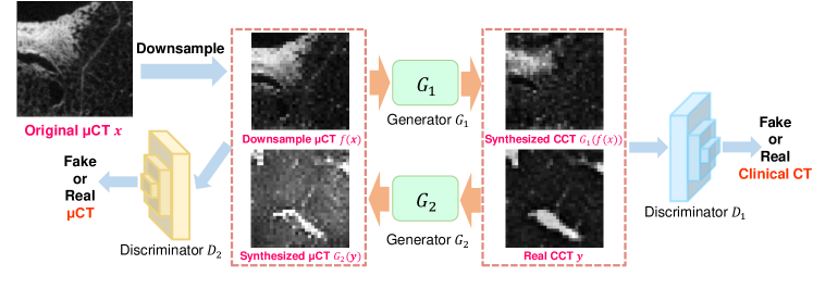
We design the mapping from the CT domain to the clinical CT domain based on a FCN. Following CycleGAN [11], we define a network as a mapping that maps an image from downsampled CT images domain to clinical CT image domain ; a network maps images from clinical CT image domain to the downsampled CT image domain . Here is defined as a gaussian pyramid downsampling function. We define a discriminator network distinguishs synthesized clinical CT-like images and real clinical CT images ; a discriminator network to distinguish CT-like images and real downsampled CT images from CT images .
Compared with the conventional CycleGAN approach, we add SSIM (structure similarity) [12] as an additional loss term. We define the SSIM loss between images and as:
| (1) |
here and denotes the average intensity of image and . and are variance of image and respectively. denotes the covariance of images and . and are constant numbers included to avoid instability.
The overall loss function can be formulated as:
| (2) |
here is the loss function of the original CycleGAN, and and are SSIM loss terms. The structure of proposed synthesize network is shown in Fig. 1.
2.2.2 Network Training
We train the synthesize network using clincial CT and CT images. Downsampled CT images ( is the downsample function) are are fed to the network . The synthesized clinical CT images which are correspond to are generated. On the other hand, clinical CT images are fed to network . The synthesized micro CT images which are correspond to are generated.
2.2.3 Building Synthesized Clinical CT - CT Dataset
We use trained generator in for synthesizing clinical CT-like images from CT images. CT images are are fed to the network to generate corresponding synthesized Clinical CT images . Large amount of corresponded and forms synthesized clinical CT - CT dataset, which is used for training SR network.
2.3 Super-resolution of Clinical CT Images using Synthesized Training Data
2.3.1 SR Network
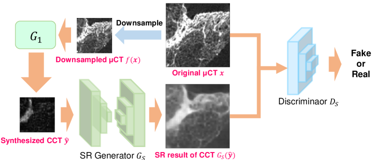
By building synthesized clinical CT - CT dataset, we are able to train a supervised SR network using synthesized clinical CT-like images and corresponding CT images . We want to learn a network that can map images in the synthesized clinical CT domain to images in CT domain .
Following the SRGAN approach [6], we also use a GAN-based network to perform SR. A super-resolution network performs SR of the input images. A discriminator network differentiates SR images and real CT images . The discriminator generator networks are trained alternatively to minimize two loss terms. The first loss term is the pixel-wise loss between a input clinical CT-like image and the desired CT output:
| (3) |
where is the distribution of the generated clinical CT-like images, and is the distribution of the micro CT images. This loss term constraints pixel-wise similarity between the input LR images and the desired output HR images. We define a second loss term that represents the adversarial loss for training the generator :
| (4) |
This loss term constraints generator for generating more realistic images that are close enough to CT domain as to fool the discriminator . The following is the total objective function for training SR generator :
| (5) |
where is the weight of adversarial loss term . The structure of the GAN-based SR network is shown in Fig. 2.
2.3.2 Network Training
We train the synthesize network using synthesized clinical CT - CT dataset. Synthesized clinical CT images are fed to network. Output is a SR image .
2.3.3 SR of clinical CT images
To perform SR of a clinical CT image , is input to trained SR generator . Then we obtain output , which is the SR image.
3 Experiments and Results
For qualitative evaluation, we applied the method to clinical CT images to obtain SR images. For quantitative evaluation, we applied the method to synthesized clinical CT images to obtain SR images, then compare SR images and corresponding CT images by SSIM.
3.1 Dataset
First, we utilize eight cases of clinical CT volumes and six cases of CT volumes for training the synthesize network. Second, we utilize five clinical CT volumes and six CT volumes for training the SR network. The CT volumes are of cancer specimens obtained after lung resection surgeries. The clinical CT volumes are acquired using a clinical CT scanner (SOMATOM Definition Flash, Siemens Inc., Munich) with a resolution of 625625600 m3 / voxel. The CT volumes were acquired using a CT scanner (inspeXio SMX90CT Plus, Shimadzu, Kyoto), with isotropic resolutions in the range of 42-52 m.
3.2 Parameter Settings
During training, we extract 2000 patches from each case. Based on the number of pixels in the lung area in clinical CT images, the size of patches extracted from the clinical CT volumes were of 3232 pixels. The size of patches extracted from the CT volumes were of 256256 pixels. Since the super-resolution always enlarged the input images to power of 2 times (2, 4, 8 times, e.g.), and comparing the resolution of the the clinical CT volumes (625m) is about ten times of the CT volumes (52m), we considered 8 times to be the most proper. For SSIM loss, we set and as 0.02 and 0.06, respectively. For wights of loss terms of synthesize network, we set to 0.5 and to 0.4. For wights of loss terms of loss SR network, we set to 0.001. Epoch number of both synthesize network and SR network is 200 with a 64-minibatch size.
3.3 Separate Training of Synthesize Network and SR Network
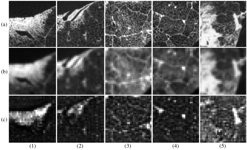
We separately train the synthesize network and the SR network. First, the synthesize network is trained to generate clinical CT-like from CT images (Synthesized clinical CT - CT pairs by are illustrated in Fig. 3.); second, the SR network is trained using synthesized clinical CT - CT images.
3.4 Results
| Method | SSIM |
|---|---|
| SR-CycleGAN [8] | 0.40 |
| Proposed Method | 0.51 |
3.4.1 Quantitative Evaluation
We used two CT volumes for quantitative evaluation. Since we do not have any corresponding clinical CT - CT pairs, we propose a novel quantitative evaluation method: first we use the trained generator network of synthesize network to generate a clinical CT-like images from a CT images , and then use trained SR generator to obtain SR image . We utilized SSIM [12] to compare the SR image and the original CT image of the conventional method (SR-CycleGAN) and our proposed method. Table 1 illustrates the quantitative results of both methods.
3.4.2 Qualitative Evaluation
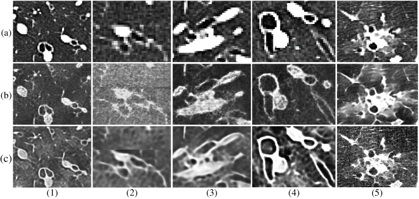
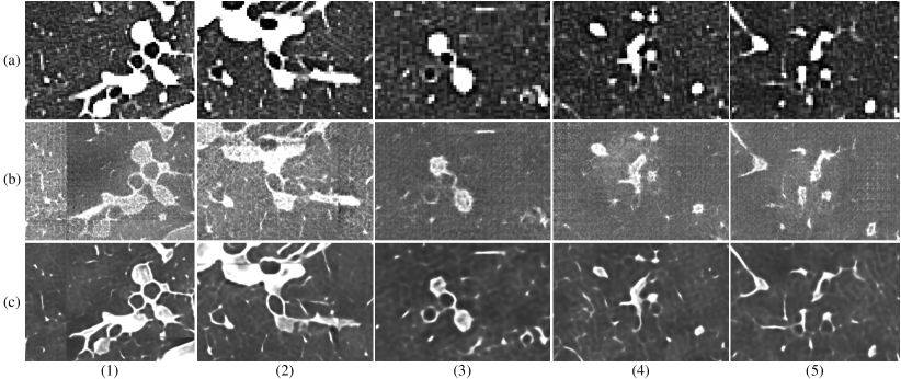
We used two clinical CT volumes for our qualitative evaluation. We utilize the synthesised clinical - CT pairs for training the SR network. We use the trained SR network for performing SR of the clinical CT images. We compare the SR results of proposed method with the SR-CycleGAN approach [8]. The results are shown in Fig. 4.
4 Discussion and Conclusion
To the best of our knowledge, our is the first study that performs SR of clinical CT images by training using synthesized clinical CT - CT pairs. Important anatomical structures such as bronchus, vein and contour of tumor are reconstructed clearly as in Fig 4: walls of bronchioles and veins become smother, and size of tumor become clearer, compared to SR-CycleGAN’s result. We consider this is because the proposed method consists of two networks for different jobs (one for modality transformation and another for SR). On the other hand, SR-CycleGAN combined modality transformation and SR in one single network, which causes training to be unstable and generate noisy results.
In this scheme, the only feasible quantitative evaluation approach is conducted by synthesizing clinical CT - CT pairs and evaluates how well synthesized clinical CT can be reconstructed to CT CT images. Identifying more convincing quantitative evaluation methods is our future work.
In this paper, we have proposed a novel unsupervised (SR) approach for performing the SR of clinical CT images. By Synthesizing clinical CT images from CT images, we solved the problem of no paired clinical CT - CT data. The proposed method outperformed conventional method qualitatively and quantitatively. The results demonstrates that our proposed method successfully performed SR of lung clinical CT images into CT level.
4.0.1 Acknowledgments
Parts of this research is supported by ********.
References
- [1] Pakzad, R., Mohammadian, A., Ghoncheh, M.: The incidence and mortality of lung cancer and their relationship to development in Asia. Transl Lung Cancer Res. 4: 763-774 (2015)
- [2] Silvestri, G.A., Gonzalez, A.V., Jantz, M.A., Margolis, M.L., Gould, M.K., Tanoue, L.T., Detterbeck, F.C.: Methods for staging non-small cell lung cancer: diagnosis and management of lung cancer. American College of Chest Physicians Evidence-Based Clinical Practice Guidelines. 143(5): 211-250 (2013)
- [3] Dong, C., Loy, C.C., He, K., Tang, X.: Image super-resolution using deep convolutional networks. IEEE Transactions on Pattern Analysis and Machine Intelligence. 38(2): 295-307 (2015)
- [4] Aggarwal, H.K., Mani, M.P., Jacob, M.: MoDL: Model-based deep learning architecture for inverse problems. IEEE Transactions on Medical Imaging. 38(2), 394-405 (2018)
- [5] Johnson, J., Alahi, A., Li F.: Perceptual losses for real-time style transfer and super-resolution. European Conference on Computer Vision. pp. 694-711 (2011)
- [6] Ledig, C., Theis, L., Huszár, F., Caballero, J., Cunningham, A., Acosta, A., Shi, W.: Photo-realistic single image super-resolution using a generative adversarial network. In Proceedings of the IEEE Conference on Computer Vision and Pattern Recognition. pp. 4681-4690 (2017)
- [7] Ravìa, D., Szczotkab, A.B., Pereirac, S.P., Vercauteren T.: Adversarial training with cycle consistency for unsupervised super-resolution in endomicroscopy. Medical Image Analysis. 53: 123-131 (2019)
- [8] Zheng, T., Oda, H., Moriya, T., Sugino, T., Nakamura, S., Oda, M., Mori, M., Takabatake, H., Natori, H., Mori, K.: Multi-modality super-resolution loss for GAN-based super-resolution of clinical CT images using micro CT image database. Prcoc. SPIE, Medical Imaging: 11313-3 (2020)
- [9] Andreas L., Danelljan, M., Timofte, R.: Unsupervised learning for real-world super-resolution. arXiv e-prints. arXiv:1909.09629 (2019)
- [10] Yuan, Y., Liu, S., Zhang, J., Zhang, Y., Dong, C., Lin, L.: Unsupervised image super-resolution using cycle-in-cycle generative adversarial networks. Proceedings of the IEEE Conference on Computer Vision and Pattern Recognition Workshops. pp. 701-710 (2018)
- [11] Zhu, J.Y., Park, T., Isola, P., Efros, A.A.: Unpaired image-to-image translation using cycle-consistent adversarial networks. Proceedings of the IEEE International Conference on Computer Vision. pp. 2223-2232 (2017)
- [12] Wang, Z., Bovik, A.C., Sheikh, H.R., Simoncelli, E.P.: Image quality assessment: from error visibility to structural similarity. IEEE Transactions on Image Processing. 13(4): 600-612 (2004)