Exploring photoelectron angular distributions emitted from molecular dimers by two delayed intense laser pulses
Abstract
We describe the results of experiments and simulations performed with the aim of extending photoelectron spectroscopy with intense laser pulses to the case of molecular compounds. Dimer frame photoelectron angular distributions generated by double ionization of N2-N2 and N2-O2 van der Waals dimers with ultrashort, intense laser pulses are measured using four-body coincidence imaging with a reaction microscope. To study the influence of the first-generated molecular ion on the ionization behavior of the remaining neutral molecule we employ a two-pulse sequence comprising of a linearly polarized and a delayed elliptically polarized laser pulse that allows distinguishing the two ionization steps. By analysis of the obtained electron momentum distributions we show that scattering of the photoelectron on the neighbouring molecular potential leads to a deformation and rotation of the photoelectron angular distribution as compared to that measured for an isolated molecule. Based on this result we demonstrate that the electron momentum space in the dimer case can be separated, allowing to extract information about the ionization pathway from the photoelectron angular distributions. Our work, when implemented with variable pulse delay, opens up the possibility of investigating light-induced electronic dynamics in molecular dimers using angularly resolved photoelectron spectroscopy with intense laser pulses.
I Introduction
A powerful method for tracing molecular dynamics induced by a pump pulse is to observe the evolution of the angular distributions of photoelectrons emitted by single- or few-photon ionization during a delayed probe pulse Stolow et al. (2004); Hertel and Radloff (2006). If combined with coincidence imaging of the photoion momenta, this technique allows for the measurement of molecular frame photoelectron angular distributions (MFPADs) of a single molecule, which can provide unique insight into the intra-molecular dynamics with femtosecond resolution Gessner et al. (2006). This method can also be implemented with an intense, elliptically polarized laser pulse as the ionizing probe Akagi et al. (2009); Staudte et al. (2009); Odenweller et al. (2011); Spanner et al. (2012); Wu et al. (2012a); Hanus et al. (2020). In that case, the rotating electric field vector of the probe pulse can be exploited for mapping laser-sub-cycle time to electron momentum, a concept known as angular streaking Maharjan et al. (2005); Eckle et al. (2008); Schöffler et al. (2016); Hanus et al. (2019); Kangaparambil et al. (2020); Hanus et al. (2020). When distortions of the such obtained MFPADs due to the parent ion’s Coulomb potential Staudte et al. (2009); Spanner et al. (2012), the field-driven electronic dynamics Odenweller et al. (2011), or the nuclear dynamics Hanus et al. (2019, 2020) that take place concomitantly with the photoelectron emission are properly accounted for, molecular dynamics can be extracted from them with sub-femtosecond resolution.
The next frontier in ultrafast intense laser science is to extend existing methods that are now routinely applied to isolated molecules, to the study of dynamics in more complicated systems such as molecules in a compound. This effort is motived by the fact that most molecular processes in nature or technical applications do not take place within a single molecule isolated in vacuum but between different molecules. A famous example for such an inter-molecular process is proton coupled electron transfer Weinberg et al. (2012), which is of key importance in biology, chemistry, and technology. A widely used approach to study photoinduced inter-molecular processes in femtosecond pump-probe experiments is to use small molecular van der Waals clusters and dimers as model systems Stolow et al. (2004); Hertel and Radloff (2006). In recent years, atomic and molecular dimers have also found increasing attention in experiments that study their ionization and fragmentation dynamics when driven by a strong laser fields Manschwetus et al. (2010); Ulrich et al. (2010); Wu et al. (2011a); Ulrich et al. (2011); Wu et al. (2011b, 2012b); Hoshina et al. (2012); Wu et al. (2012c); von Veltheim et al. (2013); Wu et al. (2014); Amada et al. (2015); Ding et al. (2017); Cheng et al. (2017); Wang et al. (2020). It was demonstrated that the analysis of the momenta of photoions or photoelectrons emitted during ionization-fragmentation of dimers can provide detailed insight into, e.g., the ionization dynamics Wu et al. (2013), the structural deformation induced by the strong-field interaction Amada et al. (2015), the influence of a nearby charge on the dissociation behaviour of molecules Ding et al. (2017), or into laser-sub-cycle electron transfer processes Wang et al. (2020). A common finding of these studies is that during multiple ionization the nearby charge from the neighbouring ion can have a decisive impact on the further ionization and/or fragmentation dynamics of the partner molecule.
In this Article we describe experiments and simulations performed with the aim of investigating to what extent photoelectron angular distributions (PADs) measured with a strong, elliptically polarized laser field can be used to extract structural and dynamical information from molecules bound in a hetero-dimer complex. To this end we studied with a reaction microscope Ullrich et al. (1997); Dörner et al. (2000); Hanus et al. (2019, 2020); Kangaparambil et al. (2020) the PADs from sequential double ionization of N2-N2 and N2-O2 dimers during two delayed intense laser pulses in coincidence with the photoions ejected during subsequent fragmentation of the dimers. Our main concern was to understand which information about the laser-molecule interaction is contained in the measured PADs and how this information can be extracted. The double-pulse approach in combination with the coincidence measurement permitted us to reconstruct PADs in the dimer frame of reference (referred to as DFPADs) specifically for the second ionization step. This allowed us to identify the influence of a nearby molecular ion on the ionization and fragmentation behaviour of a neutral molecule in measured DFPADs.
From our measurements we find that the nearby charge of the first-created molecular ion mainly leads to a rotation of the measured photoelectron angular distribution as compared to the isolated molecule case, whereas its overall shape is to a large degree preserved. Thus, one of the results of this study is that DFPADs can, with adaptations, be read in a similar way as MFPADs. Furthermore, by applying methods based on angular streaking developed previously Hanus et al. (2019); Kangaparambil et al. (2020); Hanus et al. (2020) and with the support from semi-classical trajectory simulations we are able to show that the dominant process underlying the rotation is scattering of the second-emitted electron on the neighbouring molecular ion as it is driven away by the strong laser field. This finding adds to the emerging evidence for the key importance of laser-sub-cycle electron scattering dynamics in strong-field driven dimers Wang et al. (2020) and clusters Treiber et al. (2020).
II Experiments
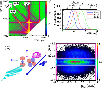
The reaction microscope used in our experiments to measure in coincidence the momenta of two molecular ions and two electrons emerging from the interaction of a sequence of two intense laser pulses with a cold jet of molecules generated by ultrasonic expansion of N2 and O2 gas is described elsewhere Hanus et al. (2019, 2020); Kangaparambil et al. (2020). The laser pulses used in the experiments had both a central wavelength of nm and a duration of about . Since the gas is strongly cooled during the ultrasonic expansion, molecular dimers, as the pre-cursors of a quantum fluid, may be formed during the ultrasonic expansion Ullrich et al. (1997); Dörner et al. (2000). When these dimers interact with the intense laser pulses, each molecule in the dimer might become singly ionized, leading to Coulomb explosion of the doubly ionized dimer complex. The signatures of these Coulomb explosions are shown in the photoion-photoion coincidence (PIPICO) histogram in Fig. 1(a) as the sharp lines. These lines reflect the momentum conservation between the two exploding fragment ions and therefore are clear signatures existence of a certain molecular species in the cold gas jet. The histogram in Fig. 1(a) shows that in our experiment N2-N2, O2-O2 and N2-O2 dimers were formed during the ultrasonic expansion and Coulomb exploded during the interaction with the strong laser pulses. The laser interaction took place in an ultra-high vacuum chamber (base pressure 0.910-10 mbar). Ions and electrons emerging from the interaction volume were guided by weak electric (19 V/cm) and magnetic fields (12 G) to two separate position-sensitive detectors. From the time-of-flight and impact position of each detected particle its momentum right after the laser interaction was calculated.
We optimized the expansion conditions for a high production rate of N2-O2 dimers in the jet, since we were interested in the influence of the molecular species on the ionization and fragmentation dynamics. Hence, O2 was set to have lower abundance in the gas jet as compared to N2. This led to low yield of O2-O2 dimers and, thus, to low statistic in their DFPADs. Therefore, in the following, we will not discuss angularly resolved data for O2-O2 dimers.
During Coulomb explosion of a dimer into two charged molecular ions with masses , the electrostatic potential energy is released as kinetic energy of the two ions with momenta . The kinetic energy released (KER) during this explosion, , is a precise measure of the intermolecular distance, , of the dimer Vager et al. (1989). The measurement revealed, cf. Fig. 1(b), that is the largest for the O2-O2 dimer, hence it exhibits the smallest intermolecular distance of 5.94 a.u. The smallest energy release was measured for the N2-N2 dimer, corresponding to a van der Waals bond length of 7.18 a.u. An intermediate distance of a.u. was measured for the heteronuclear dimer N2-O2.
The KER distributions in Fig. 1(b) were obtained when both laser pulses of the double-pulse sequence were used for Coulomb exploding the dimers. As we will describe in detail below, the combined use of a linearly polarized pump pulse and a preceding circularly polarized probe pulse, delayed to the pump by 33 ps, cf. sketch in Fig. 1(c), allowed us to distinguish the two possible ionization sequences that can initiate the fragmentations leading to the KER distributions in Fig. 1(b). These two possibilities are: (i) both ionization events take place within the duration of one of the two applied pulses (i.e., within a few tens of femtoseconds), (ii) one ionization event takes place during the pump pulse, the second one during the probe pulse (delay about 33 ps). Exploiting this opportunities we found that the measured KER distributions corresponding to the two possibilities agree within experimental errors [not shown in Fig. 1(b)]. This indicates that the dimers’ bond stretch dynamics is negligible during the 33 ps delay. Thus, the most likely scenario is that the potential energy curves of the singly charged dimers are binding and of very similar shapes as those of the neutral dimers.
Because in a two-body Coulomb explosion the two particles are ejected back-to-back, the measured ions’ momenta directly reflect the orientation of the dimer axis in the laser focus at the time of interaction, provided the explosion happens much faster than the rotational dynamics that might be induced during the laser interaction Tong et al. (2005). As for the large size and mass of the dimers this is well fulfilled, the dimer axis and therewith DFPADs can be reconstructed from the measured electron and ion momenta.
III Double-pulse approach to distinguish the two ionization steps
Our goal was to measure the DFPAD generated by an elliptically polarized laser pulse when one of the two molecules in the dimer was ionized by a preceding pulse. For such measurement it is necessary to discriminate the first emitted from the second emitted electron in the detected electrons. This discrimination cannot be simply made based on the flight times of the two electrons detected in coincidence, as the first detected could be the second emitted electron and vice versa. Therefore, we employed a technique that exploits the different shape of the electron momentum distributions produced by linearly and elliptically polarized laser pulses Song et al. (2016): Electrons released in linearly polarized light exhibit small momenta perpendicular to the light polarization direction and therefore cover regions in an electron momentum distribution that are dominantly aligned along the polarization axis. In contrast, electrons released in elliptically polarized light are angularly streaked and cover momentum regions with large perpendicular momentum that resemble circle segments Schöffler et al. (2016); Hanus et al. (2019, 2020). Thus, it becomes possible to minimize the momentum space overlap between two electrons released in a two-pulse sequence consisting of a linearly polarized pump laser pulse and a delayed, elliptically polarized probe pulse, when during each pulse only one electron is emitted, cf. sketch in Fig. 1(c). In our experiments the helicity of the elliptically polarized probe pulse was chosen such that the electric field vector rotates clockwise in the electron momentum plane. The delay between the two pulses was set constant to in order to ensure that the pulses do not overlap and that the prompt alignment of dimers or possible revivals of the alignment are avoided (Xie et al., 2014; Kumarappan et al., 2007; Wu et al., 2011b).
The pulse peak intensities of the two pulses were chosen as for both pulses. The intensities were calibrated in situ using the proton distribution from the dissociation of hydrogen, H2, from the background gas in the ultra-high vacuum chamber Alnaser et al. (2004). For these intensities, chosen because they are typical for strong-field experiments that investigate laser-ionization, an optimization of the ellipticity that produced minimal overlap of the two electron momentum distributions from each pulse yielded . Here, and are the laser peak field strengths perpendicular and parallel to the main axis of the polarization ellipse of the elliptically polarized pulse, which was aligned with the polarization direction of the linearly polarized laser pulse. Throughout the Article this axis is assumed along the horizontal direction. The electron momentum distribution measured with these pulse sequences is shown in Fig. 1(d) exemplary for N2. The distributions for the dimers look similar.
Provided that all ions and electrons are detected in coincidence, as was done in our experiment, the double-pulse approach enables the selection of dimer fragmentation events when the first electron was exclusively released by the pump pulse and the second electron exclusively by the probe pulse. To reconstruct the MFPADs respectively DFPADs corresponding to the second ionization step, we separated the measured electron momentum distributions in the laser polarization plane into three regions using , the electrons’ momentum perpendicular to the polarization direction of the linear pulse, as the decisive parameters, see colored boxes in Fig. 1(d). Electrons with a.u. [cyan box in Fig. 1(d)] were considered to be emitted during the linearly polarized first pulse. Electrons with a.u. [pink boxes in Fig. 1(d)] were considered to be emitted during the elliptically polarized second pulse. We only considered those coincidence events, where one of the two electrons was in the cyan-bordered momentum region and the other electron in one of the two pink-bordered regions. All other coincidence events were discarded. This separation works perfectly only for negligible overlap of the electron momentum distributions due to the linear and elliptical pulse, respectively. In practice, a small overlap of the wings of two momentum distributions is unavoidable. Three main types of systematic errors resulting from this overlap can be distinguished: (i) The electron emitted during the elliptical pulse is emitted with a.u., (ii) the electron emitted during the linear pulse is emitted with a.u., (iii) both electrons are emitted during the elliptical pulse, one with a.u., the other one with a.u. By fitting the three main lobes in the electron momentum distributions [that all have a similar shape as the one in Fig. 1(d)] with three Gaussians and exploiting coincidence arguments, we found that only case (iii) can result in noteworthy errors. However, even for this case this is only problematic, when the first emitted electron reaches a.u. and the second emitted only a.u. But as the first emission happens most likely during the onset and rising slope of the pulse, while the second emission is expected to happen around its peak, even this error has a small probability. In sum, we found that the double-pulse method with well-chosen relative peak intensities, as it is the case in our experiment, is relatively insensitive to the unavoidable overlap of the wings of the electron momentum distributions.
From the coincidence events that remained after demanding that the first electron was emitted during the linear pulse (with a.u.) and the second one during the elliptical pulse (with a.u.), we furthermore discarded those events where the ejection direction of the ionic fragments was perpendicular to the major axis of the polarization ellipse: We only considered those coincidence events where the internuclear axis in the case of molecules, respectively the intermolecular axis in the case of dimers, were oriented within an angle of to the major axis of the polarization ellipse. The orientation of the molecules/dimers was calculated from the fragment ions’ momentum vectors . The reason for this selection was motivated by our recent work Ref. Wang et al. (2020) in which we found that the ionization behaviour of a dimer is strongly influenced by laser-driven electron scattering and transfer processes between the two entities of the dimer. To investigate the influence of such electron scattering processes with the double-pulse method, the dimer axis should be roughly aligned along the dominant ionization direction where the field strength is largest, i.e., the major axis of the polarization ellipse. If the dimers’ axes were aligned perpendicular to this direction (i.e., along the minor axis), scattering of the ionizing electron on the opposing molecular entity is expected to be of less importance. Again, all coincidence events where the dimers/molecules were not aligned within to the major axis were discarded.
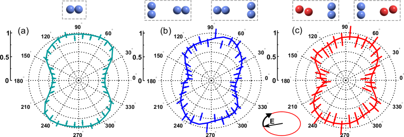
For the remaining coincidence events, one of the two detected ions (either N+, N, or O, depending on the molecule/dimer considered) was taken as the reference fragment ion and its momentum vector was rotated, together with the electron momentum vectors, into the positive horizontal axis. Then, we calculated for the electrons in the pink boxes in Fig. 1(d), that are attributed to emissions during the elliptically polarized probe laser pulse, the angle between the reference ion and the electron momentum vector, denoted by in the following. Plotting the electron yield as a function of yielded, finally, the MFPADs/DFPADs shown in Fig. 2.
As for the N2 molecule and the N2-N2 dimer the two ions are indistinguishable, we mirrored the MFPADs/DFPADs about the vertical axis. The same was done for the N2-O2 dimer in Fig. 2(c), yielding a DFPAD where both orientations (N2 ejected to the right, and N2 ejected to the left) are superposed. A DFPAD for the N2-O2 dimer, where the electron and ion momenta were rotated together such that the momentum vector of N2 exclusively points to the right, is shown in Fig. 3(e) and will be discussed in detail below.
IV Analysis of measured dimer-frame photoelectron angular distributions (DFPADs)
To understand the information contained in DFPADs, we first compare them to the more familiar case of an MFPAD measured for a Coulomb exploding doubly ionized molecule. Fig. 2(a) shows the MFPAD for the N2 molecule measured with the double-pulse sequence, where the first ionization event takes place during the linearly polarized pump pulse and the second one during the elliptically polarized probe pulse. Figs. 2(b) and 2(c) show the DFPADs for the N2-N2 and N2-O2 dimers measured with exactly the same pulse sequence. The MFPAD features a symmetric shape about the symmetry axis along , with the angular coordinate. The small ca. 5∘-rotation from that would be expected in angular streaking for an elliptically polarized pulse with its major axis of the polarization ellipse along , may be attributed to the influence of the Coulomb field on the emitted electron Spanner et al. (2012). In contrast to the symmetric MFPAD, the DFPADs feature a non-symmetric shape about the axis: For the upper hemisphere, , the DFPADs contain a larger number of events in the range . This asymmetry is even more pronounced in the DFPAD measured for N2-O2 shown in Fig. 2(c).
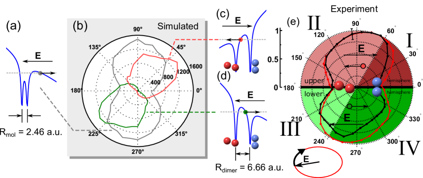
To explain the reason for the different shapes of the MFPAD and the DFPADs depicted in Fig. 2 we turn to discussing the differences in the binding potentials from which the second electron is detached. The photoelectron distributions plotted in Fig. 2 reflect the angular dependence of the second ionization step. In a simplified picture we can assume that the second electron, at the instant of its emission, is bound in a potential well that is defined by two positive charge centres. In the case of a molecule these centres are close to each other and there is no considerable barrier between them, see the sketch in Fig. 3(a). When the electron tunnels through the field-suppressed barrier it directly reaches the continuum. If the two centres are further apart, as it is the case for a dimer, two situations can be distinguished: The electron can be either released from the down-field or the up-field well. An electron released from the down-field can tunnel directly to the continuum similar to the molecular case sketched in Fig. 3(a). In contrast, when an electron tunnels from the up-field well, as sketched in Figs. 3(c) and 3(d), there is a possibility that the electron, as it is driven away by the laser electric field, scatters on the down-field charge centre.
In the following we will show by an in-depth analysis of the measured DFPADs, supported by results of semiclassical trajectory Monte Carlo simulations, that the asymmetry in the measured DFPADs in Figs. 2(b) and 2(c) is indeed caused by scattering of electrons released from the up-field well on the opposing charge center. To guide our analysis, we simulate the momentum distribution of the electron that is emitted during the elliptically polarized pulse from a model binding potential by tunnel ionization and subsequently is driven by the combined forces due to the laser electric field and a binding potential situated at some distance from the electron’s parent binding potential. The neighbouring binding potential mimics the positively charged ion created by ionization during the preceding linearly polarized pump pulse. The electron released during the linear pulse is not simulated and it is assumed that this electron has already left the interaction volume. This is perfectly justified given the delay of 33 ps between the two pulses. Thus, the potential felt by the ionizing and laser-driven electron has a Coulomb double-well shape. To avoid numerical problems due to the Coulomb singularity we used soft-core potentials of the form
| (1) |
where is the Coulomb softening parameter that was set to 0.1 for all calculations, and r is the position of the simulated electron. At position the singly charged neighbouring ion is situated, at is the parent ion of the electron. The separation of the two ions was chosen as a.u. for the N2 molecule and a.u. for the N2-O2 dimer. At every instant of the discrete temporal mesh the starting position of the electron, , i.e., the tunnel exit sketched in Figs. 3(a,c,d), was calculated by numerically solving the equation
| (2) |
using the secant method. Here, is the vectorial laser field and is the assumed ionization potential. At each instant of the temporal grid a trajectory was launched at the position with a probability given by the molecular ADK ionization theory (Tong et al., 2002). As the sole purpose of the simulations was to obtain a qualitative understanding of the ionization behaviour and the subsequent laser-driven free electron dynamics rather than obtaining quantitative agreement with the measured data, we used, for the sake of simplicity, the parameters of the helium atom in the ADK formula, where the was adapted to that of the molecules. The trajectories were propagated in three dimensions for an elliptically polarized laser pulse with the same parameters as in the experiments using the Runge-Kutta integration scheme. The helicity of the elliptically polarized laser pulse was, as in the experiments, assumed clockwise. From the final momentum value of each trajectory at time (long after the pulse) the electron momentum distribution was obtained by binning all launched trajectories.
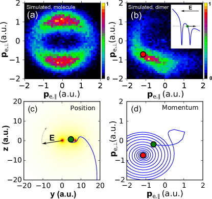
The simulation for the molecular-type potential with the shorter internuclear distance resulted in a symmetric MFPAD with its symmetry axis around [see gray line in Fig. 3(b)], similar to the measured MFPAD in Fig. 2(a). This case also applies to the dimer-case when the potoelectrons are emitted from the down-field potential well, as in both cases the electrons tunnel directly into the continuum [cf. Fig. 3(a)]. In contrast, the simulated angular distribution of photoelectrons emitted from the up-field potential well of the dimer-like potential with the larger intermolecular separation features a stronger rotation and a clearly visible distortion, in agreement with the measurement, cf. Fig. 3(b). The angular distribution in Fig. 3(b) is separated into two cases: The red line shows the DFPAD for electrons emitted to the left, the green line for electrons emitted to right [cf. the sketches Figs. 3(c) and (d)]. In both cases the DFPAD in Fig. 3(b) shows a strong rotation. The simulated photoelectron momentum distributions from which the MFPAD and the green-colored half of the DFPAD in Fig. 3(b) are calculated are shown in Figs. 4(a) and 4(b), respectively.
The simulations, thus, predict a stronger rotation and distortion of the angular distributions of photoelectrons emitted from the upper potential [red and green lines in Fig. 3(b)] as compared to photoelectrons emitted from the lower potential well [gray line in Fig. 3(b)]. By a detailed analysis of the electrons’ trajectories we found that the dominant reason for this stronger rotation is that the photoelectrons emitted from the up-field potential well, scatter off the neighbouring potential well as they are driven away by the strong laser field. To visualize this process we show in Figs. 4(c) and 4(d) the temporal evolution of an example trajectory in real space and in momentum space, respectively. The green dot depicts the trajectory’s starting conditions (space and momentum). The red dot indicates the final momentum [also indicated in Fig. 4(b)]. For trajectories that make up the red half of the simulated DFPAD in Fig. 3(b) the situation is reversed and the trajectories emitted from the right well scatter on the left potential well. Thus, based on our semiclassical trajectory simulations we attribute the rotation and distortion of the measured DFPADs in Figs. 2(b) and (c) to scattering of photoelectrons emitted from the up-field potential well on the charge in the opposing potential well of the partner molecule in the dimer.
V Information retrieval from the DFPADs
We now turn to discussing the information that is contained in DFPADs measured with strong, elliptically polarized laser fields and how it can be extracted. It was shown above that a DFPAD can be quite different from the corresponding MFPAD. Specifically we have shown that the nearby presence of a molecular ion results in a notable rotation and deformation of the photoelectron angular distribution from the dimer. In the following we will show for the example of the heterodimer N2-O2 that, despite the distortions that are introduced by the presence of the neighbouring molecular ion in the DFPAD, it is still possible to extract from it detailed information about the ionization process in the elliptically polarized pulse and the nature of the distortion due to the neighouring ion.
To start the discussion, we note that double ionization and fragmentation of the N2-O2 dimer in the pump-probe scheme of our experiment can proceed via two pathways. In the first pathway, the N2 molecule is ionized during the linearly polarized pump pulse and subsequently the O2 molecule becomes ionized by the elliptically polarized probe pulse, see Equ. (3). During the second pathway the sequence of ionization is reversed, see Equ. (4).
| (3) | |||||
| (4) |
In both pathways the ionization process during the pump pulse takes place within a neutral dimer. The ionization dynamics of the second molecule during the probe pulse, in contrast, takes place in the vicinity of a molecular ion. The presence of this ion may modify the ionization process of the second molecule. Which ionization step will be modified more strongly by the presence of the neighbouring ion as compared to the neutral case, the one of O2 or of N2?
To explore this question, one should evaluate the probability ratio of pathways (3) and (4). Is it possible to extract this ratio from the angular distribution of the second electron that is emitted from the heteromolecular dimer? As we will show in the following, the answer to this question is yes. In our experiment we measure the electrons and ions emitted in coincidence. Therefore, we can display the PAD in the dimer frame, which results in a DFPAD. The crucial point is, however, that for a heterodimer, the DFPAD can be further refined by not only fixing the dimer-axis along the, e.g., horizontal, axis, but also by orienting the dimer with respect to the molecular species. While in Fig. 2(c) the orientations (N2 ejected to the left/right or O2 ejected to the left/right) were not considered and therefore Fig. 2(c) is a superposition of both cases, the DFPAD displayed in Fig. 3(e) is plotted such that N2 is always ejected to the right.
Under this condition the oriented DFPAD can be separated into two halves by resorting to the concept of angular streaking Maharjan et al. (2005); Hanus et al. (2019, 2020). For the clockwise helicity of the elliptical laser field used in our experiment electrons are streaked into the upper half [colored in red in Fig. 3(e)] when the laser electric field vector points from O2 to N2, cf. sketch in Fig. 3(c). In contrast, the green-colored lower half in Fig. 3(e) corresponds to electrons that are emitted when the laser field vector points from N2 to O2, cf. sketch in Fig. 3(d). If we compare the shapes of the two halves in the oriented DFPAD in Fig. 3(e) we notice that they are distinctly different. To highlight this difference we overlaid in Fig. 3(e) the non-oriented DFPAD from Fig. 2(c) by a red line. By comparison of the oriented DFPAD with this red line it becomes obvious that the upper, red-colored half comprises of significantly more electrons. Thus, we can conclude that obviously the second ionization step happens with a higher probability when the laser electric field vector points from O2 site to N2 [sketch in Fig. 3(c)]. This does, however, not imply that the second electron is more likely emitted from O2 in the down-field well of the dimer. Thus, to quantify the probability ratio and to determine how many electrons are emitted from O2 respectively N2, it is not simply possible to integrate the upper and lower halves of the DFPAD in Fig. 3(e).
The reason is that for dimers the laser field driven trajectories of the emitted electrons are strongly influenced by the potential of the neighbouring molecular ion, as we have discussed above. Consquently, electrons that are emitted when the electric field vector points into a certain direction may not be detected under an angle of 90∘ to the field direction, as it is predicted by angular streaking based on the strong-field approximation (SFA) Faisal (1973); Reiss (1980) that neglects the influence of any ionic potential on the final momentum of the photoelectron. But due to scattering on the neighbouring molecular ion, in particular electrons from the up-field potential well may be streaked into a distinctively different direction. This leads to deformations, but mainly to a global rotation of the overall DFPAD as compared to the SFA prediction. This was clearly shown by the results of our simulations, cf. Fig. 3(b). Based on the finding described above that the dominant deviation from the SFA prediction is a rotation of the angular distribution of photoelectrons emitted from the up-field potential well along the clockwise rotation of the laser electric field vector, we can, however, separate the scattered from the non-scattered electrons.
To understand the rotation due to scattering, we turn to the simulated results in Fig. 3(b). This figure shows that electrons that are emitted from the down-field potential well directly into the continuum (gray line), will only be marginally affected by the Coulomb binding potential. The overall effect is that they, when emitted dominantly around the peaks of the laser field along the main axis of the laser polarization ellipse (at and ), will be deflected only by a few degrees further along the laser field vector rotation direction (clockwise in our case) than the 90∘ predicted by the SFA. As a consequence, their PAD shows peaks around and . In contrast, electrons that are emitted from the up-field potential well are scattered on the neighbouring well, leading to a much larger rotation of the angular distribution. Our model, which reproduced the measured DFPADs reasonably well, predicts that the additional rotation due to scattering is about 40∘, such that the angular distribution of these electrons shows peaks around and , see red and green lines in Fig. 3(b). Based on these values we divide each half of the DFPAD in Fig. 3(e) into two segments using 60∘ respectively 240∘ as the borders. This results in the four segments denoted I to IV, indicated by darker and lighter red and green shading in Fig. 3(e). We note that the scattered and non-scattered regions in the DFPAD certainly overlap to some extent. It is therefore intrinsically impossible to unequivocally disentangle these regions using sharp angular limits. As a result, precise quantitative results cannot be expected from such a division of the DFPAD. The purpose of the analysis described below is, however, not to provide quantitative results, but to demonstrate a possible approach to disentangling different processes in measured DFPADs. We think that this approach could be extended to a qualitative level using a more advanced division of the DFPAD.
In the following we discuss how the regions I to IV defined by the sharp angular limits can be used to estimate the emission percentage of the second electron from the up-field and down-field potential wells. And subsequently, how from this information an estimate for the branching ratio between pathways (3) and (4) can be obtained. Region I in the upper half of the DFPAD corresponds to electrons that were scattered after their release from the up-field potential well in the situation depicted in Fig. 3(c). Thus, they can be attributed to emission from N2. Electrons in region II were not scattered. Therefore, they were emitted from the down-field well, i.e., from O2. With the same logic, region III contains scattered electrons emitted from O2, while region IV corresponds to non-scattered electrons emitted from N2. With this assignment, it is now simple to estimate the percentage of the two pathways described by Equs. (3) and (4): Regions I and IV contain electrons emitted from N2 during the probe pulse (second ionization step), thus, represent pathway (4). Regions II and III contain electrons emitted from O2 during the second ionization step and, thus, represent pathway (3). By integrating the numbers in these regions we obtain that 54% of the total counts are due to pathway (3), while only 46% of all events follow pathway (4). We emphasize once more that these numbers are of no specific value. It is the demonstration of an approach that we are interested in rather than obtaining an exact result. We have, however, checked by small variations of the angular limits in the DFPAD that the numbers are relatively robust with respect to the specific choice of the limits. Hence, despite the intrinsic uncertainties we would like to conclude that in our experiment the pathway when the O2 is ionized in the second ionization step is somewhat more likely. This can be interpreted such that the two ionization steps are not completely independent of each other.
Which factors could be responsible for this ionization behaviour? An ionization process that favors electron emission from a specific potential well in a molecule exposed to a strong field is enhanced ionization (EI) Codling et al. (1989); Xie et al. (2014). It has been shown that this molecular ionization process takes place in almost the same manner also in dimers Wu et al. (2012c). However, EI favors electron emission from the up-field well, while in our experiment the more probable electron emission from O2 takes place for the down-field well. Thus, EI cannot be hold responsible for the ionization dynamics observed in our experiment. A more likely reason for the observed preponderance of pathway (3) is the influence of the structure of the heteromolecular dimer on the ionization probability during the two-step double-ionization in our double-pulse experiment. The ionization probability of an isolated molecule with a given valence electron configuration and binding energy depends mainly on the orientation of the molecule with respect to the laser field. If two molecules are bound together by van der Waals forces, a number of different dimer configurations become possible Hoshina et al. (2012); Gong et al. (2013), for example T-shaped, X-shaped, parallel, linear, etc. Thus, not only the valence structure of the two molecules and their different ionization potentials, but also the dimer geometry determines which dimers from the randomly oriented ensemble in the utrasonic gas jet are preponderantly ionized. Therefore, in a double pulse scheme, particularly when the two pulses have different polarization states, as in our experiment, the probability of a particular pathway is determined by many convoluted parameters. A detailed analysis of the hypothesis that the dimer structure is responsible for the slightly favorable ionization of O2 during the second ionization step is, however, beyond the present work and must be left for future work.
VI Summary
In summary, we described the results of experiments and simulations performed with the aim of exploring whether and how information extraction from laser-generated photoelectron angular distributions (PADs), a standard method in atomic and molecular physics, can be extended to the case of molecular compounds. To this end we have studied strong-field double-ionization of homo- and heteromolecular dimers of O2 and N2 formed by van der Waals binding forces. To distinguish the two ionization steps we applied two delayed ultrashort intense laser pulses, where the first pulse was linearly polarized and the second one elliptically polarized. In combination with four-body coincidence imaging using a reaction microscope this allowed us to measure dimer-frame photoelectron angular distributions (DFPADs). By a detailed analysis of the DFPAD for the heteromolecular O2-N2 dimer we showed that the PAD of a molecular dimer is deformed and rotated as compared to the PAD of an isolated molecule. With the help of simulations we showed that these distortions are mainly due to scattering of electrons emitted from the up-field potential well on the potential of the molecular ion formed during the first ionization step. Building on this finding, we demonstrated that by dividing the DFPAD into regions that mainly contain scattered electrons, and regions containing mainly non-scattered electrons, it becomes possible to overcome the complications due to the DFPAD-distortion and to extract information about the ionization pathway.
The results of our study point to a promising possibility of extracting information about bound electron dynamics induced by a pump pulse from a DFPAD. Such extraction has been the subject of many works on isolated molecules Lochbrunner et al. (2001); Gessner et al. (2006); Akagi et al. (2009); Staudte et al. (2009); Odenweller et al. (2011); Spanner et al. (2012); Wu et al. (2012a); Hanus et al. (2020). The underlying principle of these efforts is that the angular ionization probability reflects the intra-molecular bound electron density. Thus, the bound electronic dynamics becomes encoded in the measured MFPAD. In turn, by a time-resolved measurement of the MFPAD it becomes possible to obtain insight into the electronic dynamics in molecules.
Our work investigates the case of molecular dimers and shows that the resulting DFPAD contains valuable information about the electronic dynamics during laser-dimer interaction. Moreover, we demonstrate a possible route to extract this information. Thus, if the DFPADs are measured with a variable delay between the first and second ionization step (rather than only for one value of the delay as in our work), the femtosecond evolution of dynamics in dimer-molecules induced by the pump pulse can be extracted.
Acknowledgements.
This work was supported by the Austrian Science Fund (FWF), Grants No. P28475-N27 and P30465-N27.References
- Stolow et al. (2004) A. Stolow, A. E. Bragg, and D. M. Neumark, Chem. Rev. 104, 1719 (2004).
- Hertel and Radloff (2006) I. V. Hertel and W. Radloff, Reports Prog. Phys. 69, 1897 (2006).
- Gessner et al. (2006) O. Gessner, A. M. D. Lee, J. P. Shaffer, H. Reisler, S. V. Levchenko, a. I. Krylov, J. G. Underwood, H. Shi, a. L. L. East, D. M. Wardlaw, E. T. H. Chrysostom, C. C. Hayden, and A. Stolow, Science 311, 219 (2006).
- Akagi et al. (2009) H. Akagi, T. Otobe, A. Staudte, A. Shiner, F. Turner, R. Dörner, D. M. Villeneuve, and P. B. Corkum, Science 325, 1364 (2009).
- Staudte et al. (2009) A. Staudte, S. Patchkovskii, D. Pavičić, H. Akagi, O. Smirnova, D. Zeidler, M. Meckel, D. M. Villeneuve, R. Dörner, M. Y. Ivanov, and P. B. Corkum, Phys. Rev. Lett. 102, 033004 (2009).
- Odenweller et al. (2011) M. Odenweller, N. Takemoto, A. Vredenborg, K. Cole, K. Pahl, J. Titze, L. P. H. Schmidt, T. Jahnke, R. Dörner, and A. Becker, Phys. Rev. Lett. 107, 143004 (2011).
- Spanner et al. (2012) M. Spanner, S. Gräfe, S. Chelkowski, D. Pavičić, M. Meckel, D. Zeidler, a. B. Bardon, B. Ulrich, a. D. Bandrauk, D. M. Villeneuve, R. Dörner, P. B. Corkum, and A. Staudte, J. Phys. B At. Mol. Opt. Phys. 45, 194011 (2012).
- Wu et al. (2012a) J. Wu, L. Schmidt, M. Kunitski, M. Meckel, S. Voss, H. Sann, H. Kim, T. Jahnke, A. Czasch, and R. Dörner, Phys. Rev. Lett. 108, 183001 (2012a).
- Hanus et al. (2020) V. Hanus, S. Kangaparambil, S. Larimian, M. Dorner-Kirchner, X. Xie, M. S. Schöffler, G. G. Paulus, A. Baltuška, A. Staudte, and M. Kitzler-Zeiler, Phys. Rev. Lett. 124, 103201 (2020) .
- Maharjan et al. (2005) C. Maharjan, A. Alnaser, X. Tong, B. Ulrich, P. Ranitovic, S. Ghimire, Z. Chang, I. Litvinyuk, and C. Cocke, Phys. Rev. A 72, 041403 (2005).
- Eckle et al. (2008) P. Eckle, M. Smolarski, P. Schlup, J. Biegert, A. Staudte, M. Schöffler, H. G. Muller, R. Dörner, and U. Keller, Nat. Phys. 4, 565 (2008).
- Schöffler et al. (2016) M. S. Schöffler, X. Xie, P. Wustelt, M. Möller, S. Roither, D. Kartashov, A. M. Sayler, A. Baltuska, G. G. Paulus, and M. Kitzler, Phys. Rev. A 93, 063421 (2016).
- Hanus et al. (2019) V. Hanus, S. Kangaparambil, S. Larimian, M. Dorner-Kirchner, X. Xie, M. S. Schöffler, G. G. Paulus, A. Baltuška, A. Staudte, and M. Kitzler-Zeiler, Phys. Rev. Lett. 123, 263201 (2019) .
- Kangaparambil et al. (2020) S. Kangaparambil, V. Hanus, M. Dorner-Kirchner, P. He, S. Larimian, G. Paulus, A. Baltuška, X. Xie, K. Yamanouchi, F. He, E. Lötstedt, and M. Kitzler-Zeiler, Phys. Rev. Lett. 125, 023202 (2020) .
- Weinberg et al. (2012) D. R. Weinberg, C. J. Gagliardi, J. F. Hull, C. F. Murphy, C. A. Kent, B. C. Westlake, A. Paul, D. H. Ess, D. G. McCafferty, and T. J. Meyer, Chem. Rev. 112, 4016 (2012).
- Manschwetus et al. (2010) B. Manschwetus, H. Rottke, G. Steinmeyer, L. Foucar, A. Czasch, H. Schmidt-Böcking, and W. Sandner, Phys. Rev. A 82, 013413 (2010).
- Ulrich et al. (2010) B. Ulrich, A. Vredenborg, A. Malakzadeh, M. Meckel, K. Cole, M. Smolarski, Z. Chang, T. Jahnke, and R. Dörner, Phys. Rev. A - At. Mol. Opt. Phys. 82, 013412 (2010).
- Wu et al. (2011a) J. Wu, A. Vredenborg, B. Ulrich, L. Schmidt, M. Meckel, S. Voss, H. Sann, H. Kim, T. Jahnke, and R. Dörner, Phys. Rev. Lett. 107, 043003 (2011a).
- Ulrich et al. (2011) B. Ulrich, A. Vredenborg, A. Malakzadeh, L. P. H. Schmidt, T. Havermeier, M. Meckel, K. Cole, M. Smolarski, Z. Chang, T. Jahnke, and R. Dörner, J. Phys. Chem. A 115, 6936 (2011).
- Wu et al. (2011b) J. Wu, A. Vredenborg, B. Ulrich, L. P. H. Schmidt, M. Meckel, S. Voss, H. Sann, H. Kim, T. Jahnke, and R. Dörner, Phys. Rev. A - At. Mol. Opt. Phys. 83, 061403 (2011b).
- Wu et al. (2012b) J. Wu, M. Kunitski, L. P. H. Schmidt, T. Jahnke, and R. Dörner, J. Chem. Phys. 137, 104308 (2012b).
- Hoshina et al. (2012) K. Hoshina, H. Hagihara, and M. Tsuge, J. Phys. Chem. A 116, 826 (2012).
- Wu et al. (2012c) J. Wu, M. Meckel, L. Schmidt, M. Kunitski, S. Voss, H. Sann, H. Kim, T. Jahnke, A. Czasch, and R. Dörner, Nat. Commun. 3, 1113 (2012c).
- von Veltheim et al. (2013) A. von Veltheim, B. Manschwetus, W. Quan, B. Borchers, G. Steinmeyer, H. Rottke, and W. Sandner, Phys. Rev. Lett. 110, 023001 (2013).
- Wu et al. (2014) C. Wu, C. Wu, D. Song, H. Su, X. Xie, M. Li, Y. Deng, Y. Liu, and Q. Gong, J. Chem. Phys. 140, 141101 (2014).
- Amada et al. (2015) M. Amada, Y. Sato, M. Tsuge, and K. Hoshina, Chem. Phys. Lett. 624, 24 (2015).
- Ding et al. (2017) X. Ding, M. Haertelt, S. Schlauderer, M. S. Schuurman, A. Y. Naumov, D. M. Villeneuve, A. R. W. McKellar, P. B. Corkum, and A. Staudte, Phys. Rev. Lett. 118, 153001 (2017).
- Cheng et al. (2017) Q. Cheng, X. Xie, Z. Yuan, X. Zhong, Y. Liu, Q. Gong, and C. Wu, J. Phys. Chem. A 121, 3891 (2017).
- Wang et al. (2020) Y. Wang, X. Lai, S. Yu, R. Sun, X. Liu, M. Dorner-Kirchner, S. Erattupuzha, S. Larimian, M. Koch, V. Hanus, S. Kangaparambil, G. Paulus, A. Baltuška, X. Xie, and M. Kitzler-Zeiler, Phys. Rev. Lett. 125, 063202 (2020) .
- Wu et al. (2013) J. Wu, X. Gong, M. Kunitski, F. K. Amankona-Diawuo, L. P. H. Schmidt, T. Jahnke, A. Czasch, T. Seideman, and R. Dörner, Phys. Rev. Lett. 111, 083003 (2013).
- Ullrich et al. (1997) J. Ullrich, R. Moshammer, R. Dörner, O. Jagutzki, V. Mergel, H. Schmidt-Böcking, and L. Spielberger, J. Phys. B At. Mol. Opt. Phys. 30, 2917 (1997).
- Dörner et al. (2000) R. Dörner, V. Mergel, O. Jagutzki, L. Spielberger, J. Ullrich, R. Moshammer, and H. Schmidt-Böcking, Phys. Rep. 330, 95 (2000).
- Treiber et al. (2020) L. Treiber, B. Thaler, P. Heim, M. Stadlhofer, M. Kitzler-Zeiler, and M. Koch, submitted (2020).
- Vager et al. (1989) Z. Vager, R. Naaman, and E. P. Kanter, Science 244, 426 (1989).
- Tong et al. (2005) X. M. Tong, Z. X. Zhao, A. S. Alnaser, S. Voss, C. L. Cocke, and C. D. Lin, J. Phys. B At. Mol. Opt. Phys. 38, 333 (2005).
- Song et al. (2016) Q. Song, Z. Li, S. Cui, P. Lu, X. Gong, Q. Ji, K. Lin, W. Zhang, J. Ma, H. Pan, J. Ding, M. F. Kling, H. Zeng, F. He, and J. Wu, Phys. Rev. A 94, 053419 (2016) .
- Xie et al. (2014) X. Xie, S. Roither, M. Schöffler, H. Xu, S. Bubin, E. Lötstedt, S. Erattuphuza, A. Iwasaki, D. Kartashov, K. Varga, G. G. Paulus, A. Baltuška, K. Yamanouchi, and M. Kitzler, Phys. Rev. A 89, 023429 (2014).
- Kumarappan et al. (2007) V. Kumarappan, S. S. Viftrup, L. Holmegaard, C. Z. Bisgaard, and H. Stapelfeldt, Phys. Scr. 76, C63 (2007).
- Alnaser et al. (2004) A. Alnaser, X. Tong, T. Osipov, S. Voss, C. Maharjan, B. Shan, Z. Chang, and C. Cocke, Phys. Rev. A 70, 23413 (2004).
- Tong et al. (2002) X. Tong, Z. Zhao, and C. Lin, Phys. Rev. A 66, 033402 (2002).
- Faisal (1973) F. Faisal, J. Phys. B At. Mol. 6, L89 (1973).
- Reiss (1980) H. Reiss, Phys. Rev. A 22, 1786 (1980).
- Codling et al. (1989) K. Codling, L. J. Frasinski, and P. A. Hatherly, J. Phys. B At. Mol. Opt. Phys. 22, L321 (1989).
- Gong et al. (2013) X. Gong, M. Kunitski, L. P. H. Schmidt, T. Jahnke, A. Czasch, R. Dörner, and J. Wu, Phys. Rev. A 88, 013422 (2013).
- Lochbrunner et al. (2001) S. Lochbrunner, T. Schultz, M. Schmitt, J. P. Shaffer, M. Z. Zgierski, and A. Stolow, J. Chem. Phys. 114, 2519 (2001).