Diffusion-based height analysis reveals robust microswimmer-wall separation
Abstract
Microswimmers typically move near walls, which can strongly influence their motion. However, direct experimental measurements of swimmer-wall separation remain elusive to date. Here, we determine this separation for model catalytic microswimmers from the height dependence of the passive component of their mean-squared displacement. We find that swimmers exhibit “ypsotaxis”, a tendency to assume a fixed height above the wall for a range of salt concentrations, swimmer surface charges, and swimmer sizes. Our findings indicate that ypsotaxis is activity-induced, posing restrictions on future modeling of their still debated propulsion mechanism.
Confining surfaces, such as planar walls, have a far-reaching impact in the microswimmer world, often ensuring microswimmer function and survival Elgeti et al. (2015). Encounters with surfaces give rise to accumulation, as seen for sperm Rothschild (1963), algae Kantsler et al. (2013) and bacteria Berke et al. (2008), and enable the formation of bacterial biofilms that facilitate their spreading, cooperation, and capture of nutrients Flemming et al. (2016); Berne et al. (2016); Flemming and Wuertz (2019). Moreover, surfaces can significantly modify swimming trajectories; e.g., bacteria often exhibit circular motion with direction controlled by the boundary condition DiLuzio et al. (2005); Lauga et al. (2006); Lemelle et al. (2010); Lopez and Lauga (2014), in stark contrast to their run-and-tumble motion in bulk.
Striking surface effects are not only found in biological systems, but are also present for synthetic microswimmers Das et al. (2015); Simmchen et al. (2016); Brown et al. (2016); M. et al. (2018); Holterhoff et al. (2018); Ketzetzi et al. (2020); Heidari et al. (2020). Model catalytic colloidal swimmers exhibit autonomous directed motion due to self-generated chemical gradients Ebbens et al. (2010). Recently, neighboring walls were shown to significantly alter the magnitude of their swim speeds M. et al. (2018); Holterhoff et al. (2018); Ketzetzi et al. (2020); Heidari et al. (2020). This revealed that walls play a far greater than previously expected role on self-propulsion, providing a path towards resolving seemingly conflicting experimental observations. For example, speed differences under similar conditions may stem from the phoretic interplay between the hydrodynamic boundary condition on the wall and the out-of-equilibrium chemical species generated by the swimmer Ketzetzi et al. (2020). Current models predict a wide range of behaviors close to walls, including hovering, sliding, forward and/or backward propulsion Das et al. (2015); Uspal et al. (2016); Popescu et al. (2009); Crowdy (2013); Chiang and Velegol (2014); Uspal et al. (2015a, b); Ibrahim and Liverpool (2015); Mozaffari et al. (2016); Shen et al. (2018); Ebbens et al. (2012); Popescu et al. (2018); Spagnolie and Lauga (2012); Lintuvuori et al. (2016); Kuron et al. (2019). This diversity is partly due to the complexity of and uncertainties in the propulsion mechanism, and partly due to the hydrodynamic and numerous phoretic couplings that wall proximity can introduce. Thus, quantitative insight into swimmer-wall separation is pivotal to pinpointing missing details of the propulsion mechanism, and in turn tailoring swimming behaviors, e.g., for guiding microswimmers in complex environments.
To date, no reported experiment has directly measured swimmer-wall separations. However, based on qualitative observations, separations are anticipated to be smaller than the swimmer size Simmchen et al. (2016); Brown and Poon (2014), even as small as a few tens of nm Takagi et al. (2013, 2014). Such separations cannot be directly resolved by standard optical microscopy Takagi et al. (2013), which is why holographic microscopy has been proposed Holterhoff et al. (2018), as it yields three-dimensional positions of spherical particles with high precision Lee et al. (2007). However, fitting holograms of spheres half-coated with a metal is computationally expensive, especially when studying dynamics, since discrete dipole approximations have to be employed in the numerical calculations to obtain their positions Wang et al. (2014). Furthermore, inhomogeneities in the metal coating introduce additional fit parameters and uncertainties in determining particle positions. Another way to measure small particle-wall separations is Total Internal Reflection Microscopy (TIRM), which yields separations from the scattering of evanescent waves off of particles close to a wall Brown and Staples (1990). Here too, the asymmetric coating interferes with interpreting the result and obtaining accurate measurements. Hence, a novel measuring approach is needed.
In this Letter, we present a facile and straightforward method for obtaining microswimmer-wall separations in situ. We determine the translational diffusion coefficient of the swimmer from mean-squared displacement curves, and obtain the height from its theoretically predicted dependence on swimmer-wall separation. Our method can be applied to most synthetic microswimmers, and may be extended to a range of swimming microorganisms, moving parallel to walls. We applied it here to catalytically propelled model microswimmers. Besides the fuel concentration, we systematically varied additional parameters known to affect self-propulsion, as well as particle-wall separations in passive systems: the salt concentration in solution, swimmer size, and swimmer zeta potential. We were thereby able to gain unprecedented insights into their effect and the presence of a wall on the swimming behavior.
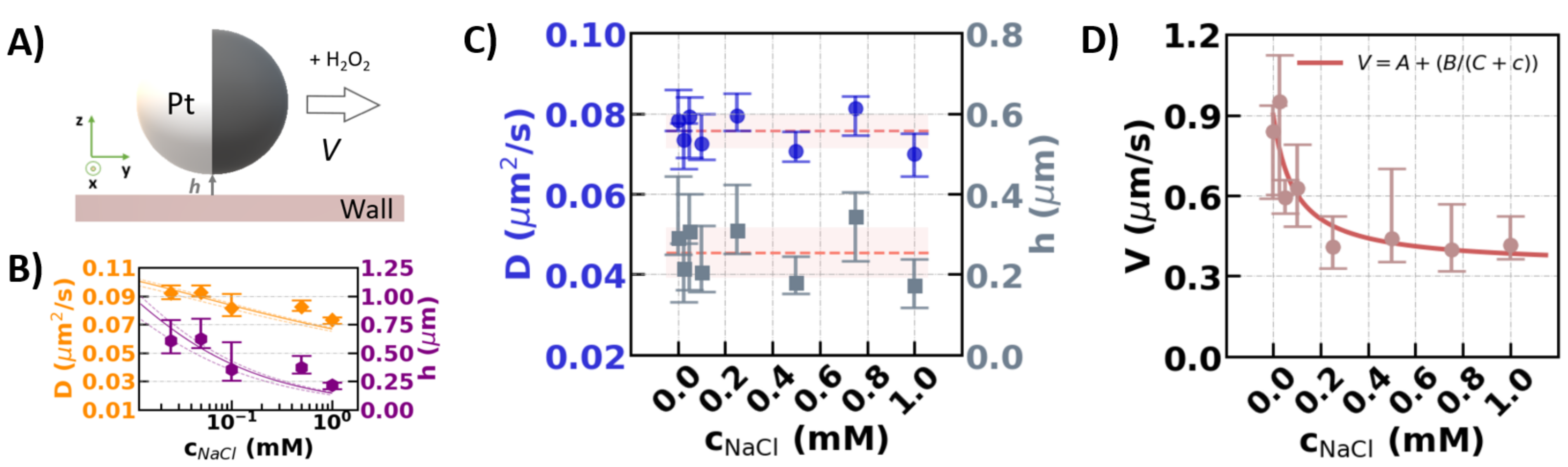
We obtained swimmer-wall separations from experimental measurements of the separation-dependent translational diffusion coefficient, , of the swimmers. as well as propulsion speeds were extracted from mean square displacements (MSDs) following Ref. Howse et al. (2007); Bechinger et al. (2016). That is, we fitted the short-time regime () of the MSDs with Howse et al. (2007); is the rotational diffusion time, , with the bulk rotational diffusion coefficient, the radius, the viscosity, the Boltzmann constant, and the absolute temperature. The first term corresponds to the passive diffusion contribution that is usually obscured by the activity-induced, short-time ballistic behavior Bechinger et al. (2016), but may be obtained with sufficient statistics. Reliable measurements require frame-rate adjustment, such that the regime where both diffusion and activity contribute to the MSD can be resolved. See the Supplemental Information (SI) sup , which additionally includes Ref. Henry (1931); de Graaf et al. (2015); Huang et al. (2008); Eloul et al. (2020); Zhou et al. (2018), for details on tracking, MSD calculation Allan et al. (2018), as well as a discussion on the consistent short-time () expansion of the MSD Howse et al. (2007); Palacci et al. (2010); Bechinger et al. (2016). To calculate the separation, , between the particle and wall, see also Figure 1A, we first consider the ratio , with the bulk diffusion constant. For 0.6, the well-known prediction by Faxén Faxén (1921); Oseen (1927); Sharma et al. (2010); Ha et al. (2013): , with , can be used to extract . For 0.4 a lubrication theory result, , is more appropriate Goldman et al. (1967); O’Neill (1964). In the intermediate (0.4 0.6) regime, applicable to most of our experiments, we interpolate the combined numerical data by O’Neill O’Neill (1964) and Kezirian Kezirian (1992), see SI Section II-A sup . The relation that follows is provided as supplement to this publication. Here, we fitted for using the interpolated expression.
In all experiments, we used 3-(trimethoxysilyl)propyl methacrylate (TPM) monodisperse colloids van der Wel et al. (2017a) half-coated with a thin Pt layer ( 4.5 0.2 nm) at dilute concentration ( v/v). In water, colloids exhibited passive Brownian motion, while dispersion in 10% H2O2 rendered them active through a catalytic process. Colloids quickly reached the lower glass wall and continued to move adjacent to it, while their motion was recorded with an inverted Nikon Eclipse Ti microscope through a 60x oil objective (NA=1.4). Swimming experiments were recorded for 30 s at 19 fps, see SI, Section I-C sup .
To demonstrate the effectiveness of our method, we first carried out control experiments in deionized water and in water at pH 3.3, equivalent to the pH in the swimming experiments, at 5 fps. In these cases, was acquired from fitting MSDs with . Figure 1B shows that the extracted separation corresponds well to a theoretical prediction based on a balance of electrostatic repulsion and gravity Flicker and Bike (1993); Rashidi and Wirth (2017), see the SI Section II-B sup . That is, we recovered the expected decrease in separation with increasing salt concentration: salt increases the solution’s ionic strength, thereby effectively screening the charge on the particle and wall. This reduces the Debye length, i.e., the distance over which surface charges act, bringing the colloids closer to the wall. To verify our method further, we compared separations resulting from our diffusion coefficient-based method to those directly measured with DIHM, for uncoated silica spheres with well-known size and refractive index Verweij et al. (2020). We found good agreement between the two methods, which confirmed that we indeed recovered colloid-wall separations, using a computed rather than a measured value of Dbulk.
Having established the validity of our method, we employed it to our catalytic microswimmers. First, we studied the effect of salt concentration in solution. For these experiments, we used TPM spheres of 2.77 0.08 m diameter half-coated with 4.4 0.2 nm Pt. Surprisingly, in active systems we found a behavior completely unlike that of passive systems in Figure 1B. For the same particles and salt concentration range, and remain constant within measurement precision, see Figure 1C. Particles propel themselves parallel to the wall at constant separations of 0.25 0.06 m.
At the same time, we found a decrease in speed with increasing salt concentration, see Figure 1D, where the line represents the least-squares fit with . This expression follows from a salt-gradient based contribution to the observed speed Brown and Poon (2014), with the remaining speed in the limit of high salt, a prefactor, and the ion concentration already present in the medium. From the fit we find the reasonable numbers 0.35 0.09 m/s and 0.09 0.07 mM, for and , respectively. The fitted C value agrees reasonably well with the background ion concentration (0.008 mM) we obtained from electrical conductivity measurements Coury (1999) for 10% H2O2 (2.7 S/cm, Ilium technology, Model 2100 Conductivity Meter) assuming hydrogen ions as the dominant ion species. We return to this salt gradient in the discussion.
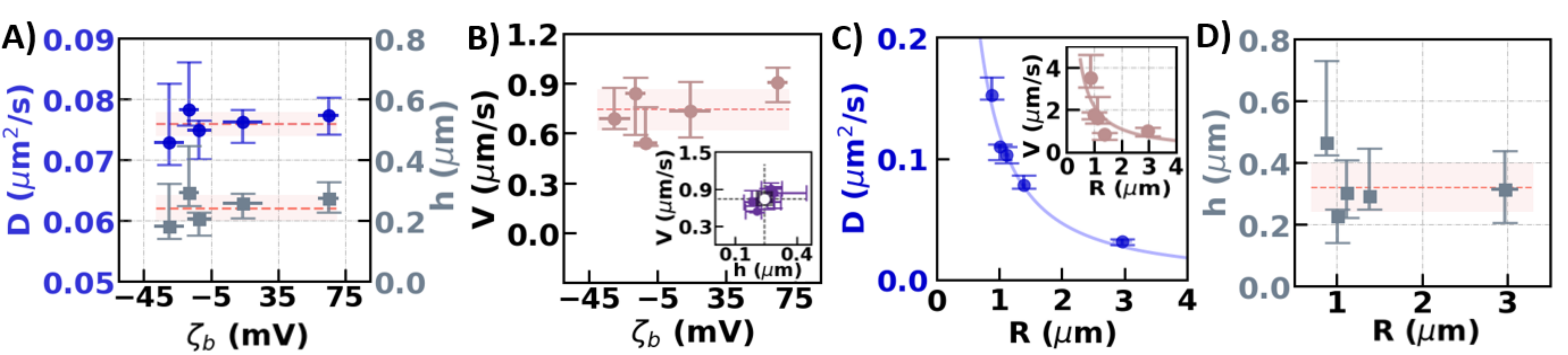
Second, we explored the effect of colloid zeta potential, , the electric potential at the colloid’s surface. We used 2.70 0.06 and 2.77 0.08 m diameter colloids with different surface functionalizations van der Wel et al. (2017b) and thus different . The reported correspond to those of the parent colloids, see SI Sections I-A and I-B sup for characterization, before adding the Pt-coating. We therefore use the adjective “base” and a subscript “b”, i.e, , to indicate that we know only the zeta potential of the uncoated colloid, and not that of the swimmer. We note that passive colloids with mV were typically stuck on the negatively charged wall, see also SI Section I-D sup .
However, for the active system, we found that wall separation remained unaffected for the entire (wide) range of under study, see Figure 2A. In all cases, particles moved at 0.24 0.04 m from the wall, which matches the separations measured for different salt concentrations. Unexpectedly, as we will return to, the colloids self-propelled not only at a constant when varying , but also at quantitatively comparable speeds , see Figure 2B. We can indeed collapse the data by plotting as a function of , see inset of Figure 2B, further demonstrating that does not affect the swimming behavior. We note that the direction of motion was away from the Pt cap both for positive and negative .
Third, we focused on swimmer size, another parameter known to affect swim speeds Ebbens et al. (2012). We performed experiments using TPM spheres with a wide range of radii, but with similar Pt coating thicknesses and , see SI Section I-A for characterization sup . We found that diffusion coefficient decreases with swimmer size, see Figure 2C, where the solid line represents the least-squares fit with (m3/s, ). Inset shows the measured swim speeds together with a fit of the expected scaling Ebbens et al. (2012) (m2/s). Strikingly, swimmer-wall separation remained relatively constant with , see Figure 2D; the dashed line shows the mean separation of 0.32 0.08 m.
The above experiments reveal that our swimmers exhibit “ypsotaxis”: a tendency to assume a specific height for a wide range of parameters. Remarkably, the height appears independent of salt concentration, , and even size, running not only counter to our intuition for passive systems but also to features of common self-propulsion mechanisms. For our Pt-coated swimmers, this robust separation distance was found to be 0.27 0.11 m on average, in line with the observation that micron-sized catalytic swimmers do not self-propel over steps of a few hundred nanometers Simmchen et al. (2016). Such a height is further consistent with wall-dependent speeds Holterhoff et al. (2018); M. et al. (2018); Ketzetzi et al. (2020); Heidari et al. (2020), for which wall separation must not substantially exceed the swimmer size to ensure strong osmotic coupling Das et al. (2015); Ibrahim and Liverpool (2015); Uspal et al. (2016). The wide range of swimmer sizes employed here showed that buoyancy is not the prime contributor to ypsotaxis. This is further underpinned by our observation of swimmers moving along the top wall, upon inversion of the sample holders, for a period of time. We hypothesize that ypsotaxis is instead primarily caused by phoretic and osmotic flows, i.e. it is activity-driven.
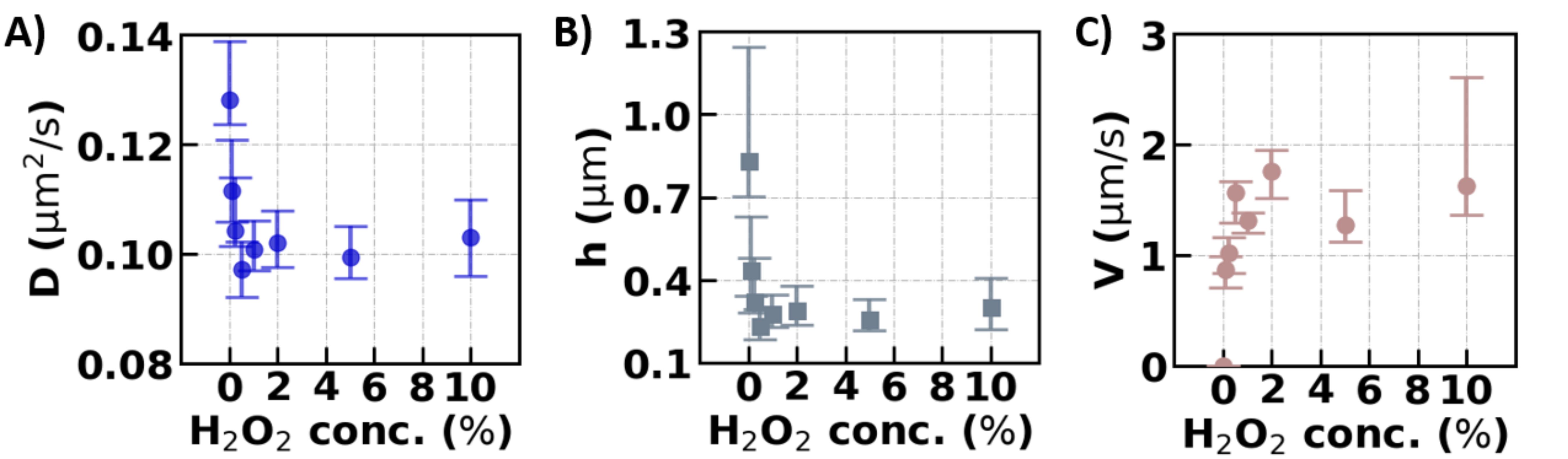
To test for this, we performed experiments using 2.23 0.11 m diameter TPM spheres with 4.5 0.2 nm Pt for various H2O2 concentrations, and hence degrees of activity. Indeed, we found that diffusion coefficient and thus swimmer-wall separation not only decreases rapidly with increasing fuel concentration from the Brownian state (0% H2O2), see Figure 3A and B, respectively, but also plateaus beyond 0.25% H2O2. Similarly, the speed also increases sharply and then plateaus above 0.25% H2O2, see Figure 3C. These observations imply that the constant separation distance is induced by the activity, thereby confirming our hypothesis on the origin of ypsotaxis. We argue that said origin also causes the active alignment of catalytic swimmers with respect to the wall Das et al. (2015); Simmchen et al. (2016); Ebbens and Howse (2011); sup , see discussion in SI Section II-D sup .
Our results provide new insights into the debated nature of the propulsion mechanisms Ebbens et al. (2014). Current thinking favors self-electrophoresis Brown and Poon (2014); Ibrahim et al. (2017), i.e., motion generated via self-generated ionic currents, as simple salts are known to greatly decrease propulsion speeds. The lack of speed variation with , however, is not commensurate with this, or other ion-based propulsion mechanisms, typically scaling with or , see Brown et al. (2017). A possible explanation is that a different at the Pt cap dominates the swimmer’s behavior.
However, speed variation with salt — typically indicative of a change in activity — is not readily reconciled with a constant which is also activity-driven, even if the cap’s dominates. Drawing upon our previous work Ketzetzi et al. (2020), we provide an alternative wall-centric explanation: Suppose that the swimmer’s bulk speed is unaffected by adding salt. The swimmer’s effective near-wall speed may still vary, provided salt impacts the osmotic counterflow induced by the swimmer-generated chemical species interacting with the wall Ketzetzi et al. (2020). Our fit in Figure 1D reveals that the osmotic contribution to the speed bears the hallmarks of ionic diffusion Anderson (1989). This requires a net-neutral gradient of ions with different electric mobilities to be involved, often referred to as a salt gradient. This salt gradient might originate from the chemical dissociation reactions in the long-range H2O2 gradient with the wall Brown et al. (2017), stemming from fuel consumption at the Pt cap. This model would have the right features to show an ionic diffusioosmosis along the wall, see SI Section II-C sup .
In summary, we established a novel method for measuring microswimmer-wall separations utilizing the height dependence of the diffusive component of their mean-squared displacement. We found that catalytic model microswimmers propel at roughly fixed heights of few hundred nanometers from planar walls. Our work further showed that nearby walls could be dominant factors in controlling swim speeds, i.e., ion-induced flow may only play a role at the wall, and not at the swimmer surface. This would necessitate a paradigm shift in modeling experimental observations and in identifying the still missing details of their propulsion mechanism. Our method can be readily applied to other types of spherical microswimmers moving parallel to walls, and may be extended to different swimmer shapes as well. We are confident that further application of our method will provide novel insights on the impact of confining surfaces in the microswimmer world, and in turn facilitate predicting swimming behaviors in complex environments.
Acknowledgements.
We gratefully acknowledge Rachel Doherty for providing TPM colloids and for discussions on colloid functionalizations. We thank Ruben Verweij and Nikos Oikonomeas for useful discussions on holographic microscopy and Aidan Brown for discussions on the propulsion mechanism and for pointing out a relevant passage in the literature. J.d.G. thanks NWO for funding through Start-Up Grant 740.018.013 and through association with the EU-FET project NANOPHLOW (766972) within Horizon 2020. D.J.K. gratefully acknowledges funding from the European Research Council (ERC) under the European Union’s Horizon 2020 research and innovation program (grant agreement no. 758383).References
- Elgeti et al. (2015) Jens Elgeti, Roland G. Winkler, and Gerhard Gompper. Physics of microswimmers – single particle motion and collective behavior. Rep. Prog. Phys., page 056601, 2015. doi: 10.1088/0034-4885/78/5/056601.
- Rothschild (1963) L. Rothschild. Non-random distribution of bull spermatozoa in a drop of sperm suspension. Nature, 198:1221–1222, 1963.
- Kantsler et al. (2013) Vasily Kantsler, Jörn Dunkel, Marco Polin, and Raymond E. Goldstein. Ciliary contact interactions dominate surface scattering of swimming eukaryotes. PNAS, 110:1187–1192, 2013. doi: 10.1073/pnas.1210548110.
- Berke et al. (2008) Allison P. Berke, Linda Turner, Howard C. Berg, and Eric Lauga. Hydrodynamic attraction of swimming microorganisms by surfaces. Phys. Rev. Lett., 101:038102, 2008. doi: 10.1103/PhysRevLett.101.038102.
- Flemming et al. (2016) Hans-Curt Flemming, Jost Wingender, Ulrich Szewzyk, Peter Steinberg, Scott A. Rice, and Staffan Kjelleberg. Biofilms: an emergent form of bacterial life. Nat Rev Microbiol, 14:563–575, 2016. doi: 10.1038/nrmicro.2016.94.
- Berne et al. (2016) C. Berne, C. K. Ellison, and A. Ducret. Bacterial adhesion at the single-cell level. Nat Rev Microbiol, 18:616–627, 2016. doi: 10.1038/s41579-018-0057-5.
- Flemming and Wuertz (2019) H. Flemming and S. Wuertz. Bacteria and archaea on earth and their abundance in biofilms. Nat Rev Microbiol, 17:247–260, 2019. doi: 10.1038/s41579-019-0158-9.
- DiLuzio et al. (2005) Willow R. DiLuzio, Linda Turner, Michael Mayer, Piotr Garstecki, Douglas B. Weibel, Howard C. Berg, and George M. Whitesides. Escherichia coli swim on the right-hand side. Nature, 435:1271–1274, 2005. doi: 10.1038/nature03660.
- Lauga et al. (2006) Eric Lauga, Willow R. DiLuzio, George M. Whitesides, and Howard A. Stone. Swimming in circles: Motion of bacteria near solid boundaries. Biophysical Journal, 90:400–412, 2006. doi: 10.1529/biophysj.105.069401.
- Lemelle et al. (2010) Laurence Lemelle, Jean-Francois Palierne, Elodie Chatre, and Christophe Place. Counterclockwise circular motion of bacteria swimming at the air-liquid interface. JOURNAL OF BACTERIOLOGY, 192:6307–6308, 2010. doi: 10.1128/JB.00397-10.
- Lopez and Lauga (2014) Diego Lopez and Eric Lauga. Dynamics of swimming bacteria at complex interfaces. Physics of Fluids, 26:071902, 2014. doi: 10.1063/1.4887255.
- Das et al. (2015) Sambeeta Das, Astha Garg, Andrew I. Campbell, Jonathan Howse, Ayusman Sen, Darrell Velegol, Ramin Golestanian, and Stephen J. Ebbens. Boundaries can steer active janus spheres. Nat. Commun., 6:8999, 2015. doi: 10.1038/ncomms9999.
- Simmchen et al. (2016) Juliane Simmchen, Jaideep Katuri, William E. Uspal, Mihail N. Popescu, Mykola Tasinkevych, and Samuel Sánchez. Topographical pathways guide chemical microswimmers. Nat. Commun., 7:10598, 2016. doi: 10.1038/ncomms10598.
- Brown et al. (2016) Aidan T. Brown, Ioana D. Vladescu, Angela Dawson, Teun Vissers, Jana Schwarz-Linek, Juho S. Lintuvuori, and Wilson C. K. Poon. Swimming in a crystal. Soft Matter, 12:131–140, 2016. doi: 10.1039/c5sm01831e.
- M. et al. (2018) Wei M., Zhou C., Tang J., and Wang W. Catalytic micromotors moving near polyelectrolyte-modified substrates: The roles of surface charges, morphology, and released ions. ACS Appl Mater Interfaces, 10:2249–2252, 2018. doi: 10.1021/acsami.7b18399.
- Holterhoff et al. (2018) A. L. Holterhoff, M. Li, and J. G. Gibbs. Self-phoretic microswimmers propel at speeds dependent upon an adjacent surface’s physicochemical properties. J. Phys. Chem. Let., 9:5023–5028, 2018. doi: 10.1021/acs.jpclett.8b02277.
- Ketzetzi et al. (2020) S. Ketzetzi, J. de Graaf, R. P. Doherty, and D. J. Kraft. Slip length dependent propulsion speed of catalytic colloidal swimmers near walls. Phys. Rev. Let., 124:048002, 2020. doi: 10.1103/PhysRevLett.124.048002.
- Heidari et al. (2020) Mojdeh Heidari, Andreas Bregulla, Santiago Muinos Landin, Frank Cichos, and Regine von Klitzing. Self-propulsion of janus particles near a brush-functionalized substrate. Langmuir, 36(27):7775–7780, 2020. doi: 10.1021/acs.langmuir.0c00461.
- Ebbens et al. (2010) Stephen Ebbens, Richard A. L. Jones, Anthony J. Ryan, Ramin Golestanian, and Jonathan R. Howse. Self-assembled autonomous runners and tumblers. Phys. Rev. E, 82:015304(R), 2010. doi: 10.1103/PhysRevE.82.015304.
- Uspal et al. (2016) W.E. Uspal, M.N. Popescu, S. Dietrich, and M. Tasinkevych. Guiding catalytically active particles with chemically patterned surfaces. Phys. Rev. Lett., 117:048002, 2016.
- Popescu et al. (2009) M. N. Popescu, S. Dietrich, and G. Oshanin. Confinement effects on diffusiophoretic self-propellers. J. Chem. Phys., 130:194702, 2009. doi: 10.1063/1.3133239.
- Crowdy (2013) D. G. Crowdy. Wall effects on self-diffusiophoretic janus particles: a theoretical study. J. Fluid Mech., 735:473–498, 2013. doi: 10.1017/jfm.2013.510.
- Chiang and Velegol (2014) T.-Y. Chiang and D. Velegol. Localized electroosmosis (leo) induced by spherical colloidal motors. Langmuir, 30:2600–2607, 2014. doi: 10.1021/la402262z.
- Uspal et al. (2015a) W. E. Uspal, M. N. Popescu, S. Dietrich, and M. Tasinkevych. Self-propulsion of a catalytically active particle near a planar wall: from reflection to sliding and hovering. Soft Matter, 11:434–438, 2015a. doi: 10.1039/c4sm02317j.
- Uspal et al. (2015b) W. E. Uspal, M. N. Popescu, S. Dietrich, and M. Tasinkevych. Rheotaxis of spherical active particles near a planar wall. Soft Matter, 11:6613–6632, 2015b. doi: 10.1039/c5sm01088h.
- Ibrahim and Liverpool (2015) Y. Ibrahim and T. B. Liverpool. The dynamics of a self-phoretic janus swimmer near a wall. Eur. Phys. Let., 111:48008, 2015. doi: 10.1209/0295-5075/111/48008.
- Mozaffari et al. (2016) Ali Mozaffari, Nima Sharifi-Mood, Joel Koplik, and Charles Maldarelli. Self-diffusiophoretic colloidal propulsion near a solid boundary. Phys. Fluids, 28:053107, 2016. doi: 10.1063/1.4948398.
- Shen et al. (2018) Zaiyi Shen, Alois Würger, and Juho S. Lintuvuori. Hydrodynamic interaction of a self-propelling particle with a wall comparison between an active janus particle and a squirmer model. Eur. Phys. J. E, 41:39, 2018. doi: 10.1140/epje/i2018-11649-0.
- Ebbens et al. (2012) Stephen Ebbens, Mei-Hsien Tu, Jonathan R. Howse, and Ramin Golestanian. Size dependence of the propulsion velocity for catalytic janus-sphere swimmers. Phys. Rev. E, 85:020401, 2012. doi: 10.1103/PhysRevE.85.020401.
- Popescu et al. (2018) M.N. Popescu, W.E. Uspal, A. Domínguez, and S. Dietrich. Effective interactions between chemically active colloids and interfaces. Acc. Chem. Res., 51:2991, 2018.
- Spagnolie and Lauga (2012) S.E. Spagnolie and E. Lauga. Hydrodynamics of self-propulsion near a boundary: predictions and accuracy of far-field approximations. J. FLuid Mech., 700:105, 2012.
- Lintuvuori et al. (2016) J.S. Lintuvuori, A.T. Brown, K. Stratford, and D. Marenduzzo. Hydrodynamic oscillations and variable swimming speed in squirmers close to repulsive walls. Soft Matter, 12:7959, 2016.
- Kuron et al. (2019) M. Kuron, P. Stärk, C. Holm, and J. de Graaf. Hydrodynamic mobility reversal of squirmers near flat and curved surfaces. Soft Matter, 15:5908, 2019.
- Brown and Poon (2014) Aidan Brown and Wilson Poon. Ionic effects in self-propelled pt-coated janus swimmers. Soft Matter, 10:4016–4027, 2014. doi: 10.1039/c4sm00340c.
- Takagi et al. (2013) Daisuke Takagi, Adam B. Braunschweig, Jun Zhang, and Michael J. Shelley. Dispersion of self-propelled rods undergoing fluctuation-driven flips. Physical Review Letters, 10:038301, 2013. doi: 10.1103/PhysRevLett.110.038301.
- Takagi et al. (2014) D. Takagi, J. Palacci, A. B. Braunschweig, M. J. Shelley, and J. Zhang. Hydrodynamic capture of microswimmers into sphere-bound orbits. Soft Matter, 10:1784, 2014. doi: 10.1039/c3sm52815d.
- Lee et al. (2007) Sang-Hyuk Lee, Yohai Roichman, Gi-Ra Yi, Shin-Hyun Kim, Seung-Man Yang, Alfons van Blaaderen, Peter van Oostrum, and David G. Grier. Characterizing and tracking single colloidal particles with video holographic microscopy. Optics Express, 15:18275, 2007. doi: 10.1364/OE.15.018275.
- Wang et al. (2014) Anna Wang, Thomas G. Dimiduk, Jerome Fung, Sepideh Razavi, Ilona Kretzschmar, Kundan Chaudhary, and Vinothan N. Manoharan. Using the discrete dipole approximation and holographic microscopy to measure rotational dynamics of non-spherical colloidal particles. Journal of Quantitative Spectroscopy and Radiative Transfer, 146:499–509, 2014. doi: 10.1016/j.jqsrt.2013.12.019.
- Brown and Staples (1990) M. A. Brown and E. J. Staples. Measurement of absolute particle-surface separation using total internal reflection microscopy and radiation pressure forces. Langmuir, 6:1260–1265, 1990. doi: 10.1021/la00097a012.
- Howse et al. (2007) Jonathan R. Howse, Richard A. L. Jones, Anthony J. Ryan, Tim Gough, Reza Vafabakhsh, and Ramin Golestanian. Self-motile colloidal particles: From directed propulsion to random walk. Phys. Rev. Lett., 99:048102, 2007. doi: 10.1103/PhysRevLett.99.048102.
- Bechinger et al. (2016) Clemens Bechinger, Roberto Di Leonardo, Hartmut Löwen, Charles Reichhardt, Giorgio Volpe, and Giovanni Volpe. Active particles in complex and crowded environments. Rev. Mod. Phys., 88:045006, 2016. doi: 10.1103/RevModPhys.88.045006.
- (42) See Supplemental Material at [URL will be inserted by publisher] for experimental details on particle preparation and MSD analysis, as well as theoretical details on height-dependent diffusion coefficients, ionic diffusiophoresis along the wall, and activity-induced ypsotaxis.
- Henry (1931) D.C. Henry. The cataphoresis of suspended particles. part i.—the equation of cataphoresis. Proc. R. Soc. London Ser. A, 133:106, 1931.
- de Graaf et al. (2015) Joost de Graaf, Toni Peter, Lukas P Fischer, and Christian Holm. The raspberry model for hydrodynamic interactions revisited. ii. the effect of confinement. The Journal of chemical physics, 143(8):084108, 2015.
- Huang et al. (2008) David M. Huang, Christian Sendner, Dominik Horinek, Roland R. Netz, and Lydéric Bocquet. Water slippage versus contact angle: A quasiuniversal relationship. Phys. Rev. Lett., 101:226101, 2008.
- Eloul et al. (2020) Shaltiel Eloul, Wilson CK Poon, Oded Farago, and Daan Frenkel. Reactive momentum transfer contributes to the self-propulsion of janus particles. Phys. Rev. Lett., 124:188001, 2020. doi: 10.1103/PhysRevLett.124.188001.
- Zhou et al. (2018) C. Zhou, H.P. Zhang, J. Tang, and W. Wang. Photochemically powered AgCl janus micromotors as a model system to understand ionic self-diffusiophoresis. Langmuir, 34:3289, 2018.
- Allan et al. (2018) Daniel B. Allan, Thomas Caswell, Nathan C. Keim, and Casper M. van der Wel. trackpy: Trackpy v0.4.1, April 2018. URL https://doi.org/10.5281/zenodo.1226458.
- Palacci et al. (2010) J. Palacci, C. Cottin-Bizonne, C. Ybert, and L. Bocquet. Sedimentation and effective temperature of active colloidal suspensions. Phys. Rev. Lett., 105:088304, 2010. doi: 10.1103/PhysRevLett.105.088304.
- Faxén (1921) Hilding Faxén. Einwirkung der Gefässwände auf den Widerstand gegen die Bewegung einer kleinen Kugel in einer zähen Flüssigkeit. Uppsala Universitet, 1921.
- Oseen (1927) Carl Wilhelm Oseen. Neuere methoden und ergebnisse in der hydrodynamik. Leipzig: Akademische Verlagsgesellschaft mb H., 1927.
- Sharma et al. (2010) P. Sharma, S. Ghosh, and S. Bhattacharya. A high-precision study of hindered diffusion near a wall. Appl Phys Let, 97:104101, 2010. doi: 10.1063/1.3486123.
- Ha et al. (2013) Chungil Ha, H. D. Ou-Yang, and Hyuk Kyu Pak. Direct measurements of colloidal hydrodynamics near flat boundaries using oscillating optical tweezers. Physica A, 392:3497–3504, 2013. doi: 10.1016/j.physa.2013.04.014.
- Goldman et al. (1967) A.J. Goldman, R.G. Cox, and H. Brenner. Slow viscous motion of a sphere parallel to a plane wall—i motion through a quiescent fluid. Chem. Eng. Sci., 22:637, 1967.
- O’Neill (1964) Michael E O’Neill. A slow motion of viscous liquid caused by a slowly moving solid sphere. Mathematika, 11(1):67–74, 1964.
- Kezirian (1992) M.T. Kezirian. Hydrodynamics with a wall-slip boundary condition for a particle moving near a plane wall bounding a semi-infinite viscous fluid. PhD thesis, Massachusetts Institute of Technology, 1992.
- van der Wel et al. (2017a) C. van der Wel, R. K. Bhan, R. W. Verweij, H. C. Frijters, Z. Gong, A. D. Hollingsworth, S. Sacanna, and D. J. Kraft. Preparation of colloidal organosilica spheres through spontaneous emulsification. Langmuir, 33(33):8174–8180, 2017a. doi: 10.1021/acs.langmuir.7b01398.
- Flicker and Bike (1993) S.G. Flicker and S.G. Bike. Measuring double layer repulsion using total internal reflection microscopy. Langmuir, 9(1):257–262, 1993.
- Rashidi and Wirth (2017) Aidin Rashidi and Christopher L Wirth. Motion of a janus particle very near a wall. Journal of chemical physics, 147:224906, 2017.
- Verweij et al. (2020) Ruben W. Verweij, Stefania Ketzetzi, Joost de Graaf, and Daniela J. Kraft. Height distribution and orientation of colloidal dumbbells near a wall. arXiv:2009.14733, 2020.
- Coury (1999) Lou Coury. Conductance measurements part 1: Theory. Current Separations and Drug Development, Bioanalytical Systems, 18(3):91–96, 1999.
- van der Wel et al. (2017b) C. van der Wel, N. Bossert, Q. J. Mank, M. G. T. Winter, D. Heinrich, and D. J. Kraft. Surfactant-free colloidal particles with specific binding affinity. Langmuir, 33(38):9803–9810, 2017b. doi: 10.1021/acs.langmuir.7b02065.
- Ebbens and Howse (2011) Stephen J. Ebbens and Jonathan R. Howse. Direct observation of the direction of motion for spherical catalytic swimmers. Langmuir, 27:12293–12296, 2011. doi: 10.1021/la2033127.
- Ebbens et al. (2014) S. Ebbens, D. A. Gregory, G. Dunderdale, J. R. Howse, Y. Ibrahim, T. B. Liverpool, and R. Golestanian. Electrokinetic effects in catalytic platinum-insulator janus swimmers. EPL, 106:58003, 2014. doi: 10.1209/0295-5075/106/58003.
- Ibrahim et al. (2017) Yahaya Ibrahim, Ramin Golestanian, and Tanniemola B. Liverpool. Multiple phoretic mechanisms in the self-propulsion of a pt-insulator janus swimmer. J. Fluid Mech., 828:318–352, 2017. doi: 10.1017/jfm.2017.502.
- Brown et al. (2017) Aidan T. Brown, Wilson C. K. Poon, Christian Holm, and Joost de Graaf. Ionic screening and dissociation are crucial for understanding chemical self-propulsion in polar solvents. Soft Matter, 13:1200–1222, 2017. doi: 10.1039/c6sm01867j.
- Anderson (1989) John L. Anderson. Colloid transport by interfacial forces. Ann. Rev. Fluid Mech., 21:61–99, 1989. doi: 10.1146/annurev.fl.21.010189.000425.