Colors from correlated disordered photonic systems - can we outperform nature?
Abstract
Living organisms have developed a wide range of appearances from iridescent to matt textures [1, 2]. Interestingly, angular independent structural colors, where isotropy in the scattering structure is present, only produce coloration in the blue wavelength region of the visible spectrum. One might, therefore, wonder if such observation is a limitation of the architecture of the palette of materials available in nature. Here, by exploiting numerical modeling, we discuss the origin of isotropic structural colors without restriction to a specific light scattering regime. We show that high color purity and color saturation cannot be reached in isotropic short-range order structures for red hues. This conclusion holds even in the case of advanced scatterer morphologies, such as core-shell particles or inverse photonic glasses - explaining recent experimental findings reporting very poor performances of visual appearance for such systems [3, 4].
1 Introduction
Structural colors are the results of constructive interference of light scattered by nanostructured, non-absorbing media. In contrast to color by pigmentation, where the color results from wavelength-selective absorption, structural colors spanning across the entire visible spectrum can be obtained using only one material. The possibility of exploiting such photonic coloration to replace traditional pigments catalyzed the efforts of several research groups [5, 6, 7, 8, 9, 10, 11]. Structural colors come with various advantages compared to conventional pigments: (i) structural pigments do not bleach, as the color formation is defined by the architecture instead of the composition; (ii) they can be constituted of environmental-friendly materials [12, 13]; (iii) they achieve unconventional color effects, from vivid metallic to isotropic optical response.
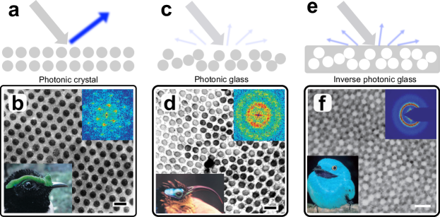
Isotropic structural color is especially appealing when it comes to replacing traditional pigmentation as they preserve an angular independent appearance. Therefore, several methods were developed to produce hierarchical [8, 14, 15], or short-range order structures with angular-independent colors [6, 4, 3]. Such studies of short-range order structures, often referred to as photonic glasses (PGs), found inspiration in analogous two-dimensional structures exploited in birds (Figure 1) [16, 17, 18, 19]. However, in nature, such PGs have been reported only to produce blue colors, as green and red coloration are usually achieved with long-range ordered structures or using pigmentation [20, 21].
In the following, we investigate the limitations of color creation by scattering from disordered, short-range correlated structures. We question whether a saturated, angular independent, color response can be achieved only in the UV-blue spectral region or whether nature has missed a trick and developed other mechanisms for red hues. In fact, while artificial PGs with red hues have been reported, their optical properties are rather poor - their color saturation and purity are limited, holding these materials back from applications. These limitations of red isotropic structural color have been attributed to single-particle resonances [22]. However, this conclusion ignored multiple scattering and coupling effects between particles, which are known to play a major role in PGs [4, 23]. Here, we exploit a numerical approach that provides direct access to the reflection spectrum of an arbitrary structure and allows us to investigate intermediate scattering regimes, i.e., in between single scattering and diffusive behavior, without further assumptions. With this method, we show that nature’s solution is actually optimal for blue production only: both high and low refractive index contrast PGs show poor color purity in the red. We demonstrate the difficulty in achieving isotropic structural color in the red spectral region with high saturation and purity even for advanced morphologies of the scatterers in both direct and inverse PGs.
2 Results
2.1 Direct vs inverse photonic glasses
Photonic glasses with tailored form and structure factors were generated using a recently developed numerical algorithm, see details in Reference [25] and Supplementary Section III. The optical properties of the generated structures were then calculated using a Finite Difference Time Domain (FDTD) method. We limited our calculations to two-dimensional structures, as the same general considerations can be extended to the three-dimensional case.
In the absence of absorption, scattering in PGs emerges from the interplay between: (i) single particle properties as size, shape and refractive index; (ii) ensemble properties as filling fraction and structural correlations. As depicted in Figure 2a, for the case of direct PGs the reflection is dominated by Mie resonances, determined by the scatterer properties. The reflected color can be, therefore, tuned in the visible by changing the scatterer dimensions. However, as the particle size increases the Mie resonance peak red-shift and a second peak appears in the blue part of the spectrum, corresponding to a higher-order resonant mode (Figure S1). In contrast, light scattering in inverse PGs is dominated by structural correlations (Figure 2b). The reflection peak, whose position corresponds well to Bragg’s law predictions, is more pronounced than in direct structures. Moreover, when the structural peak is shifted at red wavelengths the form-factor resonance at short wavelengths is less marked than in Figure 2a. The occurrence of a separated peak in the visible spectrum showcases that using inverse PGs is an effective strategy to minimize the form-factor weight on the overall optical response of a system in favor of structural contributions.
2.2 The role of refractive index (contrast)
The dependency of isotropic structural coloration on the refractive index is shown in Figure S1 and Figure S2 for direct and inverse PGs, respectively. Changing the refractive index affects the interplay between form- and structure- factor contributions. High refractive index systems are dominated by form-factor resonances preventing to reach good color purity in the red spectral region, both for direct and inverse PGs. For direct systems, even when the refractive index contrast is low, form-factor resonances lead to an enhanced reflection in the short wavelength side of the structural peak - similarly to Figure 2a. In contrast, for the case of inverse PGs, we observe that the structure-factor forms a well separated peak in the visible spectrum, even in the red wavelength region. This allows us to conclude that low refractive index, inverse PGs can outperform their direct counterpart in terms of color purity and saturation.
Reducing the refractive index contrast between the scattering matrix () and the scattering centers () can further favor structural contributions. Figure 3a shows that increasing results in a broadband reduction of the reflectance and a red-shift of the structural peak. Moreover, the structural peak decreases in width and has a higher intensity compared to its background, leading to better color purity. The reduction of the refractive index contrast lowers the role of multiple scattering, which is anyway present in disordered systems. This limits the isotropic structural colors to a light propagation regime in between diffusive scattering and ballistic transport. The multiple scattering becomes dominant when the sample thickness is increased, leading to a broadband unsaturated response (Figure S3a).
To further investigate the advantages and limitations of isotropic structural coloration in direct and inverse PGs, we studied their dependency on different ensemble parameters. Figure S3b shows the optical response of inverse PGs is more robust to variations in scatterer size distribution than the direct PGs. While for direct PGs polydispersity averages out the Mie resonances, in inverse PG polydispersity only slightly affects the structure-factor resonance peak. In inverse PGs, the polydispersity helps to reduce the Mie resonance intensity in the blue region and broadens the structural peak at long wavelengths. Similarly, reducing the filling fraction in inverse PGs leads to a red-shift of the structural peak, due to an increase of the effective refractive index of the system. In parallel, the relative intensity of the structural peak decreases compared to the Mie resonance at short wavelengths (Figure S3c). The result of these effects on the overall appearance of the system is further studied in Figure 4.
2.3 Advanced scatterer morphologies
Our observations remain valid also for complex scatterer geometries. Previous works introduced the idea of using core-shell particles to disentangle the contributions of form- and structure-factor and achieve a separated peak in the long wavelength spectral region [7, 26, 22, 27]. Figure 3b shows that reducing the scattering center (core) size while keeping the structural correlation length leads to an increase of the intensity and width of the long wavelength (structural) peak. At the same time the short wavelength contribution from Mie resonances shifts further away into the UV. In Figure 3a we showed that a lowered refractive index contrast can suppress multiple scattering, while a separation of the form- and structure-factor contributions is possible via core-shell particles, see Figure 3b. In Figure 3c both approaches are combined, to achieve higher color purity and saturation by a well separated peak in the long wavelength part of the visible spectrum.

2.4 Color saturation and purity
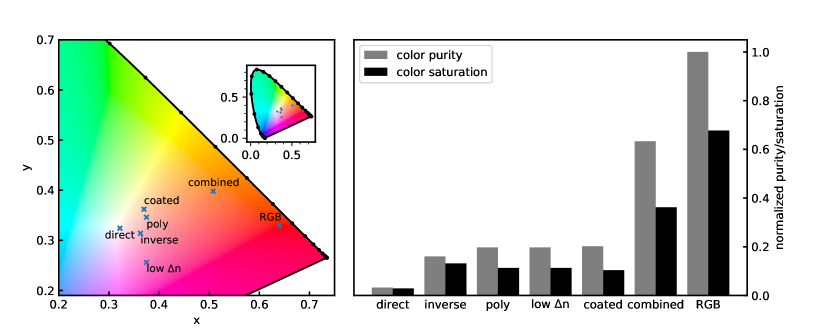
To better quantify and assess the results in terms of color purity and saturation, the reflectance spectra of direct, inverse and core-shell PGs are converted to color hues. The details of the spectrum to color conversion are reported in the Methods section. In Figure 4a the different systems for red color hues are plotted in the CIE color space chromaticity diagram (see
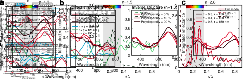
Table S1 for the xy-values). In Figure 4b the corresponding purity and saturation values are calculated. Interestingly, all the inverse PGs show a higher color purity and saturation value than the direct PGs red hues reported in Reference [4]. However, both the core-shell inverse structures as well as the low refractive index contrast (e.g. polymer filled with water) do not lead to a significant improvement compared to the standard inverse PG. Combining both approaches leads to improved purity and saturation values. Still, the achievable color purity and saturation values remain far from the ideal (RGB) red color.
3 Discussion
In summary, we demonstrated that photonic glasses have intrinsic limitations in achieving saturated red hues - reinforcing the hypothesis that isotropic structural coloration, while it can be easily achieved in the UV-blue spectral region, is challenging to obtain for larger wavelengths. In detail, we showed that inverse PGs increase color saturation and purity compared to their direct counterpart. This result is a consequence of an optical response where the structure factor contribution dominates over the form-factor scattering resonances.
While combing different scattering approaches can help improving color purity and saturation in photonic glasses, they might be more challenging to realize in natural systems than simply combing different mechanisms, such as adding absorption. Once absorption is added, more pure and saturated colors can be achieved [10, 28]. However in synthetic systems, especially in the case of metallic nanoparticles [29], one might question the need of adding a structural component, as it plays a redundant role in the overall visual response. In this context, we believe that approaches aimed at designing the scattering elements, making use of the interplay between absorption and scattering via Kramer-Kronig relations might be more suitable to increase scattering in the red spectral region [30]. Although most efforts to fabricate systems exhibiting isotropic structural colors focus on the self-assembly of colloidal particles [10], other promising approaches have recently been explored combining the purity and saturation advantages of coloration from ordered structures with the morphological isotropy of spherical systems. In this context, blockcopolymers represent a promising candidate [31]. Similarly, introducing disorder to ordered multilayer films gives rise to angle-independent structural coloration [14, 15]. In conclusion, as convincing absorption-free, angle independent, pure, and saturated red hues have not been obtained, we believe there is still space for further theoretical and experimental investigations to come up with strategies to produce viable alternatives to red pigments.
Methods
Numerical calculations
To generate the, two-dimensional, disordered structures we used a inverse-desing algorithm consisting of two steps: [25] First, hard (non-overlapping) particles are added using a random sequential approach until the desired filling fraction was reached; Second, the position of these particles are gradually changed in order to minimize the difference between the targeted S(q) and the one of the structure. This code is available from G. J. upon request.
To simulate the optical properities of the generated designed we used LUMERICAL 8.22 (Lumerical Solutions Inc., Vancouver, BC, Canada), a commercial-grade software using the finite-difference time-domain (FDTD) method. Periodic boundaries conditions in the lateral direction, i.e., perpendicular to the incoming beam, Y and perfect matching layer (PML) boundaries in the X direction were used in all the calculations. The excitation source was set as a plane wave. The simulations were performed in a purely 2D geometry and their numerical stability/convergence was ensured by choosing an adequate simulation time and boundary conditions (assuring that the electric field in the structure decayed before the end of the calculation and that all the excitation light was either reflected or transmitted). Each of the presented curves was obtained averaging the optical simulations of seven different ensembles of particles with identical parameters.
Purity and saturation of color
To quantify color purity and saturation, reflection spectra are converted in color space coordinates assuming a standard observer (CIE 1931 2∘) and a standard illuminant (Daylight ). [4, 25]
In particular, color purity can be calculated from:
| (1) |
where , are the chromaticity coordinates in the xyY color space, with , and , being the coordinates of the white point and RGB red, respectively. color saturation is calculated as:
| (2) |
where, represents the lightness, and , are the chromaticity coordinates in CIELAB space.
Acknowledgements
This work was supported in part by a BBSRC David Phillips Fellowship (BB/K014617/1), the European Research Council (ERC-2014-STG H2020 639088) to S.V and G.J and the Swiss National Science Foundation under project P2ZHP2_183998 to L.S.
Data availability
All data needed to evaluate the conclusions in the paper are present in the paper and/or the Supplementary Materials.
Conflict of interest
The authors declare that they have no competing financial interests.
References
- [1] Vukusic, P. & Sambles, J. R. Photonic structures in biology. Nature 424, 852–855 (2003).
- [2] Johansen, V. E., Onelli, O. D., Steiner, L. M. & Vignolini, S. Photonics in Nature: From Order to Disorder (Springer International Publishing, Cham, 2017).
- [3] Kim, S.-H. et al. Inverse photonic glasses by packing bidisperse hollow microspheres with uniform cores. ACS Applied Materials & Interfaces 9, 24155–24160 (2017).
- [4] Schertel, L. et al. The structural colors of photonic glasses. Advanced Optical Materials 7, 1900442 (2019).
- [5] Kolle, M. et al. Mimicking the colourful wing scale structure of the papilio blumei butterfly. Nature Nanotechnology 5, 511–515 (2010).
- [6] Forster, J. D. et al. Biomimetic isotropic nanostructures for structural coloration. Advanced Materials 22, 2939–2944 (2010).
- [7] Magkiriadou, S., Park, J.-G., Kim, Y.-S. & Manoharan, V. N. Disordered packings of core-shell particles with angle-independent structural colors. Opt. Mater. Express 2, 1343–1352 (2012).
- [8] Vogel, N. et al. Color from hierarchy: Diverse optical properties of micron-sized spherical colloidal assemblies. Proceedings of the National Academy of Sciences 112, 10845–10850 (2015).
- [9] Liang, H.-L. et al. Roll-to-roll fabrication of touch-responsive cellulose photonic laminates. Nature Communications 9, 4632 (2018).
- [10] Goerlitzer, E. S. A., Klupp Taylor, R. N. & Vogel, N. Bioinspired photonic pigments from colloidal self-assembly. Advanced Materials 30, 1706654 (2018).
- [11] Schertel, L. et al. Tunable high-index photonic glasses. Phys. Rev. Materials 3, 015203 (2019).
- [12] Wang, Y. et al. Design, fabrication, and function of silk-based nanomaterials. Advanced Functional Materials 28, 1805305 (2018).
- [13] Frka-Petesic, B. & Vignolini, S. So much more than paper. Nature Photonics 13, 365–367 (2019).
- [14] Song, D.-P. et al. Photonic resins: Designing optical appearance via block copolymer self-assembly. Macromolecules 51, 2395–2400 (2018).
- [15] Chan, C. L. C. et al. Visual appearance of chiral nematic cellulose-based photonic films: Angular and polarization independent color response with a twist. Advanced Materials 31, 1905151 (2019).
- [16] Prum, R. O., Torres, R. H., Williamson, S. & Dyck, J. Coherent light scattering by blue feather barbs. Nature 396, 28–29 (1998).
- [17] Prum, R., Torres, R., Kovach, C., Williamson, S. & Goodman, S. Coherent light scattering by nanostructured collagen arrays in the caruncles of the malagasy asities (eurylaimidae: aves). Journal of Experimental Biology 202, 3507–3522 (1999).
- [18] Prum, R. O. & Torres, R. Structural colouration of avian skin: convergent evolution of coherently scattering dermal collagen arrays. Journal of Experimental Biology 206, 2409–2429 (2003).
- [19] Saranathan, V. et al. Structure and optical function of amorphous photonic nanostructures from avian feather barbs: a comparative small angle x-ray scattering (saxs) analysis of 230 bird species. Journal of The Royal Society Interface 9, 2563–2580 (2012).
- [20] Kinoshita, S. Structural Colors in the Realm of Nature (World Scientific, 2008).
- [21] Sakoda, K. Optical Properties of Photonic Crystals. Springerseries in optical sciences (Springer, 2001).
- [22] Magkiriadou, S., Park, J.-G., Kim, Y.-S. & Manoharan, V. N. Absence of red structural color in photonic glasses, bird feathers, and certain beetles. Phys. Rev. E 90, 062302 (2014).
- [23] Hwang, V., Stephenson, A. B., Magkiriadou, S., Park, J.-G. & Manoharan, V. N. Effects of multiple scattering on angle-independent structural color in disordered colloidal materials. Phys. Rev. E 101, 012614 (2020).
- [24] Dufresne, E. R. et al. Self-assembly of amorphous biophotonic nanostructures by phase separation. Soft Matter 5, 1792–1795 (2009).
- [25] Jacucci, G., Bertolotti, J. & Vignolini, S. Role of anisotropy and refractive index in scattering and whiteness optimization. Advanced Optical Materials 7, 1900980 (2019).
- [26] Park, J.-G. et al. Full-spectrum photonic pigments with non-iridescent structural colors through colloidal assembly. Angewandte Chemie 126, 2943–2947 (2014).
- [27] Shang, G. et al. Photonic glass for high contrast structural color. Scientific Reports 8, 7804 (2018).
- [28] Kim, S.-H. et al. Solution-processable photonic inks of mie-resonant hollow carbon–silica nanospheres. Small 15, 1900931 (2019).
- [29] Lee, S.-J., Kumar, S., Choi, J. W., Kim, V. Y. & Lee, J.-S. Copolymer particles with incorporated gold and silver nanoparticles to absorb short-wavelength scattering in full-color photonic glasses. Particle & Particle Systems Characterization 36, 1900167 (2019).
- [30] Wilts, B. D., Wijnen, B., Leertouwer, H. L., Steiner, U. & Stavenga, D. G. Extreme refractive index wing scale beads containing dense pterin pigments cause the bright colors of pierid butterflies. Advanced Optical Materials 5, 1600879 (2017).
- [31] Song, D.-P., Zhao, T. H., Guidetti, G., Vignolini, S. & Parker, R. M. Hierarchical photonic pigments via the confined self-assembly of bottlebrush block copolymers. ACS Nano 13, 1764–1771 (2019).