Controlled optofluidic crystallization of colloids tethered at interfaces
Abstract
We report experiments that show rapid crystallization of colloids tethered to an oil-water interface in response to laser illumination. This light-induced transition is due to a combination of long-ranged thermophoretic pumping and local optical binding. We show that the flow-induced force on the colloids can be described as the gradient of a potential. The non-equilibrium steady state due to local heating thus admits an effective equilibrium description. The optofluidic manipulation explored in this work opens novel ways to manipulate and assemble colloidal particles.
Since their introduction Ashkin et al. (1986), optical tweezers have revolutionised the manipulation of matter at the nano- to micro-meter scale Svoboda et al. (1993); Hofkens et al. (1997); Molloy (2003); Polin et al. (2006); Mizuno et al. (2007). Tweezers have found extensive use in the trapping and assembly of micron-sized colloidal particles Chapin et al. (2006); Čižmár et al. (2010); Tkalec et al. (2011) and have enabled the formation of novel forms of colloidal matter that are held together by optical forces Burns et al. (1990); Vossen et al. (2004); Mellor and Bain (2006); Work and Williams (2015); Kudo et al. (2016); Lin et al. (2017); Liu and Li (2017). While the various mechanisms of optical trapping are well understood, much less is known about the effects of light-induced heating on the flow of the surrounding fluid medium, in particular near a liquid-liquid interface. Naively, one might think that, at a liquid-liquid interface, the fluid motion would be dominated by Marangoni flow, which should drive the fluid away from the hot spot Namura et al. (2015); Karbalaei et al. (2016). However, in our experiments, surface tethered colloids moved towards the hot spot, even when they are out of range of the direct optical binding forces Wei et al. (2016).
In this Letter, we show that the optical trapping of a single colloidal particle near a water-oil interface can set up a long-ranged, non-equilibrium force field, which causes colloidal particles that are tethered to the surface but otherwise freely diffusing, to move to the hot spot where they crystallize. The sign, magnitude, and distance-dependence of this non-equlibrium force cannot be accounted for by static colloidal interactions nor by surface-tension driven Marangoni flows. Using theory and simulation, we show that the experimentally observed colloidal motion is produced by stalled thermophoresis of a single, optically trapped colloid. In fact, to a good approximation, the stalled particle acts as a hydrodynamic monopole. For motion parallel to the interface, the force is the gradient of a potential and the particle dynamics admits an effective equilibrium description. Brownian dynamics simulations in this emergent potential, whose strength is determined by the local heating, is in excellent agreement with experiments.
Importantly, the effective attractive potential depends only on the thermophoretic mobility of the stalled colloid. It manifests itself in simple solvents such as water or oil and is of much longer range than the optical trapping potential. As a consequence, interfacially trapped colloids can be used as switchable pumps. Such addressable pumps would enable novel strategies for optofluidic manipulation and the controlled assembly of colloidal particles Whitesides and Grzybowski (2002). Below, we describe our experimental results, theoretical analyses, and numerical simulations.
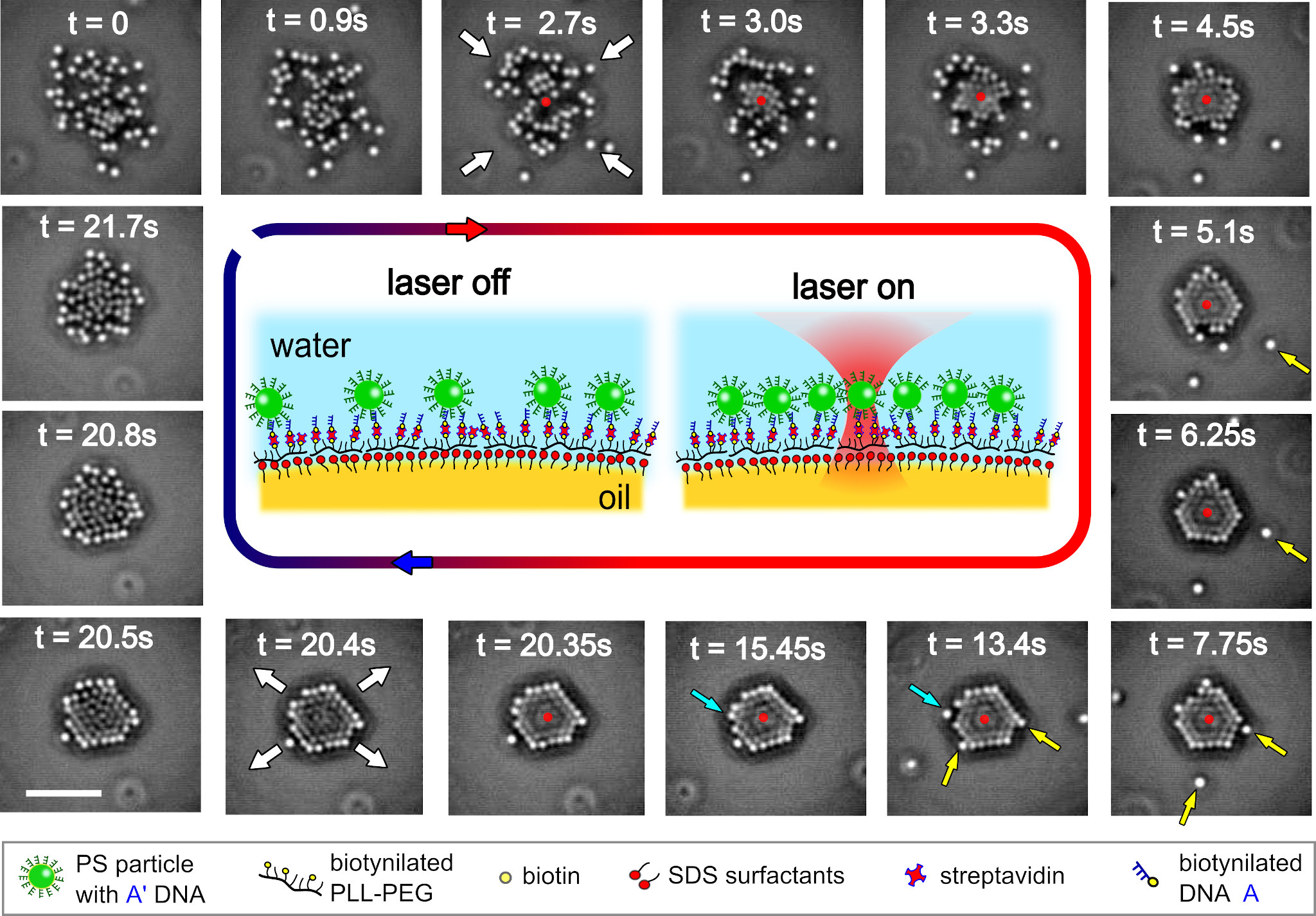
Sample geometry: The inner panels of Fig. (1) show a sketch of our sample geometry. Oil droplets with a radius between 20 and 30 were coated with a surfactant-polymer layer, following the protocol described in ref. Joshi et al. (2016). Onto this layer, we grafted a dense brush of single-stranded (ss)DNA sequences (denoted by ). Polystyrene particles (PS) of radius , functionalised by complementary ssDNA strands, were then allowed to hybridize with the chains on the surface. The DNA coating of the colloids prevents them from aggregating. As the colloids are tethered to the surface, rather than embedded in it, they are not subject to capillary forces, light-wave reflections or long-ranged electrostatic dipolar interactions that would be caused by the asymmetry of charge distributions on interfacially wetted colloids Park and Furst (2008, 2011). Moreover, the colloids do not deform the surfactant-polymer coating, as the DNA tethering keep the colloids some 50 away from the surface layer. As the oil droplets are much larger than the colloids, the interface is effectively flat on the scale probed in our experiments. Although tethered to surface polymers, the colloids are otherwise free to diffuse along the interface.
When a laser beam is focused above the oil-water surface, it will trap a single tethered colloid. This colloid will act as the thermophoretic “pump” that will recruit other tethered colloids. However, a fraction of the colloids remain untethered and diffuse freely in the bulk. These particles serve as tracers of the bulk fluid flow. Further details of the system and the calibration of the trap are provided in the SI siT .
Reversible crystallization: The outer panels of Fig. (1) show our principal experimental result. Following the frames clockwise starting from the top left, the pictures show the crystallisation and dissolution of a colloidal crystal, as the laser us switched on (red arrow) and then switched off (blue arrow). The primary optically trapped colloid is shown in red. The first two frames show free diffusion when the laser is off. When the laser is turned on in the third frame, diffusion is immediately replaced by directed motion towards the trapped particle with speeds of upto . This leads to rapid crystallization which is essentially complete within a few seconds, as shown in in the frame at . Thermal fluctuations cause small displacements in the core of the crystallite but large ones at the edges where particle rearrangements take place, as shown in the frames between and . The crystallite begins to melt as soon as the laser is turned off and a freely diffusing state is recovered within a few seconds. This cycle of freezing and melting in response to turning the laser on and off is rapid, robust and reproducible.
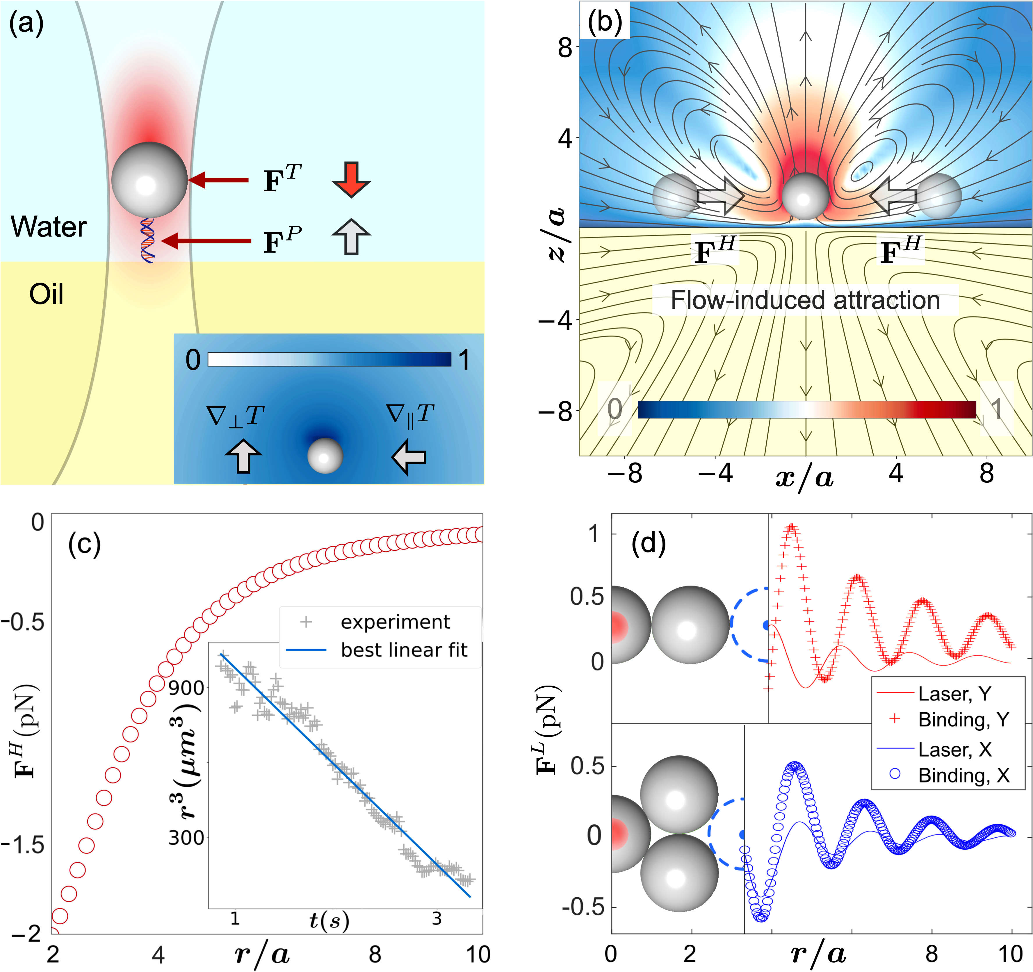
Optofluidic mechanism: What force underlies this phenomenon? By examining the motion of the colloids as the laser is turned on, it is clear that the force has a range of at least and a magnitude of the order of pN, directed radially inwards to the trapped particle. At such distances, neither the direct optical trapping, nor the optical binding forces to be discussed below, can play a role. The entrainment of colloids by Marangoni flow can be immediately ruled out, as such a flow must point outwards from the hot region surrounding the laser focus. Direct thermophoresis toward the hot colloid is also incompatible with the experimental data, as untethered colloids are seen to move first towards the heated colloid and then to move vertically away (Fig. 3(b)): thermophoresis would result in isotropic attraction. In the absence of other plausible mechanisms, we are led to postulate the following: the colloid nearest to the laser focus is optically trapped and local heating near an interface between two fluids with different thermal conductivities induces an assymmetric thermal gradient in the surrounding fluid. This gradient pushes the colloid towards the interface, where it stalls (as its Soret coefficient is positive Burelbach et al. (2017)). From that moment on, the thermal gradient along its surface drives a thermo-osmotic flow originating in a thin boundary layer around the colloid Anderson (1989); Burelbach et al. (2018a, b); Wei et al. (2020). Since the colloid remains stalled, the thermo-osmotic flow continues unabated, but produces no particle motion. This leads to a monopolar hydrodynamic counterflow in the fluid, with the monopole pointing normal to the interface and into the water phase. The long-ranged and attractive character of the flow entrains untrapped particles and draws them towards the focus. If the entrained colloids are tethered, they aggregate into crystallites under the action of the optofluidic force. The local crystalline order is enhanced by short-ranged forces, including those due to optical binding (see below). In contrast, untethered tracer colloids first move along the surface towards the trapped colloid, but then they are advected away from the surface. Such behavior is illustrated in Fig. 3(b) where an untethered particle appears and then disappears from the focal plane, consistent with the flow pattern shown in Fig. 2(b).
To make the above hypothesis quantitative and testable, we solve the equations of mass, momentum and energy conservation in the fluid with appropriate boundary conditions at the colloid surfaces and the oil-water interface (detailed in siT ). The geometry is shown schematically in Fig. 2(a). We use the boundary integral representation for the momentum (Stokes) and energy (Laplace) equations to impose boundary conditions at the colloid surfaces and use appropriate Green’s functions to satisfy the boundary conditions at the oil-water interface R. Singh et al. (2019). The integral equations are solved in a basis of irreducible tensorial harmonics to yield the temperature field and fluid flow velocity in an externally imposed temperature field (representing laser heating). These are shown in Fig. 2(b) for a single trapped colloid. From these we obtain the thermophoretic force on the trapped colloid and the optofluidic force with which free colloids are attracted to the trapped colloid as,
| (1) |
In the above, is the thermophoretic mobility, and are, respectively, the mobility perpendicular and parallel to the interface, is a Green’s function of the Stokes equation for a no-shear plane interface and indicates evaluation at the center of the trapped colloid siT . The optofluidic force, through its dependence on , varies monotonically as the inverse square of the distance from the trapped colloid. As particle motion is overdamped, the velocity scales as , and hence displacements scale with time intervals as . We test this from the experimentally measured positions to find excellent agreement, shown in the inset of Fig. 2(c).
Optofluidic potential: For motion at a constant height, the nonequilibrium optofluidic force admits a potential
| (2) |
Here , is the height of the colloid from the interface and is the ratio of the viscosities of oil and water. The optofluidic potential depends linearly on the ratio of thermal conductivity of the oil and water layers through the dependence on the temperature gradient siT . Then, the in-plane coordinate of an untrapped colloid ( obeys the overdamped Langevin equation
| (3) |
where is a potential containing the sum of all short-ranged colloid-tether and colloid-colloid interactions, is the optofluidic potential evaluated at the location of the particle, and is a zero-mean Gaussian random variable with variance . The stationary distribution of the particle positions is Gibbsian, , even though the dynamics is out of equilibrium. At an air-water interface, where , the optofluidic potential has a Coulomb form . The opposite limit of , corresponding to a no-slip wall, gives an optofluidic potential The latter form, with different prefactors, has been found in previous studies on charged Squires (2001), thermophoretic Di Leonardo et al. (2009); Weinert and Braun (2008), and active colloids Singh and Adhikari (2016); R. Singh et al. (2019); Bolitho et al. (2020). Thus, near a liquid-solid boundary, the scaling of Fig.2(b) is modified to . Thus, the optofluidic mechanism described by the monopole has a wider applicability. We believe that monopolar flow rationalises a great variety of phenomena in phoretic Wirnsberger et al. (2017) and active matter Bolitho et al. (2020) and that its relevance will be widely appreciated in due course.
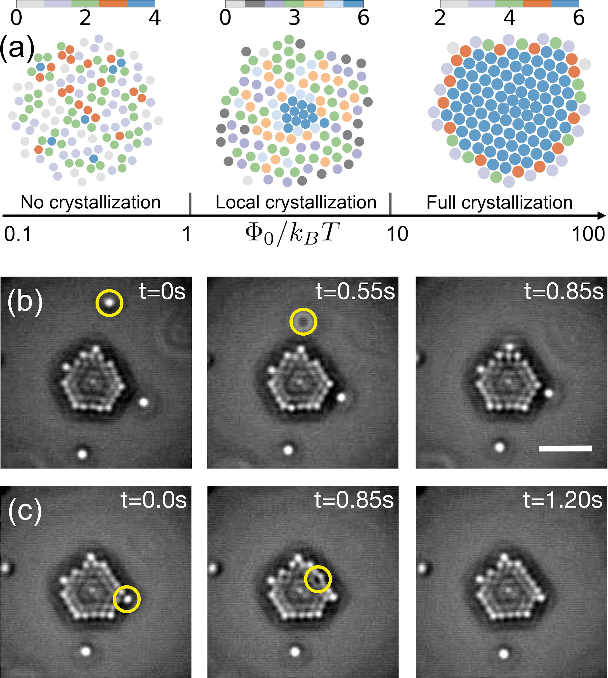
The strength of the potential when compared with the thermal energy determines the onset of crystalline order. Denoting it by
| (4) |
and using parameters , , and , we get when . Here we have used the experimentally measured positions in Fig. 2(c) to estimate the from other known parameters. The strength is proportional to the thermal gradient and leads, curiously, to freezing by heating and melting by cooling. We show this explicitly in Fig. 3(a) by direct numerical simulations of Eq.(3) as a function of the strength of the optofluidic potential.
Short-range forces: Once the long-ranged optofluidic interaction draws particles into the center of force, short-ranged optical binding forces act to enhance crystalline order. The optical binding force is obtained from a numerical solution of the Maxwell equations in the Mie approximation (detailed in siT ). It is shorter in range than the optofluidic force but, being oscillatory and anisotropic (see Fig. 2(d)), couples to both positional and bond order of the colloidal crystal. Its effect can be inferred indirectly from the rapid annealing of a defect (yellow circle) produced by the collision of a tethered particle with the crystallite, as shown in Fig. 3(c). We have not studied this coupling in detail and leave it to future work.
Conclusion: Our experiments shows how a novel non-equilibrium optofluidic force can be used to transport particles towards (or away) from an optically trapped “seed” particle. It is important to distinguish that the optofluidic force field is qualitatively different from the light-controlled thermoelectric fields generated in a medium, which contains a mixture of surfactant, ions, and micellar depletants Lin et al. (2017). It is also different from the thermo-osmotic flow generated by the differential heating of trapped Janus particles Mousavi et al. (2019). Theoretical analysis shows that the optofluidic force can be described in terms of the gradient of a potential, whose strength is proportional to the temperature gradient at the location of the seed. Untrapped particles couple to this potential regardless of their material properties, enabling the optofluidic manipulation of particles that cannot, otherwise, be optically trapped. Both the location of the potential and its strength can be modulated by the laser and its sign can be altered by changing the ratio of thermal conductivities of the liquids. We foresee this to lead to novel mechanisms of switchable, addressable transport in microfluidics, controlled self-assembly of active colloids and the meta-material synthesis.
Acknowledgements.
We thank Professors M. E. Cates, D. Frenkel, and E. J. Hinch for helpful discussions. A.C. thanks the ETN-COLLDENSE (H2020-MCSA-ITN-2014, grant no. 642774) and the Winton Programme for the Physics of Sustainability. R.S. acknowledges the support of a Royal Society-SERB Newton International Fellowship. D.J. thanks the Udayan Care-VCare grant, the Nehru Trust for Cambridge University, the Schlumberger Foundation, Faculty for the Future Program, and Hughes Hall Santander Bursary Scholarship. R.A. thanks the Isaac Newton Trust for an Early Career Grant. Work was funded in part by the European Research Council under the EU’s Horizon 2020 Program, Grant No. 740269.References
- Ashkin et al. (1986) A. Ashkin, J. M. Dziedzic, J. E. Bjorkholm, and S. Chu, Optics Letters 11, 288 (1986), 1411.1912 .
- Svoboda et al. (1993) K. Svoboda, C. F. Schmidt, B. J. Schnapp, and S. M. Block, Nature 365, 721 (1993).
- Hofkens et al. (1997) J. Hofkens, J. Hotta, K. Sasaki, H. Masuhara, and K. Iwai, Langmuir 13, 414 (1997).
- Molloy (2003) J. E. Molloy, Science 300, 2045 (2003).
- Polin et al. (2006) M. Polin, D. G. Grier, and S. R. Quake, Physical Review Letters 96 (2006), 10.1103/PhysRevLett.96.088101.
- Mizuno et al. (2007) D. Mizuno, C. Tardin, C. F. Schmidt, and F. C. MacKintosh, Science 315, 370 (2007).
- Chapin et al. (2006) S. C. Chapin, V. Germain, and E. R. Dufresne, Optics express 14, 13095 (2006).
- Čižmár et al. (2010) T. Čižmár, L. C. D. Romero, K. Dholakia, and D. L. Andrews, Journal of Physics B: Atomic, Molecular and Optical Physics 43, 102001 (2010).
- Tkalec et al. (2011) U. Tkalec, M. Ravnik, S. Čopar, S. Žumer, and I. Muševič, Science 333, 62 (2011).
- Burns et al. (1990) M. M. Burns, J.-M. Fournier, and J. A. Golovchenko, Science 249, 749 (1990).
- Vossen et al. (2004) D. L. J. Vossen, M. A. Plaisier, and A. van Blaaderen, in Optical Trapping and Optical Micromanipulation, edited by K. Dholakia and G. C. Spalding (2004) p. 755.
- Mellor and Bain (2006) C. D. Mellor and C. D. Bain, ChemPhysChem 7, 329 (2006).
- Work and Williams (2015) A. H. Work and S. J. Williams, Soft Matter 11, 4266 (2015).
- Kudo et al. (2016) T. Kudo, S. F. Wang, K. I. Yuyama, and H. Masuhara, Nano Letters 16, 3058 (2016).
- Lin et al. (2017) L. Lin, J. Zhang, X. Peng, Z. Wu, A. C. H. Coughlan, Z. Mao, M. A. Bevan, and Y. Zheng, Science Advances 3, e1700458 (2017).
- Liu and Li (2017) J. Liu and Z.-Y. Li, Photonics Research 5, 201 (2017).
- Namura et al. (2015) K. Namura, K. Nakajima, K. Kimura, and M. Suzuki, App. Phys. Lett. 106, 043101 (2015).
- Karbalaei et al. (2016) A. Karbalaei, R. Kumar, and H. J. Cho, Micromachines 7, 13 (2016).
- Wei et al. (2016) M.-T. Wei, J. Ng, C. T. Chan, and H. D. Ou-Yang, Scientific Reports 6, 38883 (2016).
- Whitesides and Grzybowski (2002) G. Whitesides and B. Grzybowski, Science 295, 2418 (2002).
- Joshi et al. (2016) D. Joshi, D. Bargteil, A. Caciagli, J. Burelbach, Z. Xing, A. S. Nunes, D. E. P. Pinto, N. A. M. Araújo, J. Brujic, and E. Eiser, Science Advances 2, e1600881 (2016).
- Park and Furst (2008) B. J. Park and E. M. Furst, Langmuir 24, 13383 (2008).
- Park and Furst (2011) B. J. Park and E. M. Furst, Soft Matter 7, 7676 (2011).
- (24) “See supplemental material at [to be inserted] which includes the details of the calculations, experiments, numerics, and movies of crystallization and includes Refs.[25-49].” .
- Caciagli et al. (2017) A. Caciagli, D. Joshi, J. Kotar, and E. Eiser, arXiv preprint arXiv:1703.08210 (2017).
- Yanagishima et al. (2010) T. Yanagishima, D. Frenkel, J. Kotar, and E. Eiser, Journal of physics: Condensed matter 23, 194118 (2010), arXiv:1010.1211 .
- Di Michele et al. (2011) L. Di Michele, T. Yanagishima, A. R. Brewer, J. Kotar, E. Eiser, and S. Fraden, Physical Review Letters 107 (2011), 10.1103/PhysRevLett.107.136101, arXiv:1106.3980 .
- Gelles et al. (1988) J. Gelles, B. J. Schnapp, and M. P. Sheetz, Nature 331, 450 (1988), arXiv:arXiv:1011.1669v3 .
- Cheezum et al. (2001) M. K. Cheezum, W. F. Walker, and W. H. Guilford, Biophysical journal 81, 2378 (2001).
- Otto et al. (2010) O. Otto, F. Czerwinski, J. L. Gornall, G. Stober, L. B. Oddershede, R. Seidel, and U. F. Keyser, Optics Express 18, 22722 (2010).
- Hell et al. (1993) S. W. Hell, G. Reiner, C. Cremer, and E. H. K. Stelzer, Journal of Microscopy 169, 391 (1993).
- Brettschneider et al. (2011) T. Brettschneider, G. G. Volpe, L. Helden, J. Wehr, and C. Bechinger, Physical Review E - Statistical, Nonlinear, and Soft Matter Physics 83, 1 (2011), arXiv:arXiv:1009.2386v1 .
- Jones et al. (2015) P. Jones, O. Maragó, and G. Volpe, Optical Tweezers: Principles and Applications (Cambridge University Press, 2015).
- Bera et al. (2017) S. Bera, S. Paul, R. Singh, D. Ghosh, A. Kundu, A. Banerjee, and R. Adhikari, Sci. Rep. 7 (2017), 10.1038/srep41638.
- Singh et al. (2018) R. Singh, D. Ghosh, and R. Adhikari, Phys. Rev. E 98, 012136 (2018).
- Anderson (1989) J. Anderson, Annual Review of Fluid Mechanics 21, 61 (1989).
- Singh and Adhikari (2018) R. Singh and R. Adhikari, J. Phys. Commun. 2, 25025 (2018).
- R. Singh et al. (2019) R. Singh, R. Adhikari, and M. E. Cates, The Journal of Chemical Physics 151, 044901 (2019).
- Aderogba and Blake (1978) K. Aderogba and J. Blake, Bulletin of the Australian Mathematical Society 19, 309 (1978).
- Pozrikidis (1992) C. Pozrikidis, Boundary Integral and Singularity Methods for Linearized Viscous Flow (Cambridge University Press, 1992).
- Singh and Adhikari (2020) R. Singh and R. Adhikari, Journal of Open Source Software 5, 2318 (2020).
- Mandel and Wolf (1995) L. Mandel and E. Wolf, Optical coherence and quantum optics (Cambridge University Press, Cambridge, 1995).
- Wolf (1959) E. Wolf, Proceedings of the Royal Society of London. Series A. Mathematical and Physical Sciences 253, 349 (1959).
- Richards and Wolf (1959) B. Richards and E. Wolf, Proceedings of the Royal Society of London. Series A. Mathematical and Physical Sciences (1959), 10.1098/rspa.1959.0200.
- A. Caciagli (2020) A. Caciagli, Ph.D. thesis, University of Cambridge (2020).
- Burelbach et al. (2017) J. Burelbach, M. Zupkauskas, R. Lamboll, Y. Lan, and E. Eiser, The Journal of Chemical Physics 147, 094906 (2017), 1705.05279 .
- Burelbach et al. (2018a) J. Burelbach, D. B. Brückner, D. Frenkel, and E. Eiser, Soft Matter 14, 7446 (2018a).
- Burelbach et al. (2018b) J. Burelbach, D. Frenkel, I. Pagonabarraga, and E. Eiser, The European Physical Journal E 41, 7 (2018b).
- Wei et al. (2020) J. Wei, S. Ramírez-Hinestrosa, J. Dobnikar, and D. Frenkel, Soft Matter 16, 3621 (2020).
- Squires (2001) T. M. Squires, Journal of Fluid Mechanics 443, 403 (2001).
- Di Leonardo et al. (2009) R. Di Leonardo, F. Ianni, and G. Ruocco, Langmuir 25, 4247 (2009).
- Weinert and Braun (2008) F. M. Weinert and D. Braun, Physical Review Letters 101, 168301 (2008).
- Singh and Adhikari (2016) R. Singh and R. Adhikari, Physical Review Letters 117, 228002 (2016).
- Bolitho et al. (2020) A. Bolitho, R. Singh, and R. Adhikari, Physical Review Letters 124, 088003 (2020).
- Wirnsberger et al. (2017) P. Wirnsberger, D. Fijan, R. A. Lightwood, A. Šarić, C. Dellago, and D. Frenkel, Proceedings of the National Academy of Sciences 114, 4911 (2017).
- Mousavi et al. (2019) S. M. Mousavi, I. Kasianiuk, D. Kasyanyuk, S. K. Velu, A. Callegari, L. Biancofiore, and G. Volpe, Soft matter 15, 5748 (2019).