Hybrid sideways/longitudinal swimming in the monoflagellate Shewanella oneidensis: from aerotactic band to biofilm
Shewanella oneidensis MR-1 are facultative aerobic electroactive bacteria, with an appealing potential for sustainable energy production and bioremediation. They gather around air sources, forming aerotactic bands and biofilms. Though accumulation is crucial to technological exploitation, their collective behaviour remains poorly reported. Here we establish a comprehensive framework for the study of aerotaxis, unveiling a novel hybrid locomotion pattern. Despite having only one flagellum, MR-1 combine motility features of mono- and multiflagellate bacteria, alternating longitudinal fast and sideways slow swimming. The adaptive tuning of the resulting bimodal velocity distributions fulfils different biological functions, such as aerotaxis and confinement. Overall, we reveal the mechanisms underlying the aerotactic collective behaviour of MR-1, in the process leading from accumulation to biofilm formation.
Introduction
The energy levels of biological cells are a crucial factor for their growth and survival. Many microorganisms are able to react to a decrease in their energy levels by moving towards a microenvironment that can replenish them (energy taxis) (?). Chemotaxis (energy taxis driven by chemical gradients) is ubiquitous in natural environments, regulating symbiotic interactions (?). Aerotaxis is the migration of living systems towards areas with favourable oxygen concentrations for their metabolism (?). Aerotactic bacteria (?, ?, ?) accumulate next to air sources forming aerotactic bands (?). The strategies used by bacteria to effectively sample space in their quest for a better environment depend on their morphology (?). Typically, bacteria with one flagellum go fast with episodic 180∘ reversals or reverse-flicks (?). Bacteria with multiple flagella swim slower, adopting a run-and-tumble strategy (?).
In the present work, we develop a comprehensive framework for the study of aerotaxis, encompassing a novel experimental setup, numerical simulations and the analysis of bacterial trajectories. Our setup reproduces a minimalistic ecological niche, where the microorganisms modify the environment and adapt their behaviour in return. We use the Gram-negative facultative aerobic Shewanella oneidensis MR-1 as model bacterial strain (?). These bacteria shift electrons from an electron donor towards an electron acceptor available in the environment (e.g. solid metals or oxygen) during their respiration (?, ?). Hence, they are presently considered the cornerstone for the development of sustainable technologies for energy production (?, ?) and wastewater treatment (?), as well as biosynthesis of metal nanoparticles (?), heavy metals reduction (?) and biosensors (?).
MR-1 are extremely versatile and responsive to available resources. Their genome has been sequenced (?), allowing intensive physiological and biochemical studies on their rich signal-transduction regulatory systems (?). Despite the big effort put in their genetic and microbiological characterisation, their collective behaviour has not been intensively investigated. The present study fills this gap by providing a quantitative characterisation of the collective strategies adopted by MR-1 to optimise resources, namely the formation of aerotactic bands (?) and air-liquid biofilms (?, ?). Understanding such strategies is the first crucial step towards technological exploitation.
Here we investigate the process leading from accumulation to biofilm formation and we clarify how each step is related to different motility types, in terms of velocities and directional changes. We assess the change of their locomotion strategies in response to different ambient conditions (oxygen concentration and gradient), discovering a surprisingly rich behaviour for a monotrichous bacterium. Most remarkably, besides the expected run-reverse (?) and run-reverse-flick patterns (?), they can transition between longitudinal (fast) and sideways (slow) swimming. This mechanism allows them to access motility schemes typical of both mono- and multiflagellate bacteria. The resulting velocity distributions are bimodal and continuously changing with the local environment. Our work discloses how the dynamic tuning of the unique hybrid locomotion patterns of MR-1 regulates their collective behaviour, from the accumulation in aerotactic bands to the inception of biofilm formation.
Results
The main setup, the ”closed setup” (Fig. S1.a), consists of an air-tight closed chamber filled with Shewanella oneidensis MR-1 liquid bacterial culture. An entrapped micrometric air bubble provides a limited oxygen supply. The air diffuses from the bubble into the liquid, where the bacteria reduce the oxygen during their respiration, depleting it. This configuration is used to create a time-variable oxygen concentration gradient in the sample.
A second setup, the ”open setup” (Fig. S1.b), consists of a microfluidic device, partially filled with the bacterial culture, open to the air on one side and closed on the other side. The air side acts as an unlimited oxygen supply. This configuration is used to create a time-constant oxygen concentration gradient.
Imaging is done with dark-field and phase-contrast microscopy.
Dynamic evolution of the aerotactic band
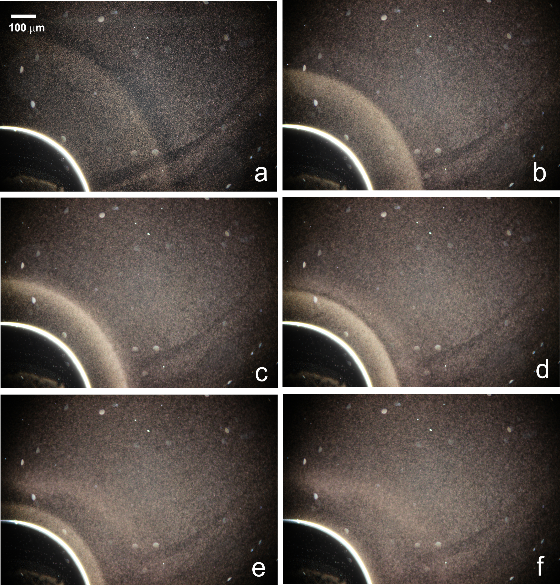
In the closed setup, MR-1 form an aerotactic band around the bubble (Movie S1, Fig. 1). We describe here the formation and evolution of the band.
Initially, the bacteria have a uniform concentration and a high motility in the whole sample. After 20 minutes, they start to aggregate around the bubble, forming a distinct band (Fig. 1.a-b). The bacteria move inside the band and in the area between the bubble and the band, while the rest of the sample enters a non-motile vibrational state (Movie S2). As the bacteria consume the oxygen, the band progressively advances towards the bubble (Fig. 1.c), leaving behind a depletion layer (Fig. 1.d) with non-motile vibrating bacteria. Eventually, the band reaches the bubble (Fig. 1.e) and disappears (Fig. 1.f). At this point, all the bacteria are in the non-motile vibrational state. Depending on initial conditions, the band forms in 5 to 40 minutes and disappears 15 to 60 minutes after closing the sample.
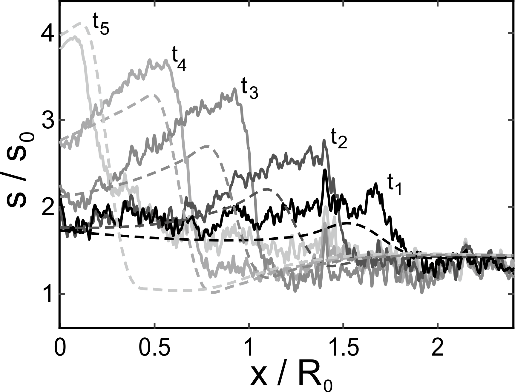
We provide a quantitative evaluation of the phenomenon, by tracking the time evolution of the concentration profiles of Shewanella around the bubble (Fig. 2). The bacterial density is estimated from dark-field images, as proportional to the local light intensity. We compare the experimental results (solid lines) with the predictions from an in-house numerical model (dashed lines) (Supplementary Text), finding good qualitative and quantitative agreement. The model captures how the maximum bacterial concentration increases, as the band moves towards the bubble. The time scales for the band formation and disappearance are reproduced within a 5% and 15% error, respectively.
In the open setup, the observed phenomenon is analogous but a steady state is reached after 2 hours: the Shewanella concentration profile becomes stable, with a band at a fixed distance from the air-liquid interface (Fig. S2.a). The bacteria in the band retain their motility and never enter the vibrational state. The observation is unaltered after 24 hours.
We conclude that the transition between motile and non-motile bacteria observed in the closed setup corresponds to the transition between aerobic and anaerobic functioning.
The oxygen concentration regulates the biofilm formation
Shewanella form a biofilm called pellicle at air-liquid interfaces (?, ?). We clarify here how the oxygen level regulates the process.
With dark-field imaging, we do not have direct access to the air-liquid interface itself, which is saturated by the high amount of scattered light. However, we can extract equivalent information by imaging the bacterial concentration in the liquid in its immediate neighbourhood. In the closed setup, such a concentration remains constant during the initial phases of the band formation (Fig. 2, curves , ); later on, it increases while the band moves towards the interface (Fig. 2, curves ). This behaviour indicates that, initially, the bacterial flux at the bubble wall is entirely adsorbed, while it gets progressively reflected as the oxygen supply decreases. Conversely, in the open setup, where the oxygen supply is unlimited and the oxygen concentration at the interface is constant, the nearby bacterial concentration remains unvaried throughout the whole process. This observation indicates that the incoming flux is entirely adsorbed by the interface.
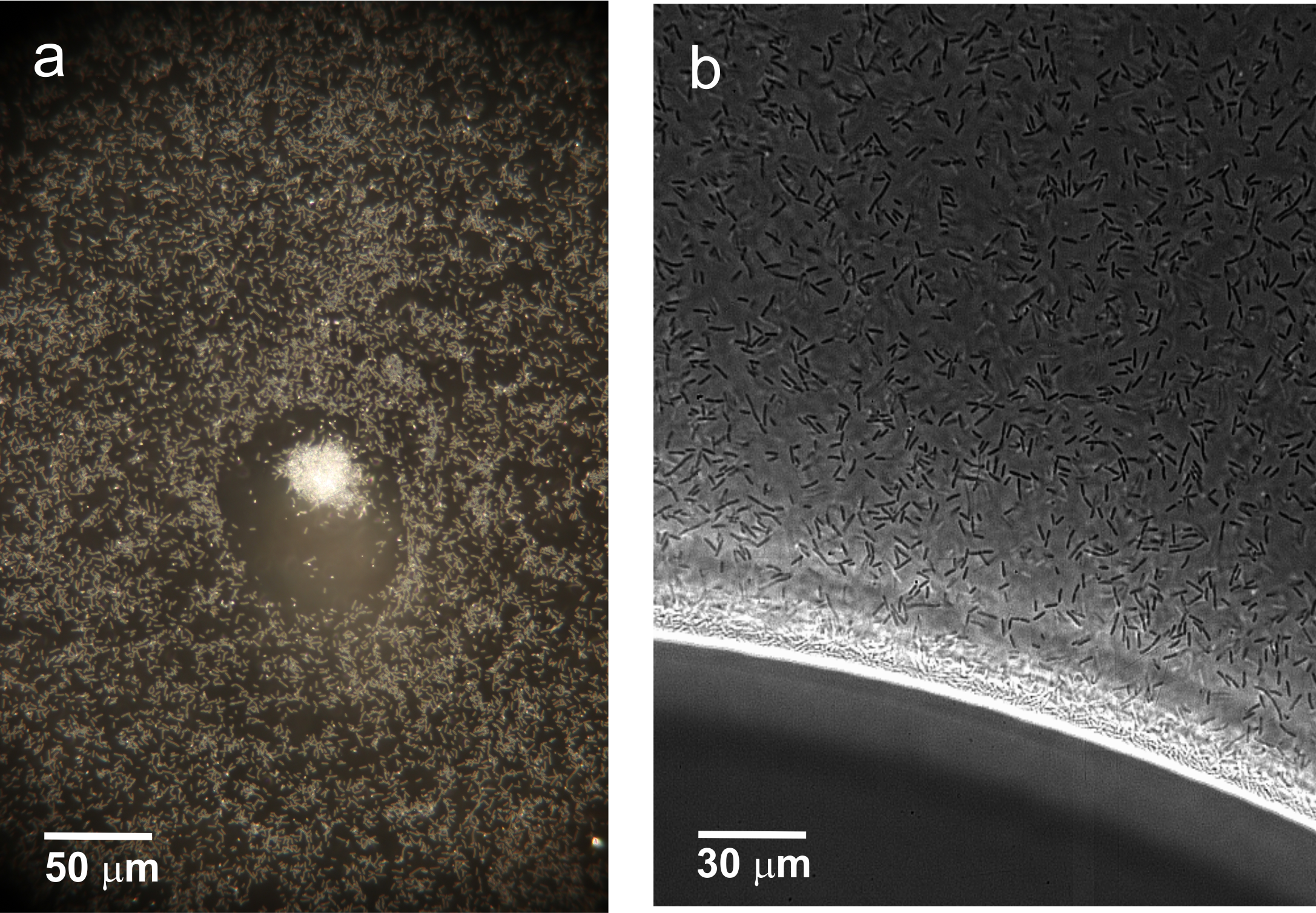
Phase-contrast microscopy reveals a layer of bacteria with nematic-like ordering, piled up at the air-liquid interface and surrounded by a depletion zone. In the closed setup, entailing a limited oxygen supply, such a layer does not increase indefinitely, but stops growing presumably below a certain oxygen concentration (Fig. 3). Conversely, with an unlimited oxygen supply, the layer keeps growing (Fig. S3). We conclude that the oxygen concentration regulates the adsorption of MR-1 at the air-liquid interface.
On occasion, small droplets are captured inside the bubble in the closed setup. Their observation allows direct imaging of the pellicle formation process (Fig. S4, Movie S3).
Here, two air-liquid interfaces are present: (i) between the bubble and the liquid,
where a moving aerotactic band appears, and (ii) between the droplet and the bubble.
On the latter interface, we observe the presence of bacterial clusters with an active motion. They progressively grow in size, by incorporating colliding swimming bacteria (Movie S4).
The process slows down in time. Eventually, the bacteria stop moving and the clusters stop growing. This happens exactly when the band surrounding (i) reaches the bubble, i.e. when the lack of oxygen induces the anaerobic transition everywhere. We infer a connection between bacterial motility and pellicle formation, both regulated by the oxygen level. This conclusion is in line with previous findings that a fully functional flagellum is a prerequisite for pellicle formation in MR-1 (?).
Hybrid locomotion patterns: alternated sideways/longitudinal swimming
We show here that MR-1 feature hybrid locomotion patterns, even in the absence of oxygen gradients. The trajectories of the motile bacteria are analysed in a variation of the closed setup where no bubble is present, immediately after closure of the sample (i.e. with high uniform oxygen concentration).
To evaluate the motility, we consider the probability distribution function (PDF) of the absolute values of instantaneous velocities, derived from the time sampling of trajectories.
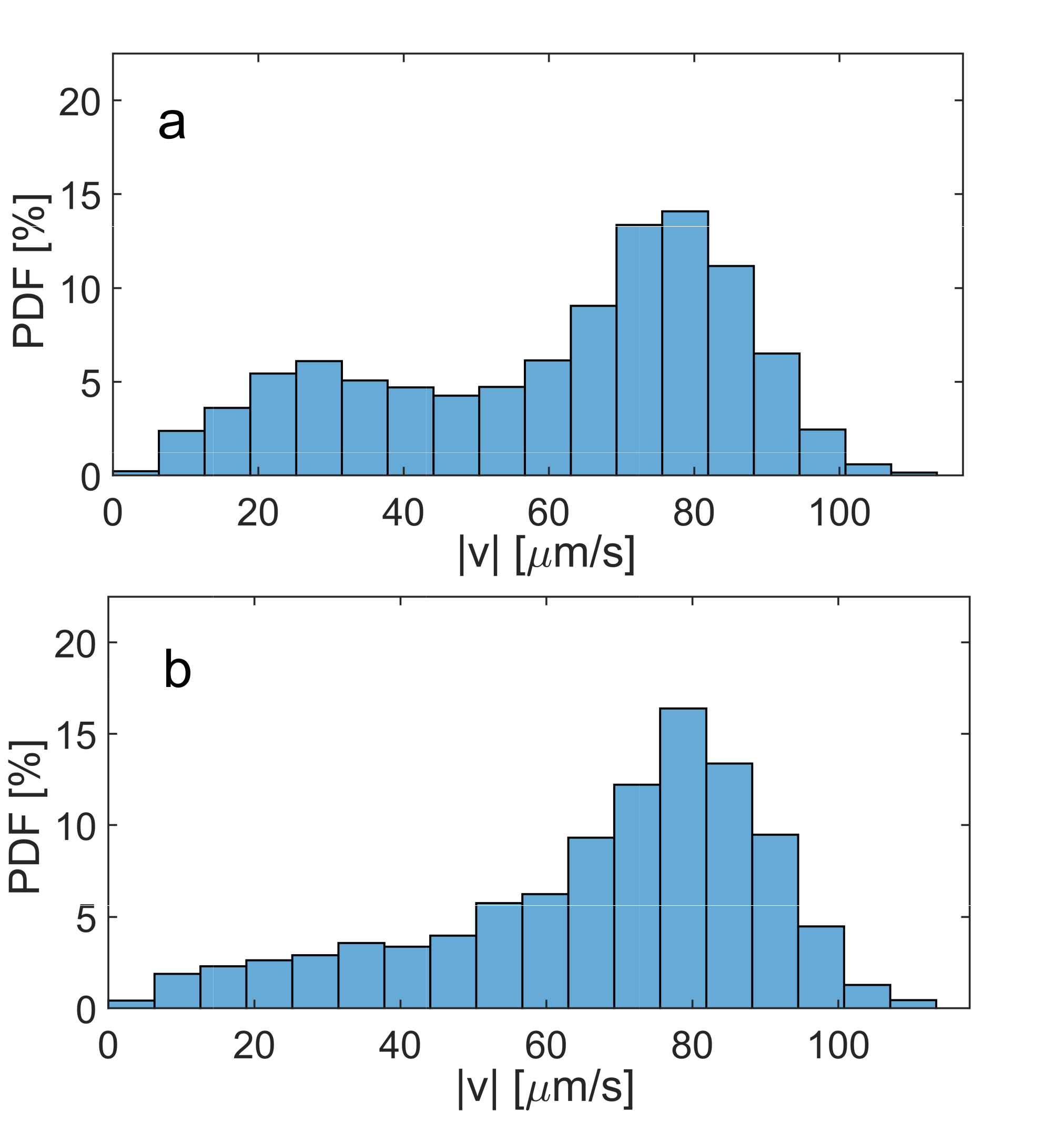
Around 15% of bacteria perform a stop-and-go type of motion: they alternate short pauses and runs (Movie S5). Hence, there is a peak in the PDF, around 0 \microm/s (Fig. S5). We will show in the next section that this is consistent with the inception of biofilm formation. By removing the pauses and accounting only for the motile parts of trajectories, the PDF becomes bimodal, with peaks around 25 \microm/s and 75 \microm/s (Fig. 4.a). A priori, this could correspond to two different scenarios: two populations of bacteria with a constant velocity (fast or slow), or bacteria changing their velocity, by alternating fast and slow runs. We compare the PDF of the instantaneous velocities with the PDF of the average velocities along the trajectories (Fig. S6). Coincidence between the two PDFs would indicate that there are two populations with constant velocity and no switching. We observe that the PDF of the average velocities along the trajectories is still bimodal but the relative amplitude of the peaks has changed. Therefore, we conclude that both scenarios occur: there are fast and slow bacteria but also bacteria changing their velocity along a single trajectory, as confirmed by visual inspection.
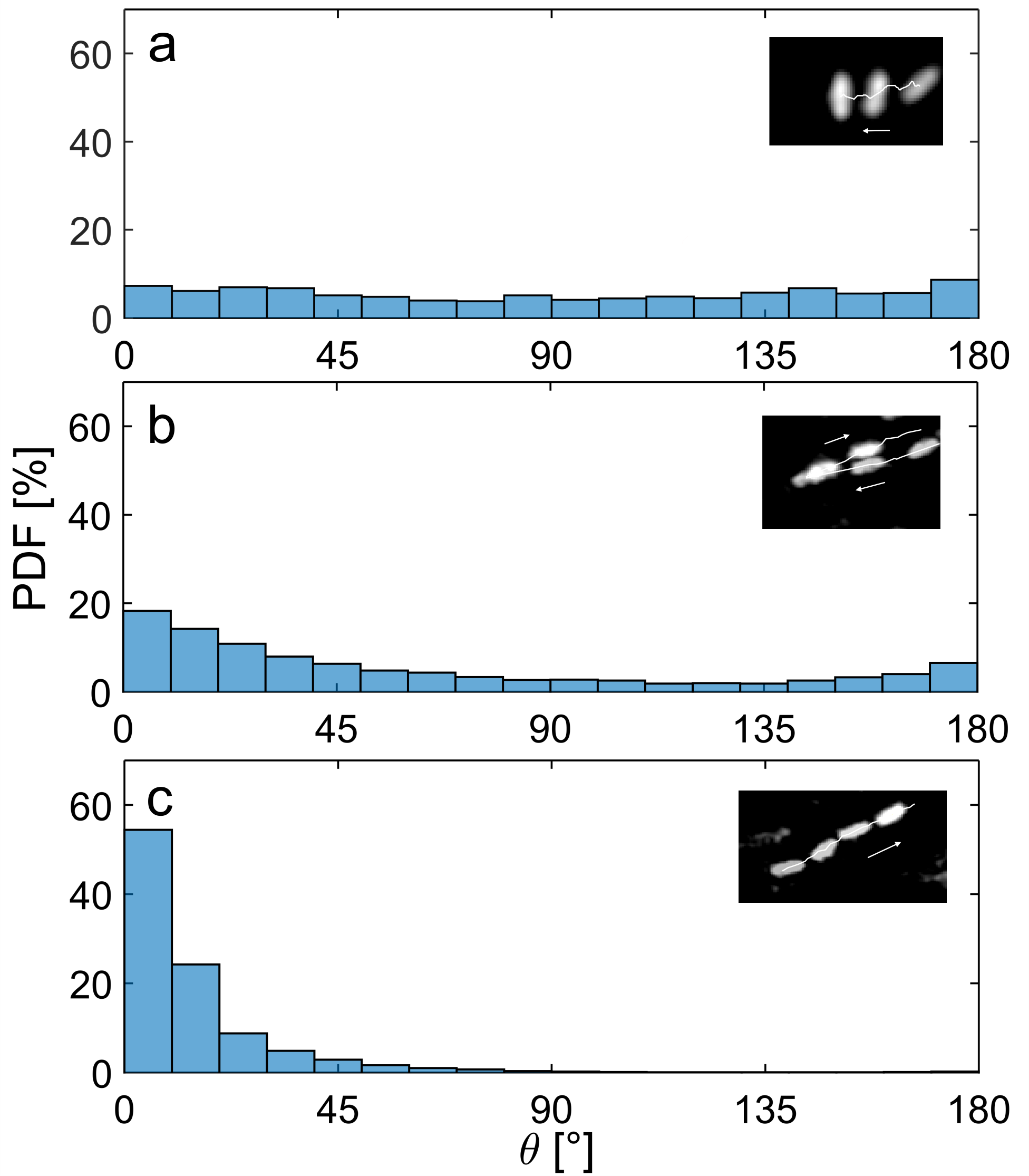
To assess how the speed is related to the reorientation strategies, we analyse the angles between consecutive portions of trajectories and we sort them based on the average speed preceding the turn (Fig. 5). We find three regimes with significant differences: slow (below 25 \microm/s), fast (above 60 \microm/s) and intermediate (25-60 \microm/s). Slow bacteria span all angles between 0∘ and 180∘; more than 80% of such bacteria swim sideways with respect to their main body axis. Fast bacteria swim with straight or continuously curving trajectories, parallel to their longitudinal axis, and turn by small angles (up to 70∘). Bacteria with intermediate velocities exhibit an intermediate behaviour, performing either small angles (0∘-80∘) or reversals (140∘-180∘); they mostly swim parallel to their main axis but can switch to sideways motion (Movie S6).
This differentiation within the same population of monoflagellate bacteria is remarkable, as it combines features of typical strategies used by morphologically different species to explore the surroundings. Monotrichous bacteria, such as 70% of marine bacteria, are typically fast (up to 75 \microm/s), moving by straight or curved trajectories (?). They change direction by inverting their flagellar rotation, inducing reversals (150∘-180∘ angles), like MR-1 in the intermediate velocity runs. Peritrichous bacteria, such as most enteric bacteria, are typically slower (30 \microm/s) and move by run-and-tumbling (?). They swim in almost straight runs, with the flagella bundled together, stopping and tumbling, when one flagellum inverts its rotation disrupting the bundle. All angles occur upon reorientation, like in the slow runs of MR-1 (?).
Hybrid locomotion mechanisms have been reported for several bacterial species, as a way to enhance direction randomisation (?).
Other monotrichous bacteria alternate 180∘ reversals with 90∘ flicks induced by the flagellum bending (run-reverse-flick) (?, ?). Though MR-1 can perform this type of motion, the absence of the peak at 90∘ shows that it is not their main strategy. The predominance of sideways swimming in the slow regime, together with the possibility to switch between sideways and longitudinal motion, suggests that they generate torque at low velocities by modulating the coiling shape of the flagellum. This is in line with the recent discovery that some bacteria, including the conspecific S. putrefaciens, can swim with the flagellum wrapped around their body (?, ?).
The adaptive tuning of sideways/longitudinal swimming shapes the collective behaviour
We study here how the ambient conditions influence the motility and the reorientation strategies of MR-1, determining different collective behaviours, namely band and biofilm formation. We perform two experiments to separately test the impact of the oxygen concentration and the oxygen gradient. To this aim, we use two variations of the closed setup: without bubble (i.e. uniform oxygen concentration, decaying in time) and with a bubble (i.e. with an oxygen gradient).
To evaluate the effect of the oxygen concentration, we examine the samples without bubbles for 15 minutes after sealing. As the bacteria deplete the oxygen, the peak at slow velocities in the PDF of instantaneous velocities progressively disappears (Fig. 4.b). The PDF becomes unimodal with a peak around 80 \microm/s. This observation is compatible with the interruption of the biofilm formation at low oxygen concentration. We conclude that, like S. putrefaciens (?), S. oneidensis adapts its speed in response to the local concentration of a chemoattractant. Hence, it does not only perform chemotaxis (reaction to a concentration gradient), but also chemokinesis (reaction to the concentration itself).
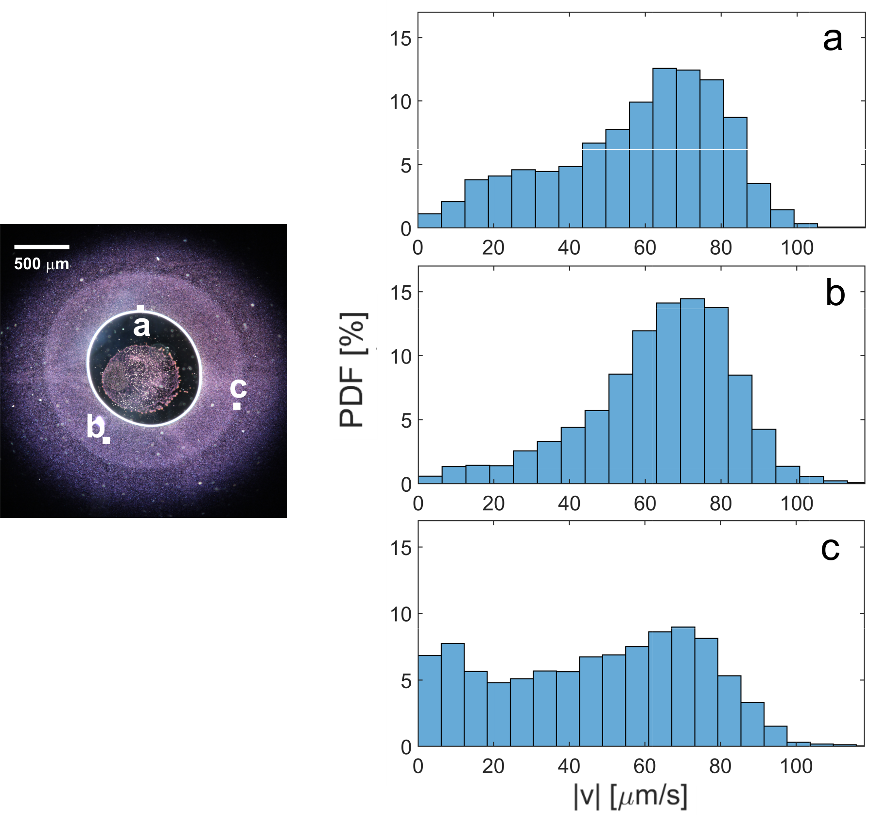
To investigate the effect of the oxygen gradient, we analyse the case with an entrapped bubble, featuring a moving band.
Fig. 6 displays the PDF of instantaneous velocities, at three different locations: (a) at the air-liquid interface, (b) between the bubble and the band (’middle’) and (c) on the band.
The numerical simulations show that the oxygen concentration is always monotonically decreasing in the radial direction. At the interface and in the middle, the PDFs reproduce the trend observed without bubbles, for decreasing oxygen concentrations: the bimodal distribution in (a) turns unimodal in (b), where the oxygen concentration is lower. On the band, however, such a trend is disrupted: the peak at slow velocities reappears and the number of particles with an intermediate speed (25-60 \microm/s) increases. The PDFs of turning angles, sorted by velocity ranges, are unvaried with respect to the case without oxygen gradient.
We conclude that different speed ranges correspond to different reorientation strategies, fulfilling specific biological functions. In particular, a peak at slow velocities, where Shewanella reorients in any direction, emerges in environmental conditions where there is no preferential direction of swimming. At the bubble wall, it relates to the inception of the biofilm formation. In the areas where the bacteria accumulate (aerotactic band), it reflects the fact that MR-1 is in its optimal oxygen concentration range. On the band, the increased number of bacteria swimming at intermediate velocities (25-60 \microm/s), favouring straight runs or reversals, guarantees the persistence in the region: when a bacterium exits the band, it either stops or immediately comes back.
In the region between the bubble and the band, high velocities are dominant (above 60 \microm/s), mostly associated to straight runs. This is typical of directional motion and reflects the aerotaxis towards the band.
Summarising, slow velocities are associated to sideways displacements and isotropic direction randomisation, intermediate velocities present run-reverse patterns producing confinement, high velocities encompass straight runs, related to directional aerotactic motion.
Conclusions
The present work is a comprehensive study of the collective behaviour of Shewanella oneidensis MR-1 next to an air source, from accumulation to the air-liquid biofilm formation. We studied the dynamic evolution of the aerotactic band gathering around a bubble, both with experiments and simulations. When the air source is not confined, the band remains at a constant distance from the air-liquid interface. Conversely, when the air source is confined and the oxygen is depleted by the bacteria, the band moves towards the interface, eventually reaching it and disappearing. The local oxygen concentration regulates the biofilm formation, by affecting the bacterial adsorption at the interface and their motility. Both are suppressed when the oxygen is entirely depleted.
The system at hand is a minimal model of an ecological niche, where the inhabitants modify the environment and their behaviour dynamically adapts in return.
We characterised the collective locomotion strategies of MR-1 in response to the environmental conditions (oxygen concentration and gradient), by tracking their trajectories. They explore the surroundings with a combination of fast and slow runs, spanning a broad range of velocities. Their velocity distributions are bimodal and dynamically change with the ambient conditions, posing a challenge to the concept of average velocity of a bacterial population. Different velocities are associated to specific reorientation strategies, realising different biological functions, such as aerotaxis or confinement. Fast runs are mostly straight or curved; their number increases when the bacteria perform aerotaxis in response to an oxygen gradient.
Slow runs correspond to a higher direction randomisation, with turns spanning all angles. They are associated to favourable conditions for the bacteria, such as their preferred oxygen concentration range in the aerotactic band, or to the inception of the biofilm formation. Surprisingly for a monotrichous bacterium, they are mostly realised through sideways swimming. At intermediate velocities the bacteria alternate straight runs and reversals. In the aerotactic band this behaviour supports confinement, correcting the trajectories leading away from the optimal oxygen concentration.
The possibility to switch between fast (longitudinal) and slow (mostly sideways) swimming allows MR-1 to access typical velocity and reorientation ranges of both mono- and multiflagellate bacteria, tremendously increasing their biological competitiveness.
The present work establishes the link between the intriguing hybrid locomotion strategies of MR-1 and their aerotactic collective behaviour, showing how the adaptive tuning of their bimodal velocity distributions regulates the process leading from confinement in aerotactic bands to biofilm formation.
References
- 1. T. Schweinitzer, C. Josenhans, Bacterial energy taxis: a global strategy?, Arch. Microbiol., 192, 507 –520 (2010).
- 2. J.-B. Raina, V. Fernandez, B. Lambert, R. Stocker, J. R. Seymour, The role of microbial motility and chemotaxis in symbiosis, Nat. Rev. Microbiol., 17, 284– 294 (2019).
- 3. B. L. Taylor, I. B. Zhulin, M. S. Johnson, Aerotaxis and other energy-sensing behavior in bacteria, Annu. Rev. Microbiol., 53, 103–128 (1999).
- 4. O. Baracchini, J. C. Sherris, The chemotactic effect of oxygen on bacteria, J. Path. Bact., 77, 565–574 (1959).
- 5. J. Adler, Chemotaxis in bacteria, Science, 153, 708–716 (1966).
- 6. J. Li, Molecular mechanisms of behavioral responses in Shewanella oneidensis MR-1, Ph.D. thesis, Johns Hopkins University, Baltimore (2008).
- 7. K. Son, D. R. Brumley, R. Stocker, Live from under the lens: exploring microbial motility with dynamic imaging and microfluidics, Nat. Rev. Microb., 13, 761–775 (2015).
- 8. R. Stocker, Reverse and flick: Hybrid locomotion in bacteria, Proc. Natl. Acad. Sci. USA, 108, 2635–.2636 (2011).
- 9. H. C. Berg, D. A. Brown, Chemotaxis in Escherichia coli analysed by three-dimensional tracking, Nature, 239, 500–504 (1972).
- 10. K. Venkateswaran, Polyphasic taxonomy of the genus Shewanella and description of Shewanella oneidensis sp. nov., Int. J. Syst. Bacteriol., 49, 705–724 (1999).
- 11. Y. A. Gorby, S. Yanina, J. S. McLean, K. M. Rosso, D. Moyles, A. Dohnalkova, T. J. Beveridge, I. S. Chang, B. H. Kim, K. S. Kim, D. E. Culley, S. B. Reed, M. F. Romine, D. A. Saffarini, E. A. Hill, L. Shi, D. A. Elias, D. W. Kennedy, G. Pinchuk, K. Watanabe, S. Ishii, B. Logan, K. H. Nealson, J. K. Fredrickson, Electrically conductive bacterial nanowires produced by Shewanella oneidensis strain MR-1 and other microorganisms, Proc. Natl. Acad. Sci. USA, 103, 11358 –11363 (2006).
- 12. P. Subramanian, S. Pirbadian, M. Y. El-Naggar, G. J. Jensen, Ultrastructure of Shewanella oneidensis MR-1 nanowires revealed by electron cryotomography, Proc. Natl. Acad. Sci. U.S.A., 115, E3246–E3255 (2018).
- 13. B. E. Logan, Exoelectrogenic bacteria that power microbial fuel cells, Nat. Rev. Microbiol., 7, 375 –381 (2009).
- 14. D. Wu, D. Xing, X. Mei, B. Liu, C. Guo, N. Ren, Electricity generation by Shewanella sp. HN-41 in microbial fuel cells, Int. J. Hydrog. Energy, 38, 15568–15573 (2013).
- 15. K. Rabaey, R. A. Rozendal, Microbial electrosynthesis - revisiting the electrical route for microbial production, Nat. Rev. Microbiol., 8, 706–716 (2010).
- 16. C. K. Ng, K. Sivakumar, X. Liu, M. Madhaiyan, L. Ji, L. Yang, C. Tang, H. Song, S. Kjelleberg, B. Cao, Influence of outer membrane c-type cytochromes on particle size and activity of extracellular nanoparticles produced by Shewanella oneidensis, Biotechnol. Bioeng., 110, 1831–1837 (2013).
- 17. C. K. Ng, T. K. C. Tan, H. Songad, B. Cao, Reductive formation of palladium nanoparticles by Shewanella oneidensis: role of outer membrane cytochromes and hydrogenases, RSC Adv., 3, 22498–22503 (2013).
- 18. B. E. Logan, Microbial fuel cells: Methodology and technology, Environ. Sci. Technol., 40, 5181 –5192 (2006).
- 19. J. F. Heidelberg, I. T. Paulsen, K. E. Nelson, E. J. Gaidos, W. C. Nelson, T. D. Read, J. A. Eisen, R. Seshadri, N. Ward, B. Methe, R. A. Clayton, T. Meyer, A. Tsapin, J. Scott, M. Beanan, L. Brinkac, S. Daugherty, R. T. DeBoy, R. J. Dodson, A. S. Durkin, D. H. Haft, J. F. Kolonay, R. Madupu, J. D. Peterson, L. A. Umayam, O. White, A. M. Wolf, J. Vamathevan, J. Weidman, M. Impraim, K. B. K. Lee, C. Lee, J. Mueller, H. Khouri, J. Gill, T. R. Utterback, L. A. McDonald, T. V. Feldblyum, H. O. Smith, J. C. Venter, K. H. Nealson, C. M. Fraser, Genome sequence of the dissimilatory metal ion-reducing bacterium Shewanella oneidensis, Nat. Biotechnol., 20, 1118–1123 (2002).
- 20. J. K. Fredrickson, M. F. Romine, A. S. Beliaev, J. M. Auchtung, M. E. Driscoll, T. S. Gardner, K. H. Nealson, A. L. Osterman, G. Pinchuk, J. L. Reed, D. A. Rodionov, J. L. M. Rodrigues, D. A. Saffarini, M. H. Serres, A. M. Spormann, I. B. Zhulin, J. M. Tiedje, Towards environmental systems biology of Shewanella, Nat. Rev. Microbiol., 6, 592–603 (2008).
- 21. Y. Liang, H. Gao, J. Chen, Y. Dong, L. Wu, Z. He, X. Liu, G. Qiu, J. Zhou, Pellicle formation in Shewanella oneidensis, BMC Microbiol., 10, 291–301 (2010).
- 22. J. Armitano, V. Méjean, C. Jourlin-Castelli, Aerotaxis governs floating biofilm formation in Shewanella oneidensis, Environ. Microbiol., 15, 3108–3118 (2013).
- 23. J. E. Johansen, J. Pinhassi, N. Blackburn, U. L. Zweifel, A. Hågström, Variability in motility characteristics among marine bacteria, Aquat. Microb. Ecol., 28, 229–237 (2002).
- 24. L. Xie, T. Altindal, S. Chattopadhyay, X. Wu, Bacterial flagellum as a propeller and as a rudder for efficient chemotaxis, Proc. Natl. Acad. Sci. USA, 108, 2246–2251 (2011).
- 25. M. Hintsche, V. Waljor, R. Grossmann, M. J. Kühn, K. M. Thormann, F. Peruani, C. Beta, A polar bundle of flagella can drive bacterial swimming by pushing, pulling, or coiling around the cell body, Sci. Rep., 7, 1–10 (2017).
- 26. M. Kühn, F. Schmidt, B. Eckhardt, K. Thormann, Bacteria exploit a polymorphic instability of the flagellar filament to escape from traps, Proc. Natl. Acad. Sci. U.S.A., 114, 6340–6345 (2017).
- 27. G. M. Barbara, J. G. Mitchell, Marine bacterial organisation around point-like sources of amino acids, FEMS Microbiol. Ecol., 43, 99–109 (2003).
Acknowledgements
We thank Laura Turco for technical support in the experiments, Gal Scholnik for performing preliminary experiments,
Andreas Kappler for providing the bacterium S. oneidensis MR-1 and Mathias Schröter for support in preliminary bacterial tracking.
We acknowledge discussions with Anupam Sengupta and Martin Kröger.
We thank Hans-Christian Öttinger, James Clewett and Martin Callies for feedback on the manuscript.
Funding We acknowledge financial support from the Deutsche Forschungsgemeinschaft (SFB 937, project A20) and from the People Programme (Marie Curie Actions) of the European
Union s Seventh Framework Programme FP7/2007-2013/ under REA grant agreement n∘[628154].
Author Contributions
I.G., L.S. and M.M. designed the experiments.
I.G. and L.S. performed the experiments.
L.S. and J.V. developed the numerical model.
T.B. and L.S. did the tracking.
L.S., I.G. and J.V. did the statistical analysis and interpretation of the data.
J.V. and M.M. contributed with comments.
L.S., I.G. and J.V. wrote the draft. All the authors contributed to subsequent revisions.
Competing Interests The authors declare that they have no competing financial interests.
Data and materials availability
All data needed to evaluate the conclusions in the paper are present in the paper and/or the Supplementary Materials.
Any additional data supporting the findings of this study as well as the in-house codes are available from the corresponding author upon request.
Correspondence and requests for materials should be addressed to L.S (email: laura.stricker@mat.ethz.ch).
Supplementary materials
Materials and Methods
Supplementary Text
Supplementary References
Fig. S1-S6
Movie S1-S6.