Medical Imaging with Deep LearningMedical Imaging with Deep Learning
\jmlrpages
\jmlryear2019
\jmlrworkshopExtended Abstract – MIDL 2019 Submission
\midlauthor\NameRaphael Meier\nametag1 \Emailraphael.meier@insel.ch
\NameMichael Rebsamen\nametag1 \Emailmichael.rebsamen@insel.ch
\NameUrspeter Knecht\nametag2 \Emailurspeter.knecht@istb.unibe.ch
\NameMauricio Reyes\nametag2,3 \Emailmauricio.reyesaguirre@insel.ch
\NameRoland Wiest\nametag1 \Emailroland.wiest@insel.ch
\NameRichard McKinley\nametag1 \EmailrichardIain.mckinley@insel.ch
\addr1 Support Center for Advanced Neuroimaging (SCAN), University Institute of Diagnostic and Interventional Neuroradiology, University of Bern, Inselspital, Bern University Hospital, Bern, Switzerland
\addr2 Institute for Surgical Technology and Biomechanics, University of Bern, Bern, Switzerland and \addr3 Healthcare Imaging A.I. Lab, Insel Data Science Center, Inselspital, Bern University Hospital, Bern, Switzerland and
Stratify or Inject: Two Simple Training Strategies to Improve Brain Tumor Segmentation
Abstract
Deep learning methods for brain tumor segmentation are typically trained in an ad hoc fashion on all available data. Brain tumors are tremendously heterogeneous in image appearance and labeled training data is limited. We argue that incorporation of additional prior information, specifically tumor grade, associated with tumor imaging phenotypes during model training can significantly improve segmentation performance. Two strategies for incorporation of tumor grade during model training are proposed and their impact on segmentation performance is demonstrated on the BRATS 2018 dataset.
1 Introduction
The segmentation of brain tumors has been a long standing problem in medical image analysis. Research on this topic has been accelerated greatly through the availability of public datasets such as the Brain Tumor Segmentation (BRATS) Challenge dataset [Menze et al.(2015)Menze, Jakab, and et al., Bakas et al.(2017a)Bakas, Akbari, and et al., Bakas et al.(2017b)Bakas, Akbari, and et al., Bakas et al.(2017c)Bakas, Akbari, and et al.]. Currently, the best performing methods for brain tumor segmentation are based on deep learning [Bakas et al.(2018)Bakas, Reyes, and et al.], with first approaches being applied in clinically critical areas such as tumor response assessment [Kickingereder et al.(2019)Kickingereder, Isensee, and et al.] or radiation therapy planning [Jungo et al.(2018)Jungo, Meier, and et al.]. A general view in deep learning is that more training data yields better generalization performance. For tasks in computer vision it was shown that model performance increases logarithmically based on volume of training data [Sun et al.(2017)Sun, Shrivastava, and et al.]. Consequently, deep learning segmentation models are trained on all available data, often neglecting peculiarities of the data at hand.
The BRATS Challenge has been concerned so far with the segmentation of glioma, which are primary tumors of the central nervous system. Glioma can be classified into different tumor grades based on the underlying molecular characteristics and histology [Louis et al.(2016)Louis, Perry, and et al.]. A higher grade reflects increasing malignancy and glioma are commonly grouped into high-grade (grade III/IV) and low-grade glioma (grade I/II). Furthermore, they exhibit a tremendous genetic and molecular heterogeneity which spans across tumor grades but also manifests itself within a particular type such as glioblastoma (grade IV) [Verhaak et al.(2010)Verhaak, Hoadley, and et al., Sottoriva et al.(2013)Sottoriva, Spiteri, and et al.]. The underlying biological configuration of a tumor has been associated with distinctively different tumor imaging phenotypes [Grossmann et al.(2016)Grossmann, Gutman, and et al.]. In general, low-grade glioma present much less or no contrast-enhancement compared to high-grade glioma [Forst et al.(2014)Forst, Nahed, and et al]. Deep learning methods are confronted with the challenge to successfully generalize across these different imaging phenotypes.
We hypothesize that brain tumor segmentation performance of deep learning methods can be improved by utilizing prior information associated with tumor imaging phenotypes during model training. Thus, we propose two simple training strategies targeted at tumor grade and evaluate their effectiveness on the BRATS 2018 dataset using a recently proposed, top-ranked method [McKinley et al.(2019)McKinley, Meier, and Wiest]. This work is part of a more extensive study currently being submitted to a journal.
2 Methods
Model architecture. The deep learning method corresponds to a shallow U-Net style model of down and upsampling connections featuring densely connected blocks of dilated convolutions. For more details on the model architecture we refer to [McKinley et al.(2019)McKinley, Meier, and Wiest].
Incorporation of tumor grade. We propose two strategies to utilize information on tumor grade at the stage of model training. The first strategy consists of stratifying the training data into high-grade glioma (HGG) and low-grade glioma (LGG) cases and training two separate models. During testing the respective model is applied to testing data with corresponding tumor grade. As a second strategy, we propose an injection of the tumor grade. In addition to feeding the model with imaging data consisting of the co-registered Magnetic Resonance (MR) sequences, we provide it with a binary input indicating if the case at hand is either a LGG or HGG. The tumor type is injected as an image volume, with dimensions identical to the MR sequences and all voxel values set either to zeros or ones. The model is then trained on this enlarged dataset (type-aware network). For both strategies, the model architecture and hyperparameter setting remains unchanged.
In the following, we utilized training data of the BRATS 2018 Challenge, which includes 75 patients with LGGs and 210 patients with HGGs. We did not include the testing data in the analysis since tumor grade is blinded for those cases. A detailed description of the imaging data can be found in [Bakas et al.(2018)Bakas, Reyes, and et al.]. Four different models were trained using five-fold cross-validation: i) a baseline model using all available cases (N=285), ii) a model trained only on HGG data (N=210), iii) a model trained only on LGG data (N=75), and iv) the type-aware network trained on all cases (N=285).
3 Results & Conclusion
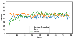
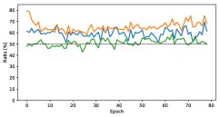
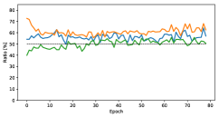
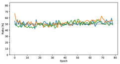
The segmentation performance of the four different deep learning models in terms of fraction of cases with improved Dice coefficient is shown in Table 1. If we look at the HGG data alone, the HGG model yields a significantly improved performance over the baseline model in 58.4% of the cases for contrast-enhancing tumor and 70.3% of the cases for the tumor core segmentation. Looking at all the data including also LGG, we see a significant improvement for the tumor core segmentation in 64.9% of the cases when using the two models trained on stratified data compared to the baseline model trained on all available data. Finally, the type-aware network yields a minor but significant improvement for the segmentation of the tumor core in 53.9 % of the cases. Figure LABEL:fig:epochs shows that observed improvements are manifested relatively consistent across all epochs of model training.
| CE | Core | Tumor | |
|---|---|---|---|
| LGG vs. Baseline | 41.7 (p=0.877) | 49.3 (p=0.454) | 54.7 (p=0.208) |
| HGG vs. Baseline | 58.4 (p=0.005) | 70.3 (p=5.659e-09) | 46.7 (p=0.877) |
| HGG/LGG vs. Baseline | 54.6 (p=0.127) | 64.9 (p=1.441e-05) | 48.8 (p=0.725) |
| Type-aware vs. Baseline | 53.4 (p=0.321) | 53.9 (p=0.028) | 52.6 (p=0.231) |
fig:epochs
We have proposed two different strategies on incorporating information of tumor grade during model training, which are straightforward to apply, and demonstrated their effectiveness on the BRATS 2018 dataset. While data stratification yields clear improvements, more advanced network architectures incorporating prior information about tumor grade beyond injecting it as an additional input should be investigated further. In addition, our strategies could also be used in conjunction with networks for tumor typing.
This work was supported by the Swiss National Foundation, grant number 169607.
References
- [Bakas et al.(2017a)Bakas, Akbari, and et al.] Spyridon Bakas, Hamed Akbari, and et al. Advancing the cancer genome atlas glioma MRI collections with expert segmentation labels and radiomic features. Scientific Data, 4:170117, September 2017a. 10.1038/sdata.2017.117. URL https://doi.org/10.1038/sdata.2017.117.
- [Bakas et al.(2017b)Bakas, Akbari, and et al.] Spyridon Bakas, Hamed Akbari, and et al. Segmentation labels for the pre-operative scans of the tcga-gbm collection, 2017b. URL https://wiki.cancerimagingarchive.net/x/KoZyAQ.
- [Bakas et al.(2017c)Bakas, Akbari, and et al.] Spyridon Bakas, Hamed Akbari, and et al. Segmentation labels for the pre-operative scans of the tcga-lgg collection, 2017c. URL https://wiki.cancerimagingarchive.net/x/LIZyAQ.
- [Bakas et al.(2018)Bakas, Reyes, and et al.] Spyridon Bakas, Mauricio Reyes, and et al. Identifying the best machine learning algorithms for brain tumor segmentation, progression assessment, and overall survival prediction in the BRATS challenge. CoRR, abs/1811.02629, 2018. URL http://arxiv.org/abs/1811.02629.
- [Forst et al.(2014)Forst, Nahed, and et al] D. A. Forst, B. V. Nahed, and et al. Low-grade gliomas. The Oncologist, 19(4):403–413, mar 2014. 10.1634/theoncologist.2013-0345. URL https://doi.org/10.1634/theoncologist.2013-0345.
- [Grossmann et al.(2016)Grossmann, Gutman, and et al.] Patrick Grossmann, David A. Gutman, and et al. Imaging-genomics reveals driving pathways of mri derived volumetric tumor phenotype features in glioblastoma. BMC Cancer, 16(1):611, Aug 2016. ISSN 1471-2407. 10.1186/s12885-016-2659-5. URL https://doi.org/10.1186/s12885-016-2659-5.
- [Jungo et al.(2018)Jungo, Meier, and et al.] Alain Jungo, Raphael Meier, and et al. Uncertainty-driven sanity check: Application to postoperative brain tumor cavity segmentation. CoRR, abs/1806.03106, 2018. URL http://arxiv.org/abs/1806.03106.
- [Kickingereder et al.(2019)Kickingereder, Isensee, and et al.] Philipp Kickingereder, Fabian Isensee, and et al. Automated quantitative tumour response assessment of MRI in neuro-oncology with artificial neural networks: a multicentre, retrospective study. The Lancet Oncology, apr 2019. 10.1016/s1470-2045(19)30098-1. URL https://doi.org/10.1016/s1470-2045(19)30098-1.
- [Louis et al.(2016)Louis, Perry, and et al.] David N. Louis, Arie Perry, and et al. The 2016 world health organization classification of tumors of the central nervous system: a summary. Acta Neuropathologica, 131(6):803–820, Jun 2016. 10.1007/s00401-016-1545-1. URL https://doi.org/10.1007/s00401-016-1545-1.
- [McKinley et al.(2019)McKinley, Meier, and Wiest] Richard McKinley, Raphael Meier, and Roland Wiest. Ensembles of densely-connected cnns with label-uncertainty for brain tumor segmentation. In Alessandro Crimi, Spyridon Bakas, and et al., editors, Brainlesion: Glioma, Multiple Sclerosis, Stroke and Traumatic Brain Injuries, pages 456–465, Cham, 2019. Springer International Publishing. ISBN 978-3-030-11726-9.
- [Menze et al.(2015)Menze, Jakab, and et al.] Bjoern H. Menze, Andras Jakab, and et al. The multimodal brain tumor image segmentation benchmark (BRATS). IEEE Transactions on Medical Imaging, 34(10):1993–2024, oct 2015. 10.1109/tmi.2014.2377694.
- [Sottoriva et al.(2013)Sottoriva, Spiteri, and et al.] Andrea Sottoriva, Inmaculada Spiteri, and et al. Intratumor heterogeneity in human glioblastoma reflects cancer evolutionary dynamics. Proceedings of the National Academy of Sciences, 110(10):4009–4014, 2013. ISSN 0027-8424. 10.1073/pnas.1219747110. URL https://www.pnas.org/content/110/10/4009.
- [Sun et al.(2017)Sun, Shrivastava, and et al.] Chen Sun, Abhinav Shrivastava, and et al. Revisiting unreasonable effectiveness of data in deep learning era. In IEEE International Conference on Computer Vision, ICCV 2017, Venice, Italy, October 22-29, 2017, pages 843–852, 2017. 10.1109/ICCV.2017.97. URL https://doi.org/10.1109/ICCV.2017.97.
- [Verhaak et al.(2010)Verhaak, Hoadley, and et al.] Roel G.W. Verhaak, Katherine A. Hoadley, and et al. Integrated genomic analysis identifies clinically relevant subtypes of glioblastoma characterized by abnormalities in pdgfra, idh1, egfr, and nf1. Cancer Cell, 17(1):98 – 110, 2010. ISSN 1535-6108. https://doi.org/10.1016/j.ccr.2009.12.020. URL http://www.sciencedirect.com/science/article/pii/S1535610809004322.