Floating Zone Growth of Sr Substituted Han Purple: Ba0.9Sr0.1CuSi2O6
Abstract
We present a route to grow single crystals of Ba0.9Sr0.1CuSi2O6 suitable for inelastic neutron studies via the floating zone technique. Neutron single crystal diffraction was utilized to check their bulk quality and orientation. Finally, the high quality of the grown crystals was proven by X-ray diffraction and magnetic susceptibility.
I Introduction
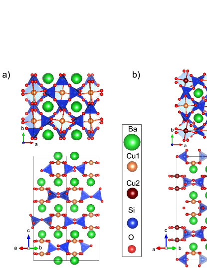
Already in the Han Dynasty, the historically known compound Han Purple Berke (2007) BaCuSi2O6 was used as a purple coloring pigment in China. The rather blue compound Berke (2007) was possibly created in ancient times with a lot of Cu2O (red) inside, thus mixing to a purple pigment. Han Purple can be found in nature, e.g., Africa, as the natural mineral Colinowensite Rieck et al. (2015). Rediscovered and reported upon in 1989 L. W. FINGER (1989), it attracted the interest of the physics community starting from 1997 Sasago et al. (1997), when the rare arrangement of the Cu ions as pairs on a two dimensional square lattice (see Figure 1) was noticed to form dimers with a singlet ground-state and triplet excited states. As the triplet state Zeeman split, in high magnetic fields, a two-dimensional Bose–Einstein condensate (BEC) of the bosonic triplet quasiparticles created a strong interest Jaime et al. (2004); Sebastian et al. (2006a) in the compound. This interest was investigated after the discovery of an incommensurately modulated low temperature structure below 100 K Rüegg et al. (2007); Sheptyakov et al. (2012); Samulon et al. (2006) complicating the model and its physics. These studies were performed on BaCuSi2O6 crystals grown by two methods, namely, the floating zone (FZ) method in an oxygen flow Jaime et al. (2004); Sparta and Roth (2004) and from an oxygen spending lithiummetaborate flux Sebastian et al. (2006b, a), both with no detailed description of the growth conditions. Recently, we reported on the substitution series of (Ba,Sr)CuSi2O6, which stabilizes the tetragonal room temperature structure of BaCuSi2O6 down to lowest temperatures already with 5% substititution Puphal et al. (2016). In a following study, we could show the growth conditions of BaCuSi2O6 single crystals and its Sr-substituted variant van Well et al. (2016) by self melt growth in oxygen pressure. The resulting crystals have a typical size of mm3. Here we report on the floating zone growth of Ba0.9Sr0.1CuSi2O6, where large, high-quality single crystals are obtained for the first time, enabling future studies by, e.g., neutron spectroscopy.
II Experimental Details
Thermogravimetric analysis was performed using a NETZSCH STA 409 analyzer. The polycrystalline rods for the floating-zone growth were pressed in a Powloka hydrostatic press. The floating-zone growth was performed in a CSC FZ-1000-H-VI-VP-PC with a 300 W halogen lamp (FZ1) and a SCIDRE HKZ equipped with a 5 kW xenon lamp (FZ2). The powder X-ray diffraction measurement was performed using a Bruker D8 Advance with a Cu cathode. Fluorescence spectra were recorded using the Orbis microXRF analyzer from EDAX. Neutron diffraction experiments were carried out on the MORPHEUS two-axis diffractometer at SINQ (PSI) at room temperature using a wavelength of Å. Magnetic susceptibility measurements were carried out in a range of 1.8–300 K at 0.1 T using a quantum design physical property measurements system (PPMS). A laboratory X-ray Laue equipped with CCD camera (Photonic Science) was used to orient the samples.
III Synthesis
Polycrystalline Ba0.9Sr0.1CuSi2O6 was prepared by sintering stoichiometric amounts of BaCO3, SrCO3, CuO, and SiO2. The powder was ground and sintered in an aluminum oxide crucible in air at 1028C for 2 months, with several intermediate grindings to remove any early stage phases in the silicate formation Berke (2007) as BaCu2Si2O7. Its phase purity was checked with laboratory X-ray diffraction, proving to be of the structure (s.g. 142) shown in Figure 1 a. The powder was then pressed into rods of a 7 mm diameter by a hydrostatic press (4000 bar) using rubber forms and subsequently annealed for 24 h in air at 1030C. The rod density was checked via dilatometry and found to be above 92%. Finally, single crystals were grown using FZ1 as described below.
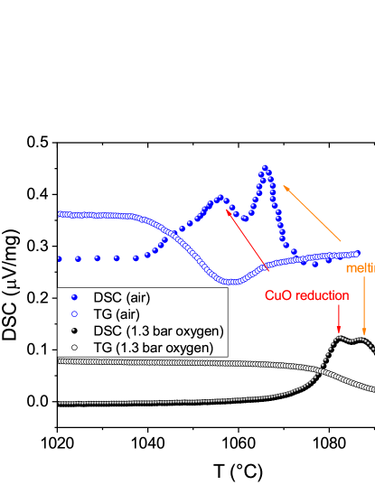
We performed differential scanning calorimetry (DSC), including a thermogravimetric (TG) analysis, on the growth conditions of Ba0.9Sr0.1CuSi2O6 and observed the reduction of Cu2+ to Cu1+ while releasing oxygen (2CuO Cu2O + O2) monitored by a mass loss in the TG signal, followed directly by the melting of the compound. Afterwards, the melting turns it to a viscous mass that glazes when cooled in air. However, upon applying oxygen pressure, the decomposition is shifted up further than the melting temperature seen in a DSC experiment performed in air compared to one in oxygen flow with a partial pressure of 1.3 bar van Well et al. (2016) (see Figure 2). Using a linear interpolation, the difference between decomposition and melting would meet at around 2 bar. Thusm one would expect optimal growth conditions above this oxygen pressure.
To obtain large (cm3-size) single crystals of Ba0.9Sr0.1CuSi2O6, we utilized the floating zone growth method using two furnaces equiped with halogen lamps (FZ1) and a xenon lamp (FZ2). As BaCuSi2O6 has a relative low melting point of around 1060 C, which is slightly lowered by Sr substitution Puphal et al. (2016), a low-power halogen lamp (FZ1) with a better focus can be applied. As a first growth attempt, we followed the short report on the floating zone growth of BaCuSi2O6 from Jaime et al. (2004), and we used the same conditions for the substituted variant attempting a growth rate of 0.5 mm/h in an oxygen flow of 200 cc/min. Stoichiometric seed and feed rods with a diameter of 7 mm and a length of 7 cm were used. With these conditions, we were unable to obtain a stable growth, as the reduction of CuO to Cu2O led to bubble formations in the liquid zone, causing the rods to disconnect (see Table 1 C1).
| Furnace | Gas | p [bar] | Power [%] | Rate [mm/h] | Comments | Crystallite Size | |
|---|---|---|---|---|---|---|---|
| C1 | FZ1 | O2 | 0 | 53.7 | - | could not start growth, immense bubbles | 0 |
| C2 | FZ1 | O2 | 0.5–3 | 54–56.3 | 2-0.5 | neck thinning and bubbles | m |
| C3 | FZ1 | O2 | 7 | 59 | - | bubbles | 0 |
| C4 | FZ2 | O2 | 30 | 16–18 | 1 | flowing down of liquid, disconnection | 0 |
| C5 | FZ2 | O2 | 100 | 22 | 2 | repeated disconnection, phase seperation | 0 |
| C6 | FZ1 | O2/Ar | 4.4 | 56 | 1 | stable growth conditions | mm |
| C7 | FZ1 | O2/Ar | 7 | 56.3 | 0.5 | stable growth conditions | mm-cm |
| C8 | FZ1 | O2/Ar | 5.4 | 54.7 | 0.5 | (seedcrystal) stable growth conditions | cm |
| CF | flux | – | 2:1 LiBO2, C slow cooling to C | mm | |||
III.1 Atmosphere
Surprisingly, an increase of pressure with attempts at 0.5, 1, 2.5, 3, 4, 5, and 7 bar led to similar problems and could not stabilize the melt and suppress the bubble formation (C2,3). In all cases, when changing the lamp power by a few percentage points, the melt was either too viscous at low lamp power or the temperature was too high, leading to a decomposition. This implies that the decomposition and melting point are still overlapping, as seen in the DSC curve. As CuO can only be grown at elevated oxygen pressures Prabhakaran and Boothroyd (2003); ITO et al. (1998), we wanted to attempt even higher pressures to ensure a clear separation of the CuO reduction and melting point following an extrapolation of Figure 2. Even upon application of 100 bar of oxygen pressure (using FZ2), we were still unable to stabilize the liquid zone (C4). Repeated disconnection complicated the growth attempt, and afterwards, we could still find orange parts of reduced copper oxide on crushed pieces of the obtained crystal. Thus, in order to find stable growth conditions with FZ1, the experience of applying an oxygen–argon mixture in the comparable compound of SrCu2(BO3)2 Zayed et al. (2014); Dabkowska et al. (2007) was used. Varying the pressure and oxygen/argon mixture leads to a liquidification of the melt and a breaking of the bubbles into smaller, not visible, ones with increasing argon content, which stabilizes the growth conditions. We found that the concentration of argon has to exceed the oxygen one for this effect. Nevertheless, in a cross section of obtained multigrain crystals, there is still some orange Cu2O phase present, proving the continuation of a decomposition at any attempted pressure and mixture of gases. Without the possibility of a full suppression of the CuO reduction, we studied the growth in stable conditions using four 300 W lamps operated at 54.5% with 5 bar pressure obtained with 0.4 L/min argon and 0.1 L/min oxygen gas-flow: Only in the case of several simultaneously growing grains is the Cu2O impurity incorporated in between the grains, while with a single grain, the impurity is pushed up with the liquid zone until the very last part of the melt. Thus, the orange Cu2O is kept in the molten zone without any influence on the single crystal formation.
III.2 Growth Speed
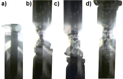
Ba0.9Sr0.1CuSi2O6 crystals grow quickly along the -direction, while the -direction develops much slower, leading to growth steps and, thus, faceted structures (terraces) of the crystal flakes grown by direct melt van Well et al. (2016). This leads to the a-axis developing as the growth direction in FZ experiments, while the -axis points perpendicular to the round surface. With growth rates of 1 mm/h, several grains develop with a tilt in the -plane limited by the c-direction growth rate (C6). We thus reduced the growth rate to 0.5 mm/h (C7) and additionally started to grow with a single crystal as a seed (C8) oriented with the c axis pointing along the growth direction (see Figure 3a,b) to prevent the formation of the misaligned grains. The seed-crystal was glued to the seed-rod by GEvarnish and then molten to it with the optical furnace by quickly melting the tip of the seed-rod, where the organic glue is fully burned away. Then the growth was started by melting the feed only and moving seed and feed together. Meanwhile, the forced growth orientation along the c-direction was not successful, as throughout the growth the growing crystal went back to the preferred orientation, the prevention of additional grains was successful in the entire grown crystal. Even with the chosen method of floating zone growth, the usually round crystal shows a shiny facet on both sides perpendicular to the c direction (see Figure 4d). In this case, as there was a reorientation, which was not completed after the end of the growth, there is an angle of 30 between the growth direction and the a-axis (see inset of Figure 5d).
IV Characterization
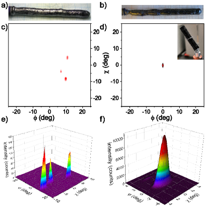
Ba0.9Sr0.1CuSi2O6 crystals cleave well perpendicular to the -direction, leaving shiny ab-planes enabling an easy orientation when breaking off the last and first part of the growth. Obtained Ba0.9Sr0.1CuSi2O6 crystals were analyzed in a neutron scattering experiment using the MORPHEUS 2-axis diffractometer at SINQ (PSI) at room temperature with a wavelength of 5 Å (see Figure 5c–f). We scanned one reflection by fixing the detector to, e.g., 53.34 for the (0 0 4) reflection shown in Figure 5 and measured while rotating the crystal to search for additional grains. As discussed above, this neutron diffraction measurement revealed that earlier growth attempts with 1 mm/h gave rise to three grains developing rather equally throughout the rod. They are slightly tilted in the ab-plane in respect of each other but share a similar c-axis orientation perpendicular to the growth direction (see Figure 5c,e).
In the seeded 0.5 mm/h growth (C8), a full 180 rotation scan of the (0 0 4) reflection showed only one grain in the entire piece, giving a sizeable single crystal of several grams and 4 cm length (see inset of Figure 5d). We found a broadening of the reflection along one angle (see Figure 5f) due to the change of the growth direction along the crystal length.
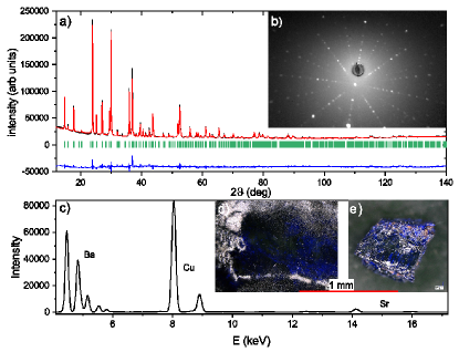
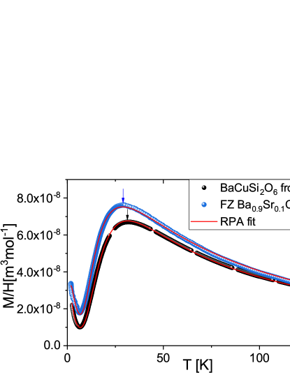
We reproduced the flux growth for non substituted (as the flux reacts with Sr) Han Purple reported in Sebastian et al. (2006b) and compared the single crystal quality of both samples. All flux grown crystals as well as crystals prepared with oxygen pressure in the way described in van Well et al. (2016) and by the floating zone growth reported here can be obtained with equal quality (in the sense of magnetic impurities). In both ways, by direct melting with oxygen pressure and with flux, the magnetic impurity amount can range from 3 to 25%. The magnetic impurities can be analyzed in this system by fitting the Curie tail arising from free spin-1/2 levels (e.g., paramagnetic BaCuSi4O10 ) at low temperatures, which is entirely dominated by impurities. In Figure 6, we show a magnetic susceptibility measurement comparing high-quality crystals of BaCuSi2O6 grown with the flux technique to the Ba0.9Sr0.1CuSi2O6 single crystal grown with the floating zone technique. Both have a similar impurity amount of less than 1% magnetic impurities. The shift of the maximum, due to a smaller unit cell, for Sr substitution is apparent (see arrows in Figure 6). For the antiferromagnetic intradimer coupling parameter, a random phase approximation (RPA) fit yields K compared to K. This hints at a full incorporation of 10% Sr following K, in agreement with the microXRF results of 9(2)% (see Figure 4c) and lattice constants of , (obtained by the Rietveld refinement shown in Figure 4a), matching the published results from a powder sample Puphal et al. (2016). The nonmagnetic Cu2O can be observed optically as an orange impurity and changes the color from blue to purple. With its sharp contrast to blue, this impurity can be seen on cleaved surfaces in the microscope. In Figure 4d, we show first a magnification of the facet on the side of the floating zone crystal C8 and, second, a broken piece of an FZ growth attempt in oxygen pressure C2. In the second case, one can clearly see orange parts of reduced copper oxide, which is not present in the final growth. The Laue image in Figure 4b was taken on the surface of C8, i.e., the facet of (d), proving the -direction orientation and crystal quality.
V Summary
Via the optical floating zone growth of Ba0.9Sr0.1CuSi2O6, we obtained large single crystals suitable for further neutron studies. With an 80%–20% argon–oxygen mixture, stable conditions could be created at 5 bar of pressure. With these conditions, using four 300 W lamps at 54.5%, a single crystal is obtained, growing with a slow rate of 0.5 mm/h. The use of a seed crystal proved crucial to get a large single grain at this growth rate. By neutron single-crystal diffraction, we proved that a single grain is obtained, with the same crystal quality as flux grown BaCuSi2O6.
Acknowledgements.
This work is partly based on experiments performed at the Swiss spallation neutron source SINQ, Paul Scherrer Institute, Villigen, Switzerland. The measurements were carried out on the PPMS/MPMS devices of the Laboratory for Multiscale Materials Experiments, Paul Scherrer Institute, Villigen, Switzerland. The authors would like to acknowledge the Swiss National Science Foundations (SNSF R’Equip, Grant No. 206021_163997 and Grant No. 206021_139082) and matching funds from Paul Scherrer Institute for purchasing the SCIDRE HKZ—high pressure high-temperature optical floating zone furnace and the MPMS.References
- Berke (2007) H. Berke, Chem. Soc. Rev. 36, 15 (2007).
- Rieck et al. (2015) B. Rieck, H. Pristacz, and G. Giester, Mineralogical Magazine 79, 1769 (2015).
- L. W. FINGER (1989) R. J. H. L. W. FINGER, R. M. HAZEN, American Mineralogist, Volume 74, pages 952-955 (1989).
- Sasago et al. (1997) Y. Sasago, K. Uchinokura, A. Zheludev, and G. Shirane, Physical Review B 55, 8357 (1997).
- Jaime et al. (2004) M. Jaime, V. F. Correa, N. Harrison, C. D. Batista, N. Kawashima, Y. Kazuma, G. A. Jorge, R. Stern, I. Heinmaa, S. A. Zvyagin, Y. Sasago, and K. Uchinokura, Physical Review Letters 93 (2004), 10.1103/physrevlett.93.087203.
- Sebastian et al. (2006a) S. E. Sebastian, N. Harrison, C. D. Batista, L. Balicas, M. Jaime, P. A. Sharma, N. Kawashima, and I. R. Fisher, Nature 441, 617 (2006a).
- Rüegg et al. (2007) C. Rüegg, D. F. McMorrow, B. Normand, H. M. Rønnow, S. E. Sebastian, I. R. Fisher, C. D. Batista, S. N. Gvasaliya, C. Niedermayer, and J. Stahn, Physical Review Letters 98 (2007), 10.1103/physrevlett.98.017202.
- Sheptyakov et al. (2012) D. V. Sheptyakov, V. Y. Pomjakushin, R. Stern, I. Heinmaa, H. Nakamura, and T. Kimura, Physical Review B 86 (2012), 10.1103/physrevb.86.014433.
- Samulon et al. (2006) E. C. Samulon, Z. Islam, S. E. Sebastian, P. B. Brooks, M. K. McCourt, J. Ilavsky, and I. R. Fisher, Physical Review B 73 (2006), 10.1103/physrevb.73.100407.
- Sparta and Roth (2004) K. M. Sparta and G. Roth, Acta Crystallographica Section B Structural Science 60, 491 (2004).
- Sebastian et al. (2006b) S. E. Sebastian, P. Tanedo, P. A. Goddard, S.-C. Lee, A. Wilson, S. Kim, S. Cox, R. D. McDonald, S. Hill, N. Harrison, C. D. Batista, and I. R. Fisher, Physical Review B 74 (2006b), 10.1103/physrevb.74.180401.
- Puphal et al. (2016) P. Puphal, D. Sheptyakov, N. van Well, L. Postulka, I. Heinmaa, F. Ritter, W. Assmus, B. Wolf, M. Lang, H. O. Jeschke, R. Valentí, R. Stern, C. Rüegg, and C. Krellner, Physical Review B 93 (2016), 10.1103/physrevb.93.174121.
- van Well et al. (2016) N. van Well, P. Puphal, B. Wehinger, M. Kubus, J. Schefer, C. Rüegg, F. Ritter, C. Krellner, and W. Assmus, Crystal Growth & Design 16, 3416 (2016).
- Prabhakaran and Boothroyd (2003) D. Prabhakaran and A. Boothroyd, Journal of Crystal Growth 250, 77 (2003).
- ITO et al. (1998) T. ITO, H. YAMAGUCHI, K. OKABE, and T. MASUMI, Journal of Materials Science 33, 3555 (1998).
- Zayed et al. (2014) M. Zayed, C. Rüegg, E. Pomjakushina, M. Stingaciu, K. Conder, M. Hanfland, M. Merlini, and H. Rønnow, Solid State Communications 186, 13 (2014).
- Dabkowska et al. (2007) H. Dabkowska, A. Dabkowski, G. Luke, S. Dunsiger, S. Haravifard, M. Cecchinel, and B. Gaulin, Journal of Crystal Growth 306, 123 (2007).