Spatiotemporal imaging of valence electron motion
Abstract
Abstract. Electron motion on the (sub-)femtosecond time scale constitutes the fastest response in many natural phenomena such as light-induced phase transitions and chemical reactions. Whereas static electron densities in single molecules can be imaged in real-space using scanning tunnelling and atomic force microscopy, probing real-time electron motion inside molecules requires ultrafast laser pulses. Here, we demonstrate an all-optical approach to imaging an ultrafast valence electron wave packet in real-time with a time-resolution of a few femtoseconds. We employ a pump-probe-deflect scheme that allows us to prepare an ultrafast wave packet via strong-field ionization and directly image the resulting charge oscillations in the residual ion. This approach extends and overcomes limitations in laser-induced orbital imaging and may enable the real-time imaging of electron dynamics following photoionization such as charge migration and charge transfer processes.
Introduction
Imaging electronic dynamics in molecules immediately following photoexcitation is of utmost interest to photochemistry as the first few femtoseconds can determine the fate of ensuing reactions Worner2017 ; Wolter2016 ; Kubel2016 . Electronic and nuclear dynamics have been probed with attosecond precision Krausz2009RMP ; Leone2014NatPhot by means of high harmonic emission Li2008 ; Haessler2010NatPhys ; Kraus2015Science , laser-induced electron diffraction Meckel2008 ; Blaga2012 ; Wolter2016 , and photoelectron holography Huismans2011 ; Porat2018 . The aforementioned techniques rely on the recollision mechanism Corkum1993 ; Krause1992 , where the photoionized electron is driven back to the parent ion by the intense laser field and probes the transient molecular or atomic structure. Recollision-free schemes, such as attosecond transient absorption Goulielmakis2010 and sequential double ionization have also been used to follow electronic Fleischer2011 ; Fechner2014 ; Calegari2014Science and nuclear dynamics Ergler2006 on a few-femtosecond time scale.
Attosecond technology not only offers unprecedented time-resolution for ultrafast processes, but also laser-based approaches to imaging electronic structure. Such images can be obtained indirectly by analyzing the high harmonic spectrum as a function of molecular alignment with respect to the laser polarization Itatani2004 ; Haessler2010NatPhys ; Vozzi2011 , or directly, by measuring the photoelectron angular distribution in the molecular frame Meckel2008 ; Staudte2009 ; Holmegaard2010 ; Comtois2013 . Photoelectron angular distributions have been studied to follow electron dynamics on a sub-picosecond time scale Hockett2011 ; Forbes2018 . However, the direct imaging of bound electron wave packets on the femtosecond time-scale has yet to be accomplished.
Some of the simplest bound electron wave packets that can be prepared by strong-field ionization are spin-orbit wave packets in noble gas ions Rohringer2009 ; Goulielmakis2010 ; Woerner2011 ; Sabbar2017 . As the spin-orbit wave packet evolves, the 3p-1 electron-hole in the noble gas ion oscillates between the state and the states ( being the magnetic quantum number). This oscillation leads to a time-dependent modulation in the angle-dependent tunnel ionization probability of the ion Fleischer2011 . Time-resolved measurements of the momentum distribution of photoelectrons, emitted from the ion, would allow for directly imaging the evolving electron-hole Woerner2011 . The main obstacle is the contamination of the signal with photoelectrons from the pump pulse Fechner2014 .
Here, we demonstrate the direct imaging of electron density variations with a temporal resolution of only a few femtoseconds. We prepare a bound wave packet in an argon ion using optical tunnel ionization by a few-cycle visible laser pulse. The resulting multi-electron wavepacket is then imaged via another tunnel ionization process induced by a second few-cycle visible laser pulse. Contamination of the probe pulse signal is avoided by superimposing a weak, orthogonally polarized, carrier-envelope phase-stable, mid-infrared streaking field Kubel2017 onto the probe pulse. This allows us to separate the primary and secondary photoelectrons spatially and thereby enables direct imaging of the valence-shell wave packet. By inverting the resulting 2D momentum spectra we obtain the autocorrelation functions of the spatial density of the bound electron wavepacket, as seen through the optical tunnel.
Results
.1 Time-resolved orbital imaging experiment
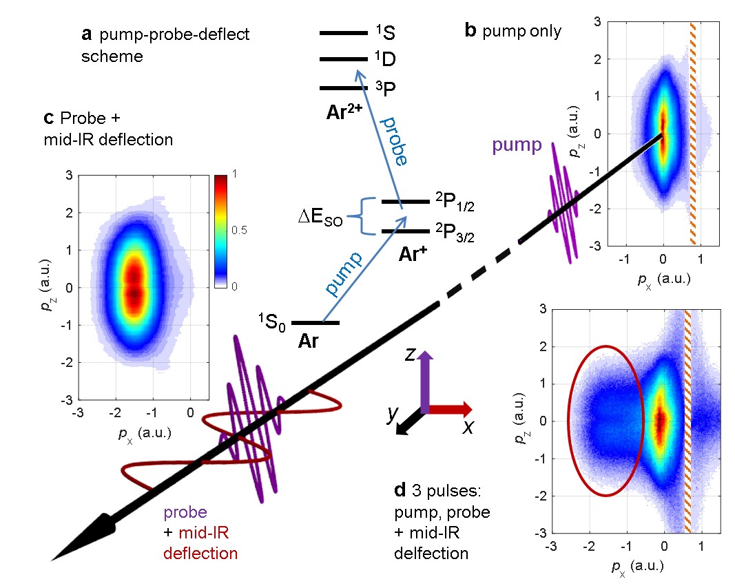
Figure 1 shows a schematic of our pump-probe experiment. In the pump step, strong field ionization of neutral Ar with a few-cycle visible laser pulse causes the coherent population of the and fine-structure states. The resulting spin-orbit wavepacket oscillates with a period and is probed at a variable time delay using strong field ionization by a second few-cycle visible laser pulse. Superimposed on the probe pulse is an orthogonally polarized, mid-infrared (mid-IR), 40 fs pulse, that deflects and thereby labels the electron created by the probe pulse.
Three-dimensional ion and electron momenta are measured in coincidence using Cold Target Recoil Ion Momentum Spectroscopy (COLTRIMS). We make use of the fact that the few cycle pulse alone produces photoelectrons with a narrow momentum distribution along centered at zero momentum (Fig. 1(b)). When the orthogonally polarized mid-IR deflection field is superimposed, the ionized electron wavepacket is shifted, as shown in Fig. 1(c). In the experiment with all three pulses (Fig. 1(d)), the probe pulse signal dominates for negative momenta along the direction of the deflection field, as marked by the red oval. In the following, we present results for electrons selected accordingly, see Methods for details.
.2 Snapshots of an electronic wave packet
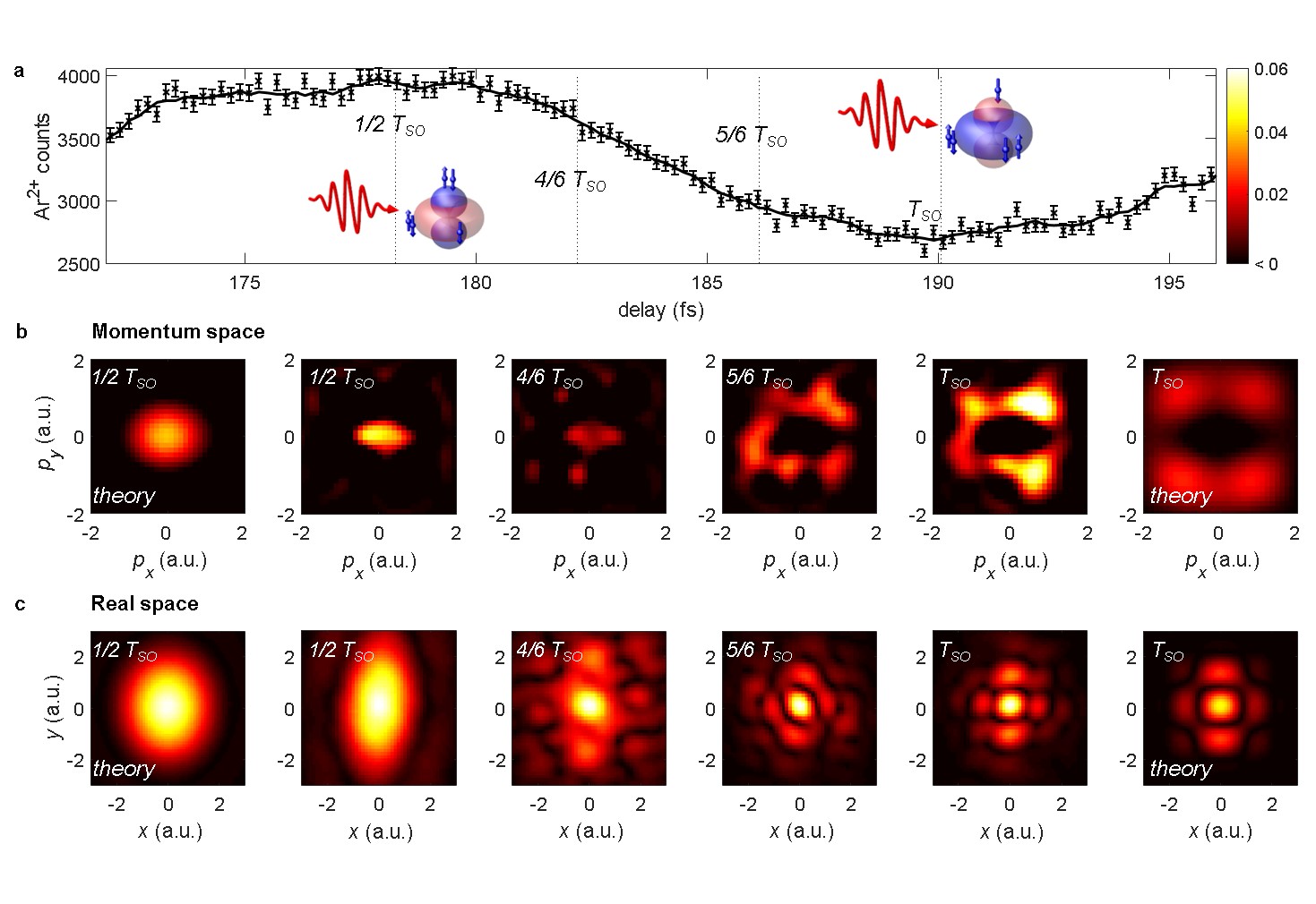
Figure 2 shows our experimental results. Figure 2 (a) shows the delay dependent Ar2+ yield for one oscillation of the valence shell wave packet. Measured data for several oscillations are shown in Supplementary Figure 1. We observe a strong modulation of the Ar2+ yield of . Ionization is favoured when the state is populated with two electrons. In this case, ionization from state is negligible Woerner2011 . On the other hand, at the yield minima, ionization from the donut shaped orbital becomes significant.
In Figure 2(b) and Supplementary Movie 1, we present a time series of measured electron density plots for the wave packet in Ar+. Each density plot is a normalized difference between the delay-dependent and delay-averaged momentum distributions. The series of snapshots shows a narrow spot in the center of the distributions for , corresponding to a yield maximum. The central spot becomes weaker for larger delays, . Eventually, the center spot disappears and a ring around the origin is established at , which appears with maximum brightness at , at the minimum of the Ar2+ yield.
The experimental images agree qualitatively with the simple calculations at and , respectively. At intermediate values , and , the images are essentially identical to the ones at , and , respectively, but exhibit a reduced contrast, see Supplementary Figure 2. For the calculated momentum distributions, we use spatial Ar+ valence orbitals for and , and calculate the transversal momentum space orbitals by Fourier transform, see Supplementary Method 2 for details. Plotted in Fig. 2 are the normalized differences between the and vacancy states.
The expected circular symmetry of the momentum distributions is broken by a noticeable stretch along the axis. This distortion arises from the mid-IR deflection field, which is used to identify the probe pulse signal. The stretch in momentum space corresponds to a contraction in the real space images. Because the and directions are equivalent, the distortion does not cause loss of information. The mean momentum shift induced by the deflection field, , has been subtracted from the presented images.
In Figure 2(c), we show the autocorrelation functions of the spatial electron density, which are obtained from the momentum distributions by Fourier transform, assuming a flat phase. The spatial distributions obtained from the experimental data qualitatively agree with the theoretical results. This indicates that our methods allows for reconstruction of real-space features of the time-dependent valence electron density.
.3 Longitudinal momentum distribution
Next, we turn our attention to the photoelectron momentum component along the ionizing laser field, the z-direction. In Fig. 3, we examine the distributions in the () plane.
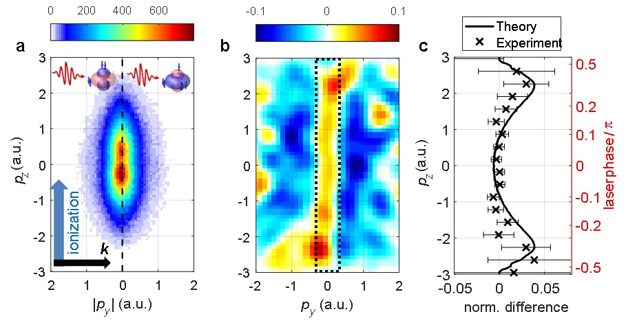
The spectra recorded at maximum () and minimum () Ar2+ yields are qualitatively indistinguishable, see Fig. 3(a). The normalized difference of the two spectra reveals the distinctions between the momentum distributions arising from ionization of and states and is presented in Fig. 3(b). A clear pattern is visible: The blue areas at larger perpendicular momenta () indicate the contribution of the donut shaped orbital at the yield minima, as seen at in Fig. 4(b). The red area at small perpendicular momenta () indicates the dominance of ionization from the orbital at the yield maxima, as seen at in Fig. 4(b).
The spectra recorded at maximum and minimum Ar2+ yields are qualitatively indistinguishable, see Fig. 3(a). However, the normalized difference of the two spectra, presented in Fig. 3(b), reveals a clear pattern: The blue areas at larger perpendicular momenta () indicate the contribution of the donut shaped orbital at the yield minima. The red area at small perpendicular momenta () indicates the dominance of ionization from the orbital at the yield maxima.
Strikingly, the normalized difference exhibits pronounced maxima for large longitudinal momenta (). Similar observations have been made in pump-probe experiments on double photodetachment from negative ions Hultgren2013 ; Eklund2013 . The maxima observed at large longitudinal momenta raise the question whether the final momentum distributions are, in fact, influenced by the momentum distribution in the bound state. Even though it is intriguing to speculate whether orbital imaging is not purely two-dimensional, as in very recent work on alignment-dependent molecular ionization Trabattoni2018 , we offer a different interpretation in Fig. 3(c). The plot shows the normalized difference of the longitudinal momentum distributions recorded at the yield maxima and yield minima. The experimental results are selected for small perpendicular momenta (indicated by the dotted box in Fig. 3(b)) and compared to the results of a computational model, similar to the one proposed in Woerner2011 , and detailed in the Methods. In the model, we calculate the instantaneous non-adiabatic tunnel ionization rates of the and vacancies.
The computational results agree very well with the experimental ones. They indicate that the maxima at large longitudinal momenta arise because the ratio of the ionization probabilities for the two vacancy states varies throughout a laser half cycle.
Specifically, large momenta are produced near the zero crossing of the laser electric field within the optical cycle. At these laser phases, the vector potential is close to its maximum and, correspondingly, the electric field is rather weak. Since the vacancy state is harder to ionize than the vacancy, its ionization probability drops faster with decreasing field strength. Hence, ionization near the zero crossing has an increased contribution from the vacancy. In the fashion of a streak camera, the laser vector potential maps the electron emission times to final momenta, leading to the observed maxima in the normalized difference at large longitudinal momenta.
Discussion
So far, we have shown that our pump-probe scheme allows us to identify double ionization events where the first and second ionization occurs in the pump and probe pulse, respectively. For these events we can separate the first from the second photoelectron, exploiting the deflection induced by the mid-IR streaking field Kubel2017 . Recording the transverse momentum distribution of the second photoelectron enables us to image the electron dynamics unfolding in the cation. We have also shown that the longitudinal momentum distribution of the second photoelectron carries information on the ionization dynamics of the cation in a non-stationary state. In the following, we address the question how quantitative information can be extracted from the measured orbital images.
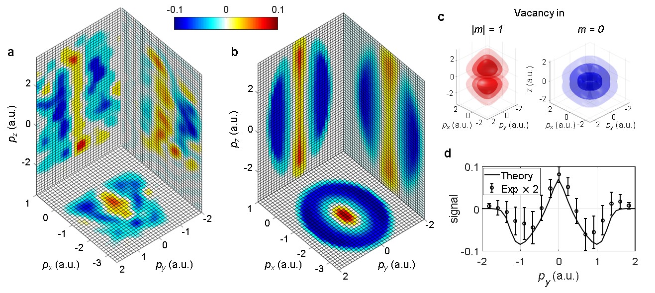
Figure 4(a) and Supplementary Movie 2 show the normalized differences of the projected 2D momentum distributions measured at maximum and minimum Ar2+ yield. Figure 4 (b) presents calculated distributions, based on a simple imaging model. The model is described in the Methods. It uses the orbitals depicted in Fig. 4(c), i.e., the Ar+ 3p orbitals with , and . These electronic density functions are multiplied with a transversal filter function, which describes the tunneling probability as a function of the perpendicular momentum Murray2011 . The acceleration in the combined laser field is simulated, and the sub-cycle streaking effect, discussed above, is taken into account.
The simulated momentum distributions exhibit reasonable agreement with the experimental results. In particular, as shown in Fig. 4(d), almost quantitative agreement is obtained for the width of the distribution along the laser propagation, which remains unaffected by the laser field after tunneling. This indicates that the model captures the essential imaging mechanism. Thus, valence electron densities in real space could be reconstructed from time-resolved orbital imaging experiments, using appropriate computational techniques. The spatial resolution of the reconstructed real-space distribution is given by the maximum perpendicular photoelectron momentum, which is limited by the transversal filter function and the signal to noise ratio. Time-resolved orbital imaging will greatly benefit from the development of intense few-cycle laser sources approaching the MHz range Krebs2013 .
Our method offers exciting prospects for time-resolved imaging of molecular orbitals. For example, strong-field ionization can induce purely electronic dynamics, such as charge migration Kraus2015Science , or correlated electron-nuclear dynamics such as dissociation or isomerization. Our pump-probe scheme will enable imaging of the electronic rearrangements that take place during such processes. At the same time, nuclear dynamics and configurations can be tracked with coulomb explosion imaging Amini2018a ; Burt2018 or laser-induced electron diffraction Meckel2008 ; Blaga2012 ; Wolter2016 ; Amini2018b .
Methods
.4 Experimental setup
The experiment relies on the set-up developed for sub-cycle tracing of ionization enabled by infrared (STIER), which has been described in Ref. Kubel2017 . Briefly, the output of a 10 kHz, 2 mJ titanium:sapphire laser (Coherent Elite) is split in two parts to obtain 5 fs, few-cycle pulses, centered at 730 nm, from a gas-filled hollow core fiber, and 40 fs, phase-stable mid-IR idler pulses at 2330 nm from an optical parametric amplifier. We extend STIER to pump-probe experiments by further splitting the few-cycle pulses and recombining them in a Mach-Zehnder interferometer in order to obtain pump and probe pulses with adjustable time delay. The few-cycle pulses pass through a broadband half wave plate and are recombined with the mid-IR pulses. In order to avoid overlap between the mid-IR pulse and the visible pump pulse, the pump-probe delay was offset by , much larger than the duration of the mid-IR pulses. The offset is not included in the delay values given in the main text. Choosing this large offset is legitimate, as it has been demonstrated that no notable dephasing of the spin orbit wave packet occurs over the course of several nanoseconds Fleischer2011 . We have tested in separate experiments that no significant overlap between visible and mid-IR pulse occurs for pump probe delays larger than 50 fs.
The laser pulses (pulse energies of for each of the visible pulses, and for the mid-IR pulse) are focused () into a cold () argon gas jet in the center of a COLTRIMS Ullrich2003 . We estimate the focal spot sizes (1/e2 width) as for the visible pulses and for the mid-IR pulse. Photoelectrons and ions arising from the interaction are detected in coincidence, and their three-dimensional momenta are measured using time and position sensitive detectors. The polarization of the mid-IR deflection field is along the axis, which is defined by the spectrometer axis of the COLTRIMS. The laser propagates along the axis and the ionizing few-cycle pulses are polarized along . The electron count rate was kept below electron per laser pulse to limit the number of false coincidences. The laser intensity of the visible pulses of was estimated from the carrier-envelope phase-dependent momentum spectra along the laser polarization Kubel2018 . The intensity of the mid-IR pulses was estimated from the deflection amplitude as , low enough to not cause notable ionization of Ar or Ar+.
.5 Data analysis
To obtain images of the transient electron density in the Ar+ valence shell, it is crucial to identify the electrons produced in the second ionization step, by the probe pulse. This is accomplished as follows. First, recorded electron spectra with and without the deflection field present are compared, see Fig. 1(b) and (d). This shows that for momenta , the (deflected) probe pulse signal clearly dominates. We estimate that the contribution of the pump pulse to the signal in the red oval in Fig. 1(d) as less than 10% at , and approximately 1% at . Next, we select events for which an Ar2+ ion has been detected in coincidence with one electron. The momentum component of the other electron along the deflection field is calculated using momentum conservation. The events in which the second ionization step occurs in the probe pulse are selected with the following conditions,
| (1) | ||||
| (2) |
where is the measured electron momentum component along the IR polarization, and is the momentum component calculated from momentum conservation. The rationale for the above conditions is outlined in Supplementary Method 1 and visualized in Supplementary Figure 3.
Having identified the electrons produced in the second ionization step, the electron density plots are obtained by calculating normalized differences of signal, , and reference, , photoelectron momentum distribution:
| (3) |
.6 Orbital effect in the longitudinal momentum spectra
To calculate the ionization rates for the two orbitals we build on the model described in Ref. Woerner2011 . However, we ignore some ionization pathways with low transition probability. For the simulations, a 5-fs (full width at half maximum of the gaussian intensity envelope) laser pulse with frequency and intensity is used. The ionization probability for either orbital is calculated at every point in time using the rates for non-adiabatic tunneling proposed in Ref. Yudin2001 . Longitudinal momentum spectra are obtained from the vector potential of the laser pulse, appropriately weighing each contribution with the calculated rate. The calculations are repeated for 16 different values of the carrier-envelope phase (CEP), and the results are averaged over the CEP. The normalized difference of the spectra calculated for and vacancies is calculated and plotted in Fig. 3(c). Usage of ADK formula Ammosov1986 instead of the non-adiabatic tunneling formula Yudin2001 , leads to very similar results for the normalized difference of the longitudinal spectra.
.7 Imaging model
Here, we give a short description of the procedure used to generate the theoretical images shown in Fig. 4(b). A detailed description can be found the SI. Real space wave functions for the Ar+ valence orbital are taken from the computational chemistry software GAMESS. Momentum-real space wave functions are calculated by partial Fourier transform, as described in Ref. Murray2011 , and plotted in Fig. 4(c).
The wavefunctions squared are multiplied with a “tunnel filter” Murray2011 to obtain the transversal momentum distribution at the tunnel exit. The tunnel filter suppresses large momenta perpendicular to the direction of tunneling. In order to obtain the momentum distributions after propagation in the laser field, the momentum distributions at the tunnel exit are convoluted with Gaussian functions, representing the ionizing visible, and mid-IR deflection fields. The orbital effect in the longitudinal direction is taken into account.
The momentum distribution that correspond to and vacancies are given by appropriate linear combination of the spectra calculated for the and wave functions. To calculate the normalized differences in the three momentum planes, the spectra are integrated over the third dimension.
Data Availability
The data for Figures 1b-d, 2a-c, 3a-c and 4a-d; and Supplementary Figures 1, 2a-c, and 3 are provided as a Source Data file. The data that support the findings of this study are available from the corresponding author upon reasonable request.
References
References
- (1) Wörner, H. J. et al. Charge migration and charge transfer in molecular systems. Struct. Dyn. 4, 61508 (2017).
- (2) Wolter, B. et al. Ultrafast electron diffraction imaging of bond breaking in di-ionized acetylene. Science 354, 308–312 (2016).
- (3) Kübel, M. et al. Steering Proton Migration in Hydrocarbons Using Intense Few-Cycle Laser Fields. Phys. Rev. Lett. 116, 193001 (2016).
- (4) Krausz, F. & Ivanov, M. Attosecond physics. Rev. Mod. Phys. 81, 163–234 (2009).
- (5) Leone, S. R. et al. What will it take to observe processes in real time? Nat. Photonics 8, 162 (2014).
- (6) Li, W. et al. Time-Resolved Dynamics in N2O4 Probed Using High Harmonic Generation. Science 322, 1207–1211 (2008).
- (7) Haessler, S. et al. Attosecond imaging of molecular electronic wavepackets. Nat. Phys. 6, 200–206 (2010).
- (8) Kraus, P. M. et al. Measurement and laser control of attosecond charge migration in ionized iodoacetylene. Science 350, 790–795 (2015).
- (9) Meckel, M. et al. Laser-induced electron tunneling and diffraction. Science 320, 1478–82 (2008).
- (10) Blaga, C. I. et al. Imaging ultrafast molecular dynamics with laser-induced electron diffraction. Nature 483, 194–197 (2012).
- (11) Huismans, Y. et al. Time-Resolved Holography with Photoelectrons. Science 331, 61–64 (2011).
- (12) Porat, G. et al. Attosecond time-resolved photoelectron holography. Nat. Commun. 9, 2805 (2018).
- (13) Corkum, P. B. Plasma Perspective on Strong-Field Multiphoton Ionization. Phys. Rev. Lett. 71, 1994–1997 (1993).
- (14) Krause, J., Schafer, K. & Kulander, K. High-order harmonic generation from atoms and ions in the high intensity regime. Phys. Rev. Lett. 68, 3535–3538 (1992).
- (15) Goulielmakis, E. et al. Real-time observation of valence electron motion. Nature 466, 739–743 (2010).
- (16) Fleischer, A. et al. Probing Angular Correlations in Sequential Double Ionization. Phys. Rev. Lett. 107, 113003 (2011).
- (17) Fechner, L., Camus, N., Ullrich, J., Pfeifer, T. & Moshammer, R. Strong-field tunneling from a coherent superposition of electronic states. Phys. Rev. Lett. 112, 213001 (2014).
- (18) Calegari, F. et al. Ultrafast electron dynamics in phenylalanine initiated by attosecond pulses. Science 346, 336–339 (2014).
- (19) Ergler, T. et al. Spatiotemporal Imaging of Ultrafast Molecular Motion: Collapse and Revival of the D Nuclear Wave Packet. Phys. Rev. Lett. 97, 193001 (2006).
- (20) Itatani, J. et al. Tomographic imaging of molecular orbitals. Nature 432, 867–71 (2004).
- (21) Vozzi, C. et al. Generalized molecular orbital tomography. Nat. Phys. 7, 822–826 (2011).
- (22) Staudte, A. et al. Angular Tunneling Ionization Probability of Fixed-in-Space H2 Molecules in Intense Laser Pulses. Phys. Rev. Lett. 102, 033004 (2009).
- (23) Holmegaard, L. et al. Photoelectron angular distributions from strong-field ionization of oriented molecules. Nat. Phys. 6, 428–432 (2010).
- (24) Comtois, D. et al. Laser-induced orbital projection and diffraction of O2 with velocity map imaging. J. Mod. Opt. 60, 1395–1408 (2013).
- (25) Hockett, P., Bisgaard, C. Z., Clarkin, O. J. & Stolow, A. Time-resolved imaging of purely valence-electron dynamics during a chemical reaction. Nat. Phys. 7, 612–615 (2011).
- (26) Forbes, R. et al. Quantum-beat photoelectron-imaging spectroscopy of Xe in the VUV. Phys. Rev. A 97, 63417 (2018).
- (27) Rohringer, N. & Santra, R. Multichannel coherence in strong-field ionization. Phys. Rev. A 79, 053402 (2009).
- (28) Wörner, H. J. & Corkum, P. B. Imaging and controlling multielectron dynamics by laser-induced tunnel ionization. J. Phys. B At. Mol. Opt. Phys. 44, 041001 (2011).
- (29) Sabbar, M. et al. State-resolved attosecond reversible and irreversible dynamics in strong optical fields. Nat. Phys. 13, 472 (2017).
- (30) Kübel, M. et al. Streak Camera for Strong-Field Ionization. Phys. Rev. Lett. 119, 183201 (2017).
- (31) Hultgren, H., Eklund, M., Hanstorp, D. & Kiyan, I. Y. Electron dynamics in the ground state of a laser-generated carbon atom. Phys. Rev. A - At. Mol. Opt. Phys. 87, 031404(R) (2013).
- (32) Eklund, M., Hultgren, H., Hanstorp, D. & Kiyan, I. Y. Orbital alignment in atoms generated by photodetachment in a strong laser field. Phys. Rev. A - At. Mol. Opt. Phys. 88, 023423 (2013).
- (33) Trabattoni, A. et al. Setting the clock of photoelectron emission through molecular alignment (2018). URL http://arxiv.org/abs/1802.06622. eprint 1802.06622.
- (34) Murray, R., Spanner, M., Patchkovskii, S. & Ivanov, M. Y. Tunnel ionization of molecules and orbital imaging. Phys. Rev. Lett. 106, 173001 (2011).
- (35) Krebs, M. et al. Towards isolated attosecond pulses at megahertz repetition rates. Nat. Photonics 7, 555–559 (2013).
- (36) Amini, K. et al. Photodissociation of aligned CH3I and C6H3F2I molecules probed with time-resolved Coulomb explosion imaging by site-selective extreme ultraviolet ionization. Structural Dynamics 5, 14301 (2018).
- (37) Burt, M. et al. Communication: Gas-phase structural isomer identification by Coulomb explosion of aligned molecules. The Journal of Chemical Physics 148, 91102 (2018).
- (38) Amini, K. et al. Imaging ultrafast skeletal deformations in polyatomic molecules using laser-induced electron diffraction. ArXiv e-prints arXiv:1805.06793 (2018). eprint 1805.06793.
- (39) Ullrich, J. et al. Recoil-ion and electron momentum spectroscopy: reaction-microscopes. Reports Prog. Phys. 66, 1463–1545 (2003).
- (40) Kübel, M. et al. Phase- and intensity-resolved measurements of above threshold ionization by few-cycle pulses. J. Phys. B At. Mol. Opt. Phys. 51, 134007 (2018).
- (41) Yudin, G. & Ivanov, M. Nonadiabatic tunnel ionization: Looking inside a laser cycle. Phys. Rev. A 64, 6–9 (2001).
- (42) Ammosov, M. V., Delone, N. B. & Krainov, V. P. Tunnel ionization of complex atoms and of atomic ions in an alternating electromagnetic field. Sov Phys JETP 64, 1191–1194 (1986).