[firstpage,color=gray!90,angle=0,scale=0.18,xpos=0,ypos=130, align=center,width=30in]This manuscript has been authored by UT-Battelle, LLC under Contract No. DE-AC05-00OR22725 with the U.S. Department of Energy. The United States Government retains and the publisher, by accepting the article for publication, acknowledges that the United States Government retains a non-exclusive, paid-up, irrevocable, world-wide license to publish or reproduce the published form of this manuscript, or allow others to do so, for United States Government purposes. The Department of Energy will provide public access to these results of federally sponsored research in accordance with the DOE Public Access Plan (http://energy.gov/downloads/doe-public-access-plan) 11affiliationtext: Computational Sciences and Engineering Division, Oak Ridge National Laboratory, Oak Ridge, TN, USA 22affiliationtext: Materials Sciences and Technology Division, Oak Ridge National Laboratory, Oak Ridge, TN, USA ++affiliationtext: Present Address: School of Chemistry, Cardiff University, Cardiff, UK **affiliationtext: laanaitn@ornl.gov
Reconstruction of 3-D Atomic Distortions from Electron Microscopy with Deep Learning
Abstract
Deep learning has demonstrated superb efficacy in processing imaging data, yet its suitability in solving challenging inverse problems in scientific imaging has not been fully explored. Of immense interest is the determination of local material properties from atomically-resolved imaging, such as electron microscopy, where such information is encoded in subtle and complex data signatures, and whose recovery and interpretation necessitate intensive numerical simulations subject to the requirement of near-perfect knowledge of the experimental setup. We demonstrate that an end-to-end deep learning model can successfully recover 3-dimensional atomic distortions of a variety of oxide perovskite materials from a single 2-dimensional experimental scanning transmission electron (STEM) micrograph, in the process resolving a longstanding question in the recovery of 3-D atomic distortions from STEM experiments. Our results indicate that deep learning is a promising approach to efficiently address unsolved inverse problems in scientific imaging and to underpin novel material investigations at atomic resolution.
Keywords Complex Oxide Perovskites Octahedral Rotations Scanning Transmission Electron Microscopy Gaussian Processes Deep Learning
1 Introduction
Deep learning has made tremendous progress in the past few years[RN1], and is poised to enable paradigm-changing breakthroughs encompassing the technology industry and the sciences [RN1, RN2]. Of particular interest are deep learning systems geared towards the analysis of imaging data, comprised chiefly of convolutional neural networks[RN3] and variants thereof [RN4, RN5]. These deep artificial neural networks have demonstrated unmatched accuracy and performance at learning to capture the salient and abstract features present in image data and to use such features to perform specific tasks in various fields from clinical pathology [RN6] to astrophysics [RN7, RN8]. Given the minimal assumptions deep learning makes about the nature of the information present in the imaging data [RN9], it can have broad applicability to imaging studies of materials but has hitherto remained underutilized [RN10]. Here, we demonstrate a deep learning-based solution to a long-standing question in electron microscopy, namely, the ability to infer three-dimensional material structural properties from a single two-dimensional image.
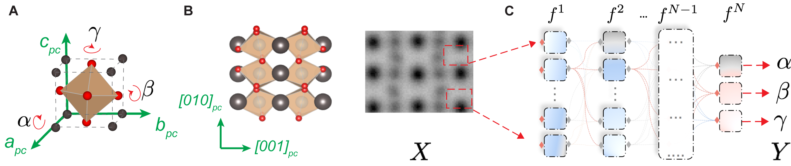
In our study, we target the materials class of complex oxide perovskites whose members host fascinating physical phenomena from correlated electron behavior [RN11, RN12] to quantum magnetism [RN13], and underpin novel device components such as ferroelectric tunnel junctions [RN14]. One of the defining structural properties of complex oxides perovskites are oxygen octahedral rotations[RN15] (Fig. 1A). The latter play a central role in the electronic configuration of these materials and consequently their properties via crystal field splitting. Moreover, as symmetry-lowering distortions, oxygen octahedral rotations readily couple to electronic and spin degrees of freedom during phase transitions[RN16]. Research in the field of tilt-driven engineering of electronic and magnetic properties has blossomed (e.g. [RN17, RN18]) and a deeper theoretical understanding of the coupling of tilts to other materials properties has been developed [RN19]. Validation of theoretical predictions of octahedral tilts in strain-engineered heterostructures is primarily carried out against synchrotron surface diffraction measurements, which quantitatively measure the 3-D symmetry and angular magnitudes of octahedral tilts[RN20], albeit in an ensemble-averaged fashion. In light of the seminal role that local structural states play in influencing the properties and responses of oxides, especially in strain-engineered heterostructures, the need for experimental access to the full local 3-D symmetry and magnitudes of octahedral tilts is indispensable, yet this goal has so far remained elusive.
Direct imaging of abrupt changes in tilts that occur at interfaces or induced by defects has only been possible for a decade [RN21, RN22] due to advances in aberration-corrected electron microscopy. Compared to other structural distortions in perovskites (e.g. strain and polarization), octahedral tilts are more difficult to quantitatively characterize, especially at the local unit cell level, as they are associated with zone-boundary modes and more subtle changes in symmetry. Scanning transmission electron microscopy (STEM) via the annular bright field mode (ABF) can readily resolve atomic columns of light elements such as oxygen. The ABF image, however, is a complex pattern of coherent scattering and interference of electrons through the material [RN23] that is often treated, for simplicity, as a two-dimensional projection of the atomic lattice. While information beyond the projection geometry contributes to the image formation, extracting additional parameters, such as the three-dimensional rotation angles, is a complex inverse problem (Fig. 1B). Underlying this complexity is the nature of the ABF image contrast which is overwhelmingly dominated by projected information; additionally, atomic columns which produce the most prominent image contrast do not participate in most distortions of the overall crystal structure.
Despite these challenges, there has been progress towards extracting 3D local information of octahedral rotations. In particular, an approach was developed for classifying ABF images based on oxygen column shapes [RN24]; it required expert and manual visual inspection of ABF micrographs to identify oxygen columns, thereby isolating the related signal from that of the contrast-dominating cations, followed by a dimensionality-reduction technique (i.e. principal component analysis) to relate the shape of adjacent oxygen columns to the local tilt symmetry. Besides the need for (subjective) input from an expert electron microscope scientist, the main limitation of the previous approach is its inability to associate quantitative 3D octahedral rotation information but for a subset of manually identified ABF oxygen column shapes.
In this work, we construct an end-to-end deep learning model to infer from a single ABF-STEM image full and quantitative 3D information of oxygen octahedral rotations (see Fig.1C). We find that by training a custom deep convolutional neural network (DCNN) on dynamical electron scattering simulations of perovskite structures, it can extract both symmetry and magnitudes of octahedral rotations from experimental data with unit-cell resolution and sub-degree angular rotations. Our model successfully generalizes what it learned to accurately predict these structural distortions to new material classes it did not see during training, over the entire range of rotation parameter space. The new interpretation of ABF-STEM imaging enabled by a DCNN allows us to quantitatively address the coupling of octahedral distortions, across engineered interfaces of thin-films and superlattices, with atomic resolution, directly from experiments for any oxide system. Furthermore, testing the DCNN on experimental data permits us to identify some of the inherent limitations in extracting 3-D structural information from ABF-STEM experiments.
Structural Refinement in Electron Microscopy with Deep Learning
In materials with a perovskite structure (ABO-’missinABO ’missin’missing, A, B: cations, O: oxygen), rotations of oxygen octahedra (BO-’missinBO ’missin’missing) disrupt the perfect alignment of oxygen atoms present in the parent cubic structure and result in oxygen column splitting in projection (Fig. 1B). While separate oxygen columns can sometimes be visualized, most rotation angles are small (, often ), placing the corresponding oxygen-oxygen separation beyond the resolving power of modern scanning transmission electron microscopes. However, imperfect alignment of oxygen atoms and the associated perturbation of scattering wave-fronts that forms ABF STEM images makes atomic column shapes of oxygen appear distorted (Fig. 1B). Consequently, the distinct shapes of oxygen columns in an ABF image encode all the information one can access regarding the BO-’missinBO ’missin’missing rotations[RN24]. Three angles uniquely determine the rotation pattern, denoted by , each indicating a rotation about the principal crystallographic axes , respectively (we use a pseudo-cubic unit cell throughout and omit the subscript hereafter). To predict the symmetry and magnitude of a BO-’missinBO ’missin’missing octahedron from an ABF image, we need to construct a model that maps column shapes to for every ABO-’missinABO ’missin’missing unit cell, whereby the absolute values of these angles give the magnitude, while the symmetry is fixed once the signs of for two neighboring ABO-’missinABO