Observation of Spider Silk by Femtosecond Pulse Laser Second Harmonic Generation Microscopy
Surf. Interface Anal., 51, 56-60 (2019). DOI:10.1002/sia.6545)
Abstract
An asymmetric \textbeta-sheet structure of spider silk is said to induce optical second harmonic generation. In this paper, using an in-house non-scanning type femtosecond pulse laser second harmonic generation microscope, we characterized the behavior of the \textbeta-sheet of spider silk under an applied external force. The orientation of the \textbeta-sheets was more unidirectional when the silk was extended. One of the origins of the high mechanical strength of the dragline is suggested to be the physical arrangement of its \textbeta-sheets.
1 Introduction
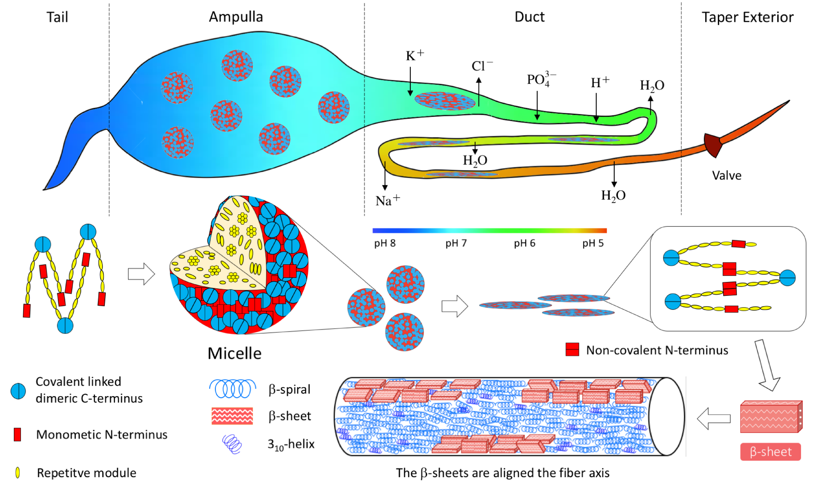
Spider silk has always been an intriguing material, with a unique combination of properties. It is important to analyze the molecular configuration of the spider silk. Traditional analytical methods are NMR, micro-Raman spectroscopy and micro X-ray diffraction, etc. Helical and \textbeta-sheet structure of spider silk were reported g3-30-13 ; g3-31 ; g3-20 ; g12 ; g3-30 ; g12-41 ; g12-39 , but the material property in macroscopic conformation for such structures was not fully clarified, in spite of the high strength and high toughness of the spider silk. An orb-weaver spider has seven types of secretory glands. The seven different glands secrete seven different silks with different compositions and properties for different uses. The dragline is a lifeline for the spider and also used for the frame and radii of an orb web. The dragline and radial line form the structural skeleton, and the spiral lines are woven on the structural skeleton. Only the spiral lines can capture the prey because only the spiral line has a sticky ball coating.
Among them, the dragline has high strength and the radial line gives the strength of the skeleton constituting the orb web. The high strength characteristic of the dragline and the radial line is given by the \textbeta-sheet structure g8-39 ; g8-47 of fibroin molecules g3-30-13 ; g3-31 ; g3-20 ; g12 ; g3-30 ; g12-41 ; g12-39 . The dragline and radial line are secreted by the ampullate gland inside the spider’s body. The ampullate gland is composed of four parts as shown in Fig. 1g38 ; g37 ; Zhao2017 ; g36 ; g30 . In the process of dragline production, the primary structure protein is secreted first from secretory granules in the tail g8 . In the ampullate (neutral environment, pH = 7), the proteins form a soft micelle of several tens of nanometers by self-organization because the hydrophilic terminals are excluded g3-29 . In ampullate, the concentration of the protein is very high g8-47 ; g36-24 . Then, the micelles are squeezed into the duct. The long axis direction of the molecules is aligned parallel to the duct by a mechanical frictional force and partially oriented g8 ; g3-29 ; g3-16 . The continuous lowering of pH from 7.5 to 8.0 in the tail to presumably close to 5.0 occurs at the end of the duct g38 ; g38-9 ; g38-13 . Ion exchange, acidification, and water removal all happen in the duct g37 . The shear and elongational forces lead to phase separation g37 . In the acidic bath of the duct, the molecules attain a high concentration liquid crystal state g8-49 . Finally, the silk is spun from the taper exterior. The molecules become more stable helixes and \textbeta-sheets from the liquid crystal.
The second-order nonlinear optical effect is sensitive to highly oriented structures BNO ; 12 . In our previous report Zhao2017 , the dragline and the radial line have been found to show an optical second-harmonic generation (SHG) response. However, no SHG signal was observed from the spiral line. Because the only difference between the constituents in the dragline, radial line, and the spiral line is the presence and absence of the \textbeta-sheets of the protein, and only the spiral line does not contain \textbeta-sheets, the origin of the SHG of the radial line and the dragline was inferred to be the oriented \textbeta-sheets. The SHG intensity of the dragline was found to depend on the polarization of the incident light. An anisotropic structure is the origin of the SHG. As shown in the bottom of Fig. 1, a model Zhao2017 was proposed that the randomly rotating arrangement around the fiber axis of the \textbeta-sheets should give the anisotropy.
On the other hand, we hypothesize that the molecules of the dragline may be further aligned by an external force. The SHG from the dragline should respond to the changes of the molecular alignment. Hence, in this study, we measured the change of the SHG response by applying an external force to the dragline in the fiber axis direction.
2 Method and Sample
The collection of the sample and the observation method for observing SHG were the same as that described in our previous report Zhao2017 . The excitation light pulse energy density was 16 µJ/mm2. We confirmed that the breaking of the draglines in Figs. 2 and 3 is not caused by the incident light. Namely, no damage in draglines was observed after 1 hour irradiation with this energy density. In the tensile testing as shown in Fig. 2(a), a dragline was pulled and stretched by 1 mm steps from the natural length until it finally breaks. After the onset of the breaking, the dragline is separated into several thinner fibers and some of them broke first. One measurement in Fig. 2(b) took less than 10 minutes, and the one in Fig. 3 less than 30 minutes.
3 Results
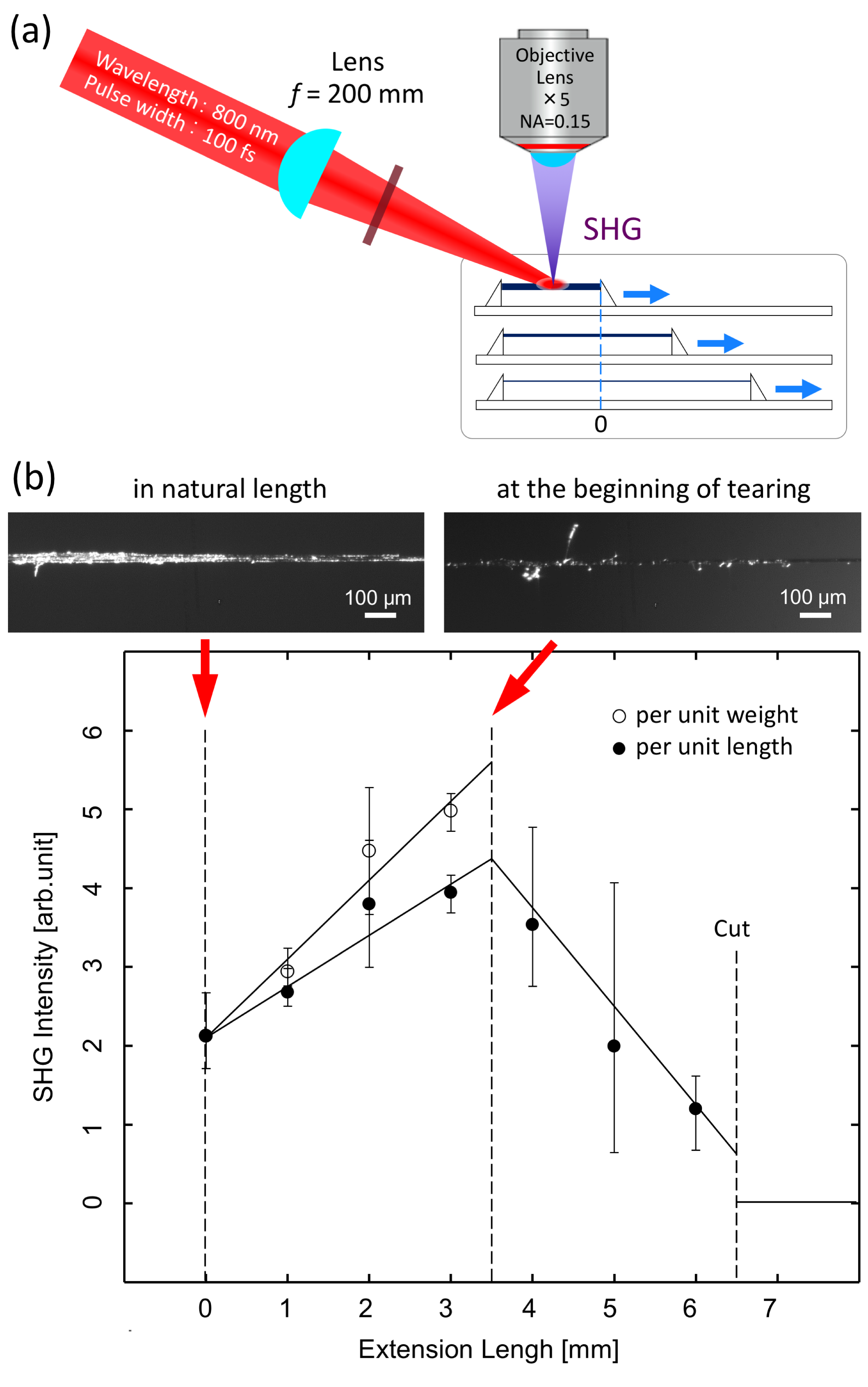
As shown in Fig. 2(a), the dragline was pulled in its length direction from a natural state (L=14.3 mm). The elongation length \textDeltaL is defined as 0 in the natural state. The length of the natural state is nearly equal to the one when the line is pulled by the force of the weight of the spider. The dragline was pulled until it broke. The incident light polarization was perpendicular to the fiber axis. The polarization of the detected SHG was not chosen. The SHG intensity was measured by microscope optics with an image intensified charge coupled device camera. The intensity of the SHG became stronger with extension until a part of the dragline began to break at \textDeltaL=3.4 mm. The diameter of the dragline became thinner as it was stretched until it was broken apart. The slope of SHG enhancement per unit mass as a function of \textDeltaL is larger than that per unit length as shown in Fig. 2(b). Breaking began when the dragline was stretched longer than \textDeltaL=3.4 mm. The microscopic images in Fig. 2(b) show a dragline in the natural length (\textDeltaL=0) and at the beginning of breaking stage (\textDeltaL=3.5 mm). The dragline at the breaking position gradually became thinner and was cut. The SHG weakened at the breaking part. The total SHG intensity decreased while the dragline was broken continuously until it breaks apart.
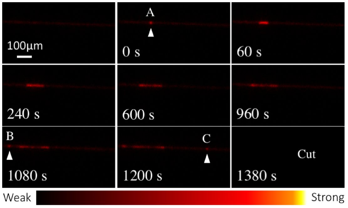
Next, we stretched the dragline until it began to break and stopped stretching at that length and observed the SHG image as a function of time. At a length near the breaking threshold, the dragline is slowly broken. A strong SHG spot A as shown in Fig. 3 appeared at some point of time. The length of the strong SHG domain grew along the fiber axis from the initial location as the time elapsed. Then, a new strong SHG spot B appeared at time 1080 seconds. Another strong SHG spot C appeared at time 1200 seconds. Then the dragline broke at time 1380 seconds.
4 Discussion
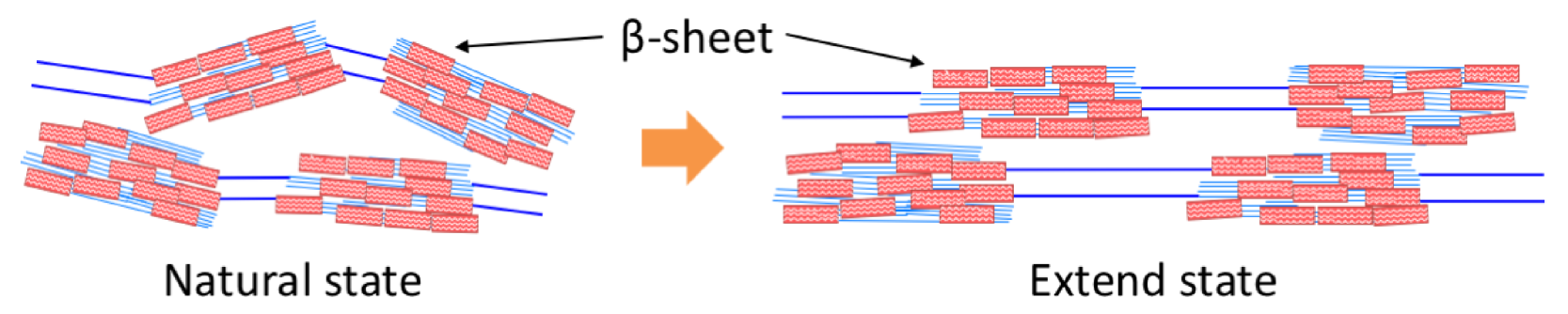
The \textbeta-sheets are aligned due to mechanical frictional force when passing through a thin duct in the spider’s body and partial ordering occurs along the fiber axis g8 ; g3-29 ; g3-16 ; g36 ; g30 . The origins of the elastic properties of dragline are similar to that of a synthetic elastomer g12-39 . As shown in the model in Fig. 4, the flexible chain segments are stretched in the strained direction due to an external force and the hard crystal regions become more highly aligned. The orientation of the \textbeta-sheet becomes more unidirectional. The origin of the observed SHG enhancement effect is suggested to be the orientation of the \textbeta-sheet induced by stretching the dragline. From the linear relationship of the stress and strain of the dragline in previous reports g1 ; g41 , the result of Fig. 2 is suggested to be a phenomenon seen in the stress increase stage before the breaking of the dragline.
The intensity change in Fig. 2 for the extension length \textDeltaL less than 3.5 mm is uniform and originates from an increase of the orientation of molecules in the whole dragline. On the other hand, Fig. 3 is in the plastic deformation yield phase of the dragline slightly above the breaking threshold. Hence, we suggest that the SHG enhancements presented in Figs. 2 and 3 are from different origins. The phenomenon in Fig. 3 can be explained by the following model. The alignment of the \textbeta-sheets in the stretched dragline is suggested to provide a stronger SHG intensity. The strong SHG spot A in Fig. 3 was located at the mechanically weakest part in the dragline. The strong SHG intensity at spot A indicates well-aligned \textbeta-sheets. There the \textbeta-sheets get oriented and the dragline becomes mechanically stronger and sustained the structure under the strain nearby the spot A. The domain of high orientation of the \textbeta-sheet extended as a function of time until 1080 seconds. From another reportg40 , it is inferred that the helixes seen in the model figure at the bottom of Fig. 1 broke first, while the \textbeta-sheet nanocrystals bore the strain until the dragline broke. When the part of the dragline near the spot A reaches the limit of flexibility, the \textbeta-sheets at other weak points B and C begin to be oriented one after another. The partial weakening of the SHG intensity in the A domain after 600 seconds in Fig. 4 is due to partial breaking of the dragline. Eventually, the dragline broke near A because it could not withstand the stress. Namely, the highly oriented \textbeta-sheets had a function of prolonging the time until the dragline broke down by the pulling force.
5 Summary
It has been discovered that the SHG response of the dragline of a spider is enhanced until it begins to break by a stretching force. The \textbeta-sheets in the dragline are suggested to be oriented more unidirectionally by the stretching force. After the onset of the breaking, the dragline is separated into several thinner fibers and some of them broke first. Here the SHG intensity integrated along the length direction decreased. We suggest that the highly unidirectional \textbeta-sheet orientation prolonged the time before breaking of the dragline. We also monitored the SHG image of the dragline as a function of time before it finally breaks down by the stretching force. After the onset of the breaking, the dragline is separated into several thinner fibers and some of them broke first by the stretching force. We found that the SHG intensity is enhanced at different positions at different times. In the dragline breaking, the \textbeta-sheets are suggested to begin to be highly oriented from the weakest location of the silk strength. This local orientation growth may be preventing the breaking of the dragline. The high strength of the dragline may be partly due to a physical structure of the \textbeta-sheets arranged in one direction.
Acknowledgements
This research was financially supported by a JAIST Research Grant (Grant for Fundamental Research in 2017) from Japan Advanced Institute of Science and Technology Foundation (JAIST Foundation).
References
- (1) J. O. Warwicker, “Comparative studies of fibroins: II. The crystal structures of various fibroins”, J. Mol. Biol. 2, IN1—350 (1960).
- (2) B. L. Thiel and C. Viney, “\textbeta sheets and spider silk”, Science 273, 1477–1480 (1996).
- (3) J. M. Gosline, P. A. Guerette, C. S. Ortlepp, and K. N. Savage, “The mechanical design of spider silks: from fibroin sequence to mechanical function”, J. Exp. Biol. 202, 3295–3303 (1999).
- (4) L. Römer and T. Scheibel, “The elaborate structure of spider silk”, Prion 2, 154–161 (2008).
- (5) J. P. O’Brien, S. R. Fahnestock, Y. Termonia, and K. H. Gardner, “Nylons from nature: synthetic analogs to spider silk”, Adv. Mater. 10, 1185–1195 (1998).
- (6) C. Y. Hayashi, N. H. Shipley, and R. V. Lewis, “Hypotheses that correlate the sequence, structure, and mechanical properties of spider silk proteins”, Int. J. Biol. Macromol. 24, 271–275 (1999).
- (7) S. Kubik, “High-Performance Fibers from Spider Silk”, Angew. Chem. Int. Ed. 41, 2721–2723 (2002).
- (8) A. H. Simmons, C. A. Michal, and L. W. Jelinski, “Molecular orientation and two-component nature of the crystalline fraction of spider dragline silk”, Science 271, 84–87 (1996).
- (9) D. H. Hijirida, K. G. Do, C. Michal, S. Wong, D. Zax, and L. W. Jelinski, “13C NMR of Nephila clavipes major ampullate silk gland”, Biophys. J. 71, 3442–3447 (1996).
- (10) A. Rising and J. Johansson, “Toward spinning artificial spider silk”, Nat. Chem. Biol. 11, 309–315 (2015).
- (11) L. Eisoldt, C. Thamm, and T. Scheibel, “The role of terminal domains during storage and assembly of spider silk proteins”, Biopolymers 97, 355–361 (2012).
- (12) Y. Zhao, K. T. T. Hien, G. Mizutani, and H. N. Rutt, “Second-order nonlinear optical microscopy of spider silk”, Appl. Phys. B 123, 188 (2017).
- (13) L. Eisoldt, A. Smith, and T. Scheibel, “Decoding the secrets of spider silk”, Mater. Today 14, 80–86 (2011).
- (14) O. Tokareva, M. Jacobsen, M. Buehler, J. Wong, and D. L. Kaplan, “Structure–function–property–design interplay in biopolymers: Spider silk”, Acta Biomater. 10, 1612–1626 (2014).
- (15) F. Vollrath and D. P. Knight, “Liquid crystalline spinning of spider silk”, Nature 410, 541–548 (2001).
- (16) J. A. Kluge, O. Rabotyagova, G. G. Leisk, and D. L. Kaplan, “Spider silks and their applications”, Trends Biotechnol. 26, 244–251 (2008).
- (17) T. Lefvre, S. Boudreault, C. Cloutier, and M. Pézolet, “Conformational and orientational transformation of silk proteins in the major ampullate gland of Nephila clavipes spiders”, Biomacromolecules 9, 2399–2407 (2008).
- (18) R. V. Lewis, “Spider silk: Ancient ideas for new biomaterials”, Chem. Rev. 106, 3762–3774 (2006).
- (19) M. Andersson, G. Chen, M. Otikovs, M. Landreh, K. Nordling, N. Kronqvist, P. Westermark, H. Jörnvall, S. Knight, Y. Ridderstråle, and Others, “Carbonic anhydrase generates CO2 and H+ that drive spider silk formation via opposite effects on the terminal domains”, PLoS Biol. 12, e1001921 (2014).
- (20) N. Kronqvist, M. Otikovs, V. Chmyrov, G. Chen, M. Andersson, K. Nordling, M. Landreh, M. Sarr, H. Jörnvall, S. Wennmalm, and Others, “Sequential pH-driven dimerization and stabilization of the N-terminal domain enables rapid spider silk formation”, Nat. Commun. 5, 3254 (2014).
- (21) D. P. Knight and F. Vollrath, “Liquid crystals and flow elongation in a spider’s silk production line”, Proc. R. Soc. B 266, 519–523 (1999).
- (22) N. Bloembergen, Nonlinear Optics (Addison-Wesley World Publishing Co., Inc., Redwood City, California, 1992).
- (23) I. Freund and M. Deutsch, “Second-harmonic microscopy of biological tissue”, Opt. Lett. 11, 94–96 (1986).
- (24) S. Osaki, “Spider silks: protein science [in Japanese]”, Protein, Nucleic Acid Enzym. 41, 2108–2116 (1996).
- (25) M. Kitagawa, H. Sasagawa, and T. Kitayama, “Mechanical properties of spider silk [in Japanese]”, J. Soc. Mat. Sci. Japan 44, 153–158 (1995).
- (26) A. Nova, S. Keten, N. M. Pugno, A. Redaelli, and M. J. Buehler, “Molecular and nanostructural mechanisms of deformation, strength and toughness of spider silk fibrils”, Nano Lett. 10, 2626–2634 (2010).