![[Uncaptioned image]](/html/1811.02678/assets/x1.png)
Protein Hydration Waters are Susceptible to Unfavorable Perturbations
Abstract
The interactions of a protein, its phase behavior, and ultimately, its ability to function, are all influenced by the interactions between the protein and its hydration waters. Here we study proteins with a variety of sizes, shapes, chemistries, and biological functions, and characterize their interactions with their hydration waters using molecular simulation and enhanced sampling techniques. We find that akin to extended hydrophobic surfaces, proteins situate their hydration waters at the edge of a dewetting transition, making them susceptible to unfavorable perturbations. We also find that the strength of the unfavorable potential needed to trigger dewetting is roughly the same, regardless of the protein being studied, and depends only the width of the hydration shell being perturbed. Our findings establish a framework for systematically classifying protein patches according to how favorably they interact with water.
keywords:
hydrophobic effect, collective dewetting, chemical patterns, enhanced sampling1 Introduction
Biomolecular binding processes involve disrupting protein-water interactions and replacing them with direct interactions between the binding partners. Thus, protein-water interactions influence the thermodynamics and kinetics of protein interactions 1, 2, 3, 4, 5, 6, 7, 8, 9, 10, as well as the stability and phase behavior of protein solutions 11, 12, 13, 14, 15, 16, 17, 18. In this article, we characterize the overall interactions between proteins and their hydration waters by using specialized molecular simulations that employ an unfavorable potential to displace water molecules from the vicinity of a protein. Because displacing interfacial waters disrupts surface-water interactions, the less favorable those interactions (e.g., for hydrophobic surfaces), the easier it is to displace the interfacial waters 19, 20, 21. Indeed, both theory 22, 23, 24, 25 and molecular simulations 26, 27 have shown that the rare, low-density fluctuations, which are accessed when interfacial waters are displaced, are substantially more probable adjacent to a hydrophobic surface than at a hydrophilic surface. Moreover, water molecules near a hydrophobic surface are susceptible to unfavorable perturbations, and undergo a collective dewetting transition in response to such a perturbation 22, 26, 27, 21. Proximity to such a dewetting transition is also reflected in other collective interfacial properties, such as compressibility, transverse density correlations, and the distribution of water dipole orientations, among others 28, 22, 29, 30, 31, 26, 32, 19, 27, 33, 34, 35.
In contrast with simple hydrophobic or hydrophilic surfaces, proteins display nanoscopic chemical and topographical patterns, which influence their interactions with water in non-trivial ways 36, 37, 38, 39, 40, 41, 42, 20, 43, 44, 45, 46. By interrogating how protein hydration waters respond to an unfavorable potential, here we find that the hydration shells of diverse proteins are also situated at the edge of a dewetting transition. Such a resemblance of protein hydration shells to extended hydrophobic surfaces appears to arise from the fact that – even for protein surfaces that are enriched in polar and charged residues – roughly half the surface consists of hydrophobic atoms. Our findings, obtained by studying proteins across a broad range of sizes, chemistries, and functions, suggest that susceptibility to unfavorable perturbations is a common feature of soluble proteins with well-defined folded structures.
We also find that the strength of the unfavorable potential needed to trigger dewetting is roughly the same across all proteins, and depends only on the width of the hydration shell we choose to perturb. Our findings lay the groundwork for systematically disrupting protein-water interactions, and uncovering regions of proteins that have the weakest (hydrophobic) and the strongest (hydrophilic) interactions with water. A knowledge of the most hydrophobic protein regions could enable the prediction of the interfaces through which protein interact with one another 47, 48, 49, 50. Similarly, uncovering the most hydrophilic protein patches could result in the discovery of novel super-hydrophilic chemical patterns 51.
2 Results and Discussion
2.1 Water near uniform, flat surfaces
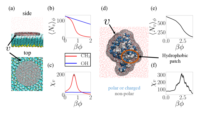
To illustrate the molecular signatures of surface hydrophobicity, we first review the contrasting behavior of water near CH3-terminated (hydrophobic) and OH-terminated (hydrophilic) self-assembled monolayer (SAM) surfaces. The SAM surfaces are not only flat and uniform, and thereby considerably simpler than proteins, but their hydrophobicity can also be defined unambiguously using a macroscopic measure, such as the water droplet contact angle. We focus on water molecules in a cylindrical observation volume, , at the SAM-water interface, as shown in Figure 1a. We choose a radius, nm, and a width, nm, for the cylindrical ; with this choice, at either SAM surface contains an average of roughly 120 waters. Following previous work 54, 27, 20, 21, we then perturb the interfacial waters in by applying an unfavorable biasing potential, , where represents the strength of the potential, and is the number of coarse-grained waters in ; a more precise definition of is included in the . The potential imposes an energetic penalty that increases linearly with , so that as is increased, waters are displaced from , resulting in a decrease in the average water numbers, , next to both SAM surfaces; see Figure 1b. The decrease in with increasing is linear for the hydrophilic SAM. In comparison, the corresponding -values for the hydrophobic SAM are smaller for every , highlighting the relative ease of displacing waters. Moreover, the decrease in with increasing is sensitive (or sigmoidal) near the hydrophobic surface rather than gradual (and linear) as it is near the hydrophilic surface. This contrast can be seen even more clearly in Figure 1c, which shows the susceptibility, , as a function of ; here, , is the Boltzmann constant, and is the system temperature. The susceptibility is nearly constant for the hydrophilic surface. However, it shows a pronounced peak for the hydrophobic surface, suggesting that a collective dewetting of the interfacial waters can be triggered when a sufficiently strong unfavorable potential is applied.
2.2 Perturbing the protein hydration shell
In contrast with the uniform SAM surfaces, proteins are heterogeneous, rugged, and amphiphilic. Their surfaces tend to have polar and charged residues to ensure that the protein is soluble in water, as well as hydrophobic residues to drive protein interactions. Given the amphiphilicity of proteins, how might their hydration waters respond to an unfavorable potential? Should we expect its hydration waters to be displaced gradually like the hydrophilic SAM surface? Or should we expect the protein hydration waters to undergo collective dewetting like the hydrophobic SAM surface? To address these questions, we first study ubiquitin, a highly-conserved protein involved in numerous signaling pathways, including protein degradation 55. Although it is a relatively small protein (76 amino acid residues), ubiquitin displays many of the characteristic features of a soluble globular protein, including a stable folded structure, a chemically and topographically heterogeneous surface, and interactions with diverse molecules that are crucial to its function (Figure 1d) 53. Many of these interactions are mediated by a well-documented hydrophobic patch, which is also shown in Figure 1d.
To characterize the overall strength of protein-water interactions, we again apply a biasing potential, , where is now the number of coarse-grained waters in the entire protein hydration shell, . The hydration shell, , is defined as the union of spherical sub-volumes centered on all the protein heavy atoms, with each sub-volume chosen to have the same radius, . Such a definition allows to capture the ruggedness of the underlying protein surface, with the width of the hydration shell determined by . Here we choose nm so that only waters in the first hydration shell of the protein are included in (Figure 1d). The decrease in the average number of ubiquitin hydration waters, , in response to the unfavorable potential, , is shown in Figure 1e. Interestingly, displays a sigmoidal dependence on , akin to that for the hydrophobic CH3-terminated SAM surface (Figure 1b). Correspondingly, a clear peak is also observed in the susceptibility around (Figure 1f). Thus, the hydration shell of the inherently amphiphilic and incredibly complex surface of the ubiquitin protein dewets collectively in response to an unfavorable perturbation.
2.3 How the hydration shells of diverse proteins respond to unfavorable potentials
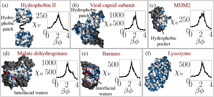
Is ubiquitin unique? If not, how general is the susceptibility of protein hydration waters to an unfavorable potential? To address this question, we studied six additional proteins, spanning a range of sizes, chemical patterns, and functional roles; see Figure 2 as well as Figure S2 in the . Given the importance of hydrophobic surface moieties in situating the interfacial waters at the edge of a dewetting transition, we first considered other proteins with well-defined hydrophobic patches. First, we consider the fungal protein, Hydrophobin II, which is highly surface-active, and is known to self-assemble at water-vapor interfaces 58. Although Hydrophobin II is charge neutral overall, the protein surface displays 10 charged residues. In Figure 2a, we show both the hydrophobic face of Hydrophobin II, which is enriched in hydrophobic residues, as well as the remainder of the protein, which has been shown to be super-hydrophilic 20. As with ubiquitin, the susceptibility, , for Hydrophobin II also displays a marked peak. Next, we consider the human hepatitis B viral capsid protein, which has a net charge of -6, but displays an even larger hydrophobic patch than the one on Hydrophobin II (Figure 2b). That patch drives the binding of two capsid proteins to form a dimer that further assembles into a 240-protein capsid shell 59. The viral capsid protein also displays a clear peak in . Does the collective dewetting seen in the above proteins stem from the presence of extended hydrophobic patches on their surfaces? Although many proteins possess such patches, not all of them do; instead, most protein surfaces display chemical patterns that are amphiphilic, and feature only smaller hydrophobic regions. The signaling protein MDM2 contains such a modest hydrophobic groove, which is nevertheless important from a functional standpoint; it enables MDM2 to exercise control over cellular senescence by binding to the transactivation domain of the tumor-suppresor protein p53 60. As shown in Figure 2c, MDM2 also displays a peak in susceptibility, .
Might the large or small, but well-defined hydrophobic patches on ubiquitin, Hydrophobin II, the capsid sub-unit, and MDM2 be responsible for rendering their hydration shell waters susceptible to unfavorable perturbation? To address this question, we study proteins that are known for being anomalously hydrophilic or charged. Malate dehydrogenase is a large hydrophilic protein with 61 charged surface residues. The protein dimerizes into a metabolic enzyme, and plays an important role in the citric acid cycle. The interface through which the protein monomers bind is fairly hydrophilic, featuring 5 charged residues and several other polar residues. In fact, regions of the binding interface are so hydrophilic that they hold on their waters even in the bound state. In other words, the binding interface features structured waters that bridge the two interacting proteins; such bridging waters, which are observed in the crystal structure of the malate dehydrogenase dimer, are shown in Figure 2d 61, 62. The proteins discussed previously, in contrast, feature binding interfaces that are entirely dry. Interestingly, even for the largely hydrophilic malate dehydrogenase protein, we observe a peak in susceptibility, , in response to an unfavorable perturbation (Figure 2d).
Another fairly hydrophilic protein that features a charged interaction interface is barnase, a bacterial RNase that interacts with its inhibitor, barstar, in one of the strongest known protein-protein interactions 63. The high-affinity sub-picomolar binding between barnase and barstar is facilitated by the formation of electrostatic contacts between five positively charged residues on barnase and five negatively charged residues on barstar 63. Remarkably, a clear peak in susceptibility is also observed for barnase (Figure 2e). Finally, we study T4 lysozyme, a bacteriophage protein that catalyzes the hydrolysis of the peptidoglycan layer of bacterial cell walls 64, and has 45 charged surface residues with an overall charge of +9. Lysozyme does not appear to participate in interactions with proteins other than its substrates, or to possess a clear hydrophobic patch; rather, it displays a checkered pattern of hydrophobic and hydrophilic atoms (Figure 2f). As with all of the proteins studied, the hydration shell of lysozyme also displays a marked peak in susceptibility. Our results, obtained across proteins with a diversity of sizes, biological functions, and surface chemistries, thus suggest that susceptibility to an unfavorable potential is a general property of protein hydration waters.
2.4 Characterizing protein surfaces: residues vs atoms
Although collective dewetting in response to an unfavorable potential may be expected for proteins that are fairly hydrophobic, it is somewhat surprising that even the more hydrophilic and charged proteins display such behavior. To better characterize the similarities and differences in the surface chemistries of the seven proteins discussed above, we plot the fraction of their surface residues that are charged and hydrophobic in Figures 3a and 3b, respectively. As expected, the more hydrophobic proteins have a smaller fraction of charged residues and a larger fraction of hydrophobic residues, with the charge fraction ranging from 0.15 to 0.35, and the hydrophobic fraction varying from 0.5 to 0.2. To interrogate whether the surface chemistries of these seven proteins are representative of the larger class of folded, globular proteins, we additionally estimated these quantities for an expanded set containing a total of 20 proteins. The results are included in Figure S4 of the , and highlight that the characteristics of proteins studied here are indeed representative of typical proteins. How then do we understand the sensitivity of such diverse protein hydration shells to unfavorable potentials, and their resemblance to extended hydrophobic surfaces?
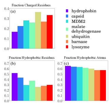
To answer this question, we draw inspiration from work by Kapcha and Rossky, who highlighted that amino acid residues are not monolithic, but are instead heterogeneous, and are composed of both hydrophobic and hydrophilic atoms 52. Kapcha and Rossky thus advocate adopting an atom-centric, rather than a residue-centric view of the protein surface. They further suggest that an atom be classified as hydrophobic only if the magnitude of its partial charge is less than 0.25 in the OPLS force field, and hydrophilic otherwise. Following these authors, we plot the fraction of surface atoms (not residues) that are hydrophobic in Figure 3c. Interestingly, we find that the fraction of surface atoms that are hydrophobic is not only larger than the corresponding fraction of surface residues, but that roughly half the protein surface (or more) consists of hydrophobic atoms. Importantly, the fraction of surface atoms that are hydrophobic is uniformly high for all proteins studied here. To better understand these results, we analyzed the atomic composition of the surface residues; as shown in Figure S5 of the , roughly 80% of atoms belonging to hydrophobic residues are hydrophobic, but nearly 50% of atoms belonging to hydrophilic (polar or charged) residues are also hydrophobic. Thus, although polar and charged surface residues do not contribute as heavily to hydrophobic surface atoms, they nevertheless have substantive contributions. We note that following Kapcha and Rossky, we classify not just the protein heavy atoms, but also hydrogen atoms as being either hydrophobic or hydrophilic 52. If the protein hydrogen atoms are excluded from the analysis, the fraction of hydrophobic surface atoms is somewhat lower, but remains close to 0.5 as shown in Figure S4 of the . Our results thus suggest that for a wide variety of proteins, roughly half the surface consists of hydrophobic atoms; these atoms situate the protein hydration waters at the edge of a dewetting transition, making them particularly susceptible to unfavorable potentials.
2.5 Strength of unfavorable potential needed to trigger dewetting
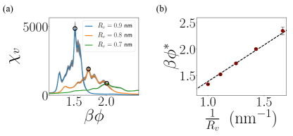
Interestingly, not only do all of proteins studied here show a peak in , the location of the characteristic peak in susceptibility, , is observed to be roughly in all cases (Figure 2). To understand this observation, we first recognize that for , the biasing potential, , favors configurations with lower -values and thereby lower densities. As a result, the biasing potential effectively lowers the pressure in the protein hydration shell to: , where is the system pressure and is the molar density of liquid water. For a sufficiently large , the tension (negative pressure) exerted on the protein hydration waters prompts the nucleation of vapor in certain regions of . The biasing potential thus stabilizes a water-vapor interface; the pressure drop across this interface is related to the corresponding interfacial tension, , and the mean interfacial curvature, , according to the Young-Laplace equation, . Moreover, because the biasing potential is only experienced by waters in , the interfacial curvature is determined by the radius, , of the spherical sub-volumes defining , such that . Thus, the biasing potential strength needed to trigger vapor nucleation ought to be: . By systematically varying , and repeating our calculations for ubiquitin, we find that as is increased, the peak in susceptibility indeed shifts to lower values (Figure 4a). Furthermore, as shown in Figure 4b, also varies linearly with as predicted; the best fit line through the origin has a slope of 1.38 nm, which is comparable to nm, estimated using nm-3 and mJ/nm2 for SPC/E water.
3 Conclusions and Outlook
Proteins employ intricate topographical and chemical patterns, which have evolved to facilitate their many biological functions. Although the space of such patterns is immense, it is likely constrained by common characteristics that all proteins must possess in order to function properly. For example, all proteins must have favorable interactions with water to be soluble, which requires the presence of hydrophilic groups on their surfaces. Conversely, protein surfaces must also feature hydrophobic regions that interact poorly with water, and provide a driving force for proteins to interact with other molecules. In this article, we shed light on how proteins accomplish these competing goals by balancing their overall interactions with their hydration waters. We show that roughly half the atoms on the protein surface are hydrophobic – a fact that can be obfuscated by focusing on surface residues rather than surface atoms. We also find that the hydration shells of diverse proteins – even those with highly charged surfaces and amphiphilic interaction interfaces – are highly susceptible to an unfavorable potential. Our results thus suggest that hydrophobic atoms on the protein surface situate its hydration waters at the edge of a dewetting transition, which can be triggered by an unfavorable perturbation. Consistent with our results, signatures of collective transitions have also been observed in studies of partially hydrated proteins. For example, Cui et al. found that partially hydrated proteins undergo a percolation transition at a critical value of protein hydration 65. Similarly, in studying the uptake of water from the vapor phase by proteins, Debenedetti and co-workers found protein hydration to display hysteresis – a hallmark of collective transitions – between the adsorption and desorption branches of the isotherm 16, 66. These authors also found that polar and charged residues contributed to the collective wetting of a dry protein (and the associated hysteresis) 67; correspondingly, here we find that non-polar regions of the protein give rise to the collective dewetting of a hydrated protein.
We also find that the biasing potential strength needed to trigger dewetting is inversely proportional to the width of the hydration shell, but does not depend meaningfully on the particular protein being perturbed. Thus, our results not only suggest that susceptibility to an unfavorable perturbation is a general feature of the hydration shells of proteins, but also highlight that the strength of the perturbation needed to trigger dewetting is remarkably similar across different proteins. This finding suggests a near-universal calibration of the perturbation strength across diverse proteins, i.e., by considering relative to . It also establishes a framework for systematically classifying how favorable the interactions between water and different parts of the protein surface are. In particular, we expect that locations on the protein surface that dewet at low -values (relative to ) will correspond to the most hydrophobic regions on the protein surface, whereas regions that retain their waters even at high -values will be the most hydrophilic. Given the importance of hydrophobicity in driving protein interactions, regions of the protein surface that dewet most readily may correspond closely with patches on the protein that participate in interactions 47, 68, 50, 6. We are investigating whether such hydrophobic protein regions could serve as predictors of protein interaction sites, and plan to report our findings in a future study. Similarly, identification of the most hydrophilic regions of the protein could facilitate the discovery of novel super-hydrophilic chemical patterns; an understanding of what enables such patterns to have strong interactions with water could also serve as the basis for the rational design of protein non-fouling surfaces or surfaces that display super-oleophobicity underwater 51, 69.
A.J.P. gratefully acknowledges financial support from the National Science Foundation (CBET 1652646, CHE 1665339, and UPENN MRSEC DMR 11-20901), and a fellowship from the Alfred P. Sloan Research Foundation. N.B.R. was supported by the National Science Foundation grant CBET 1652646.
In the , we include details of our simulations, information on enhanced sampling techniques that we use, plots supporting Figure 2, and additional analysis pertaining to the diversity of the proteins studied here.
References
- Ashbaugh and Hatch 2008 Ashbaugh, H. S.; Hatch, H. W. Natively Unfolded Protein Stability as a Coil-to-Globule Transition in Charge/Hydropathy Space. J. Am. Chem. Soc. 2008, 130, 9536–9542
- Krone et al. 2008 Krone, M. G.; Hua, L.; Soto, P.; Zhou, R.; Berne, B. J.; Shea, J.-E. Role of Water in Mediating the Assembly of Alzheimer Amyloid- A16-22 Protofilaments. J. Am. Chem. Soc. 2008, 130, 11066–11072
- Levy and Onuchic 2006 Levy, Y.; Onuchic, J. N. Water Mediation in Protein Folding and Molecular Recognition. Annu. Rev. Biophys. Biomol. Struct. 2006, 35, 389–415
- Berne et al. 2009 Berne, B. J.; Weeks, J. D.; Zhou, R. Dewetting and Hydrophobic Interaction in Physical and Biological Systems. Annu. Rev. Phys. Chem. 2009, 60, 85–103
- Jamadagni et al. 2011 Jamadagni, S. N.; Godawat, R.; Garde, S. Hydrophobicity of Proteins and Interfaces: Insights from Density Fluctuations. Annu. Rev. Chem. Biomol. Eng. 2011, 2, 147–171
- Vashisth and Abrams 2013 Vashisth, H.; Abrams, C. F. All-Atom Structural Models of Insulin Binding to the Insulin Receptor in the Presence of a Tandem Hormone-Binding Element. Proteins: Struct., Funct., Bioinf. 2013, 81, 1017–1030
- Baron and McCammon 2013 Baron, R.; McCammon, J. A. Molecular Recognition and Ligand Association. Annu. Rev. Phys. Chem. 2013, 64, 151–75
- Tiwary et al. 2015 Tiwary, P.; Limongelli, V.; Salvalaglio, M.; Parrinello, M. Kinetics of Protein–Ligand Unbinding: Predicting Pathways, Rates, and Rate-Limiting Steps. Proc. Natl. Acad. Sci. U.S.A. 2015, 112, E386–E391
- Tiwary et al. 2015 Tiwary, P.; Mondal, J.; Morrone, J. A.; Berne, B. Role of Water and Steric Constraints in the Kinetics of Cavity–Ligand Unbinding. Proc. Natl. Acad. Sci. U.S.A. 2015, 112, 12015–12019
- Bellissent-Funel et al. 2016 Bellissent-Funel, M.-C.; Hassanali, A.; Havenith, M.; Henchman, R.; Pohl, P.; Sterpone, F.; van der Spoel, D.; Xu, Y.; Garcia, A. E. Water Determines the Structure and Dynamics of Proteins. Chem. Rev. 2016, 116, 7673–7697
- Jaenicke 2000 Jaenicke, R. Stability and Stabilization of Globular Proteins in Solution. J. Biotechnol. 2000, 79, 193–203
- Liu et al. 2004 Liu, S. J. S.; Shahrokh, Z.; Jun, Challenges in the Development of High Protein Concentration Formulations. J. Pharm. Sci. 2004, 93, 1390–1402
- Shen et al. 2006 Shen, V. K.; Cheung, J. K.; Errington, J. R.; Truskett, T. M. Coarse-Grained Strategy for Modeling Protein Stability in Concentrated Solutions. II: Phase Behavior. Biophys. J. 2006, 90, 1949–60
- Trevino et al. 2008 Trevino, S. R.; Scholtz, J. M.; Pace, C. N. Measuring and Increasing Protein Solubility. J. Pharm. Sci. 2008,
- Thirumalai et al. 2012 Thirumalai, D.; Reddy, G.; Straub, J. E. Role of Water in Protein Aggregration and Amyloid Polymorphism. Acc. Chem. Res. 2012, 45, 83–92
- Debenedetti and Pablo 2012 Debenedetti, J. C. P.; Pablo, Computer Simulation of Water Sorption on Flexible Protein Crystals. J. Phys. Chem. Lett. 2012, 3, 2713–2718
- Chong and Ham 2014 Chong, S.-H.; Ham, S. Interaction with the Surrounding Water Plays a Key Role in Determining the Aggregation Propensity of Proteins. Angew. Chem., Int. Ed. 2014, 53, 3751–3751
- Remsing et al. 2018 Remsing, R. C.; Xi, E.; Patel, A. J. Protein Hydration Thermodynamics: The Influence of Flexibility and Salt on Hydrophobin II Hydration. J. Phys. Chem. B 2018, 122, 3635–3646
- Patel et al. 2011 Patel, A. J.; Varilly, P.; Jamadagni, S. N.; Acharya, H.; Garde, S.; Chandler, D. Extended Surfaces Modulate Hydrophobic Interactions of Neighboring Solutes. Proc. Natl. Acad. Sci. U.S.A. 2011, 108, 17678–17683
- Patel and Garde 2014 Patel, A. J.; Garde, S. Efficient Method To Characterize the Context-Dependent Hydrophobicity of Proteins. J. Phys. Chem. B 2014, 118, 1564–1573
- Xi et al. 2016 Xi, E.; Remsing, R. C.; Patel, A. J. Sparse Sampling of Water Density Fluctuations in Interfacial Environments. J. Chem. Theory Comput. 2016, 12, 706–713
- Lum et al. 1999 Lum, K.; Chandler, D.; Weeks, J. D. Hydrophobicity at Small and Large Length Scales. J. Phys. Chem. B 1999, 103, 4570–4577
- Varilly et al. 2011 Varilly, P.; Patel, A. J.; Chandler, D. An Improved Coarse-Grained Model of Solvation and the Hydrophobic Effect. J. Chem. Phys. 2011, 134, 074109
- Vaikuntanathan et al. 2016 Vaikuntanathan, S.; Rotskoff, G.; Hudson, A.; Geissler, P. L. Necessity of Capillary Modes in a Minimal Model of Nanoscale Hydrophobic Solvation. Proc. Natl. Acad. Sci. U.S.A. 2016, 113, E2224–E2230
- Xi and Patel 2016 Xi, E.; Patel, A. J. The Hydrophobic Effect, and Fluctuations: The Long and the Short of It. Proc. Natl. Acad. Sci. U.S.A. 2016, 113, 4549–4551
- Patel and Chandler 2010 Patel, A. J.; Chandler, D. Fluctuations of Water near Extended Hydrophobic and Hydrophilic Surfaces. J. Phys. Chem. B 2010, 114, 1632–1637
- Patel et al. 2012 Patel, A. J.; Varilly, P.; Jamadagni, S. N.; Hagan, M. F.; Chandler, D.; Garde, S. Sitting at the Edge: How Biomolecules Use Hydrophobicity to Tune Their Interactions and Function. J. Phys. Chem. B 2012, 116, 2498–2503
- Rossky et al. 1984 Rossky, C. Y. L.; McCammon, J. A.; J., P. The Structure of Liquid Water at an Extended Hydrophobic Surface. J. Chem. Phys. 1984, 80, 4448–4455
- Mittal and Hummer 2008 Mittal, J.; Hummer, G. Static and Dynamic Correlations in Water at Hydrophobic Interfaces. Proc. Nat. Acad. Sci. 2008, 105, 20130–20135
- Sarupria and Garde 2009 Sarupria, S.; Garde, S. Quantifying Water Density Fluctuations and Compressibility of Hydration Shells of Hydrophobic Solutes and Proteins. Phys. Rev. Lett. 2009, 103, 037803
- Godawat et al. 2009 Godawat, R.; Jamadagni, S. N.; Garde, S. Characterizing Hydrophobicity of Interfaces by Using Cavity Formation, Solute Binding, and Water Correlations. Proc. Natl. Acad. Sci. U.S.A. 2009, 106, 15119–15124
- Willard and Chandler 2010 Willard, A. P.; Chandler, D. Instantaneous Liquid Interfaces. J. Phys. Chem. B 2010, 114, 1954–1958
- Heyden and Tobias 2013 Heyden, M.; Tobias, D. J. Spatial Dependence of Protein-Water Collective Hydrogen-Bond Dynamics. Phys. Rev. Lett. 2013, 111, 218101
- Sosso et al. 2016 Sosso, G. C.; Caravati, S.; Rotskoff, G.; Vaikuntanathan, S.; Hassanali, A. On the Role of Nonspherical Cavities in Short Length-Scale Density Fluctuations in Water. J. Phys. Chem. A 2016, 121, 370–380
- Shin and Willard 2018 Shin, S.; Willard, A. P. Water’s Interfacial Hydrogen Bonding Structure Reveals the Effective Strength of Surface-Water Interactions. J. Phys. Chem. B 2018,
- Giovambattista et al. 2008 Giovambattista, N.; Lopez, C. F.; Rossky, P. J.; Debenedetti, P. G. Hydrophobicity of Protein Surfaces: Separating Geometry from Chemistry. Proc. Natl. Acad. Sci. U.S.A. 2008, 105, 2274–2279
- Giovambattista et al. 2009 Giovambattista, N.; Debenedetti, P. G.; Rossky, P. J. Enhanced Surface Hydrophobicity by Coupling of Surface Polarity and Topography. Proc. Natl. Acad. Sci. U.S.A. 2009, 106, 15181–15185
- Acharya et al. 2010 Acharya, H.; Vembanur, S.; Jamadagni, S. N.; Garde, S. Mapping Hydrophobicity at the Nanoscale: Applications to Heterogeneous Surfaces and Proteins. Faraday Discuss. 2010, 146, 353–365
- Daub et al. 2010 Daub, C. D.; Wang, J.; Kudesia, S.; Bratko, D.; Luzar, A. The Influence of Molecular-Scale Roughness on the Surface Spreading of an Aqueous Nanodrop. Faraday Discuss. 2010, 146, 67–77
- Mittal and Hummer 2010 Mittal, J.; Hummer, G. Interfacial Thermodynamics of Confined Water near Molecularly Rough Surfaces. Faraday Discuss. 2010, 146, 341–352
- Wang et al. 2011 Wang, J.; Bratko, D.; Luzar, A. Probing Surface Tension Additivity on Chemically Heterogeneous Surfaces by a Molecular Approach. Proc. Natl. Acad. Sci. U.S.A. 2011, 108, 6374–6379
- Fogarty and Laage 2014 Fogarty, A. C.; Laage, D. Water Dynamics in Protein Hydration Shells: The Molecular Origins of the Dynamical Perturbation. J. Phys. Chem. B 2014, 118, 7715–7729
- Harris and Pettitt 2014 Harris, R. C.; Pettitt, B. M. Effects of Geometry and Chemistry on Hydrophobic Solvation. Proc. Natl. Acad. Sci. U.S.A. 2014, 111, 14681–14686
- Xi et al. 2017 Xi, E.; Venkateshwaran, V.; Li, L.; Rego, N.; Patel, A. J.; Garde, S. Hydrophobicity of Proteins and Nanostructured Solutes Is Governed by Topographical and Chemical Context. Proc. Natl. Acad. Sci. U.S.A. 2017, 201700092
- Monroe and Shell 2018 Monroe, J. I.; Shell, M. S. Computational Discovery of Chemically Patterned Surfaces that Effect Unique Hydration Water Dynamics. Proc. Natl. Acad. Sci. U.S.A. 2018, 115, 8093–8098
- 46 Heyden, M. Disassembling solvation free energies into local contributions—Toward a microscopic understanding of solvation processes. Wiley Interdiscip. Rev.: Comput. Mol. Sci. e1390
- Bogan and Thorn 1998 Bogan, A. A.; Thorn, K. S. Anatomy of Hot Spots in Protein Interfaces. J. Mol. Biol. 1998, 280, 1–9
- DeLano 2002 DeLano, W. L. Unraveling Hot Spots in Binding Interfaces: Progress and Challenges. Curr. Opin. Struct. Biol. 2002, 12, 14–20
- Nooren and Thornton 2003 Nooren, I. M.; Thornton, J. M. Diversity of Protein–Protein Interactions. EMBO J. 2003, 22, 3486–3492
- White et al. 2008 White, A. W.; Westwell, A. D.; Brahemi, G. Protein-Protein Interactions as Targets for Small-Molecule Therapeutics in Cancer. Expert Rev. Mol. Med. 2008, 10, e8
- Chen et al. 2009 Chen, S.; Cao, Z.; Jiang, S. Ultra-Low Fouling Peptide Surfaces Derived from Natural Amino Acids. Biomaterials 2009, 30, 5892–5896
- Kapcha and Rossky 2014 Kapcha, L. H.; Rossky, P. J. A Simple Atomic-Level Hydrophobicity Scale Reveals Protein Interfacial Structure. J. Mol. Biol. 2014, 426, 484–498
- Winget and Mayor 2010 Winget, J. M.; Mayor, T. The Diversity of Ubiquitin Recognition: Hot Spots and Varied Specificity. Mol. Cell 2010, 38, 627–635
- Patel et al. 2011 Patel, A. J.; Varilly, P.; Chandler, D.; Garde, S. Quantifying Density Fluctuations in Volumes of All Shapes and Sizes Using Indirect Umbrella Sampling. J. Stat. Phys. 2011, 145, 265–275
- Hershko and Ciechanover 1982 Hershko, A.; Ciechanover, A. Mechanisms of Intracellular Protein Breakdown. Annu. Rev. Biochem. 1982, 51, 335–364
- Hakanpää et al. 2006 Hakanpää, J.; Linder, M.; Popov, A.; Schmidt, A.; Rouvinen, J. Hydrophobin HFBII in Detail: Ultrahigh-Resolution Structure at 0.75 Å. Acta Crystallogr., Sect. D: Biol. Crystallogr. 2006, 62, 356–367
- Wösten and de Vocht 2000 Wösten, H. A. B.; de Vocht, M. L. Hydrophobins, the Fungal Coat Unravelled. Biochim. Biophys. Acta, Rev. Biomembr. 2000, 1469, 79–86
- Hakanpaa et al. 2006 Hakanpaa, J.; Linder, M.; Popov, A.; Schmidt, A.; Rouvinen, J. Hydrophobin HFBII in Detail: Ultrahigh-Resolution Structure at 0.75 A. Acta Crystallogr., Sect D: Biol. Crystallogr. 2006, 62, 356–367
- Wynne et al. 1999 Wynne, S. A.; Crowther, R. A.; Leslie, A. G. The Crystal Structure of the Human Hepatitis B Virus Capsid. Mol. Cell 1999, 3, 771–780
- Kussie et al. 1996 Kussie, P. H.; Gorina, S.; Marechal, V.; Elenbaas, B.; Moreau, J.; Levine, A. J.; Pavletich, N. P. Structure of the MDM2 Oncoprotein Bound to the P53 Tumor Suppressor Transactivation Domain. Science 1996, 274, 948–953
- Rodier et al. 2005 Rodier, F.; Bahadur, R. P.; Chakrabarti, P.; Janin, J. Hydration of Protein-Protein Interfaces. Proteins: Struct., Funct., Bioinf. 2005, 60, 36–45
- Zaitseva et al. 2009 Zaitseva, J.; Meneely, K. M.; Lamb, A. L. Structure of Escherichia Coli Malate Dehydrogenase at 1.45 A Resolution. Acta Crystallogr., Sect. F: Struct. Biol. Cryst. Commun. 2009, 65, 866–869
- Schreiber and Fersht 1995 Schreiber, G.; Fersht, A. R. Energetics of Protein-Protein Interactions: Analysis Of the Barnase-Barstar Interface by Single Mutations and Double Mutant Cycles. J. Mol. Biol. 1995, 248, 478–486
- Shoichet et al. 1995 Shoichet, B. K.; Baase, W. A.; Kuroki, R.; Matthews, B. W. A Relationship between Protein Stability and Protein Function. Proc. Natl. Acad. Sci. U.S.A. 1995, 92, 452–456
- Cui et al. 2014 Cui, D.; Ou, S.; Patel, S. Protein-Spanning Water Networks and Implications for Prediction of Protein–Protein Interactions Mediated through Hydrophobic Effects. Proteins: Struct., Funct., Bioinf. 2014, 82, 3312–3326
- Kim et al. 2015 Kim, S. B.; Palmer, J. C.; Debenedetti, P. G. A Computational Study of the Effect of Matrix Structural Order on Water Sorption by Trp-Cage Miniproteins. J. Phys. Chem. B 2015, 119, 1847–1856
- Kim et al. 2017 Kim, S. B.; Sparano, E. M.; Singh, R. S.; Debenedetti, P. G. Microscopic Origin of Hysteresis in Water Sorption on Protein Matrices. J. Phys. Chem. Lett. 2017, 8, 1185–1190
- Keskin et al. 2005 Keskin, O.; Ma, B.; Nussinov, R. Hot Regions in Protein–Protein Interactions: The Organization and Contribution of Structurally Conserved Hot Spot Residues. J. Mol. Biol. 2005, 345, 1281–1294
- Si et al. 2018 Si, Y.; Dong, Z.; Jiang, L. Bioinspired Designs of Superhydrophobic and Superhydrophilic Materials. ACS Cent. Sci. 2018,