Active prestress leads to an apparent stiffening of cells through geometrical effects
Abstract
Tuning of active prestress e.g. through activity of molecular motors constitutes a powerful cellular tool to adjust cellular stiffness through nonlinear material properties. Understanding this tool is an important prerequisite for our comprehension of cellular force response, cell shape dynamics and tissue organisation.
Experimental data obtained from cell-mechanical measurements often show a simple linear dependence between mechanical prestress and measured differential elastic moduli. While these experimental findings could point to stress-induced structural changes in the material, we propose here a surprisingly simple alternative explanation in a theoretical study. We show how geometrical effects can give rise to increased cellular force response of cells in the presence of active prestress. The associated effective stress-stiffening is disconnected from actual stress-induced changes of the elastic modulus and should therefore be regarded as an apparent stiffening of the material. We argue that new approaches in experimental design are necessary to separate this apparent stress-stiffening due to geometrical effects from actual nonlinearities of the elastic modulus in prestressed cellular material.
*Correspondence: elisabeth.fischer-friedrich@tu-dresden.de
I Introduction
Cells need to deform themselves during many physiological processes in the body such as cell division, cell migration or the complex events of morphogenesis. To understand dynamic cell deformation from a physical point of view, we need to quantify cell material properties and its active regulation through the underlying molecular cell biology.
Cells are a complex viscoelastic material whose material properties are time-scale dependent and nonlinear beyond strains of few percent Kollmannsberger and Fabry (2011).
Mechanical stress therefore alters cell-mechanical properties making cells stiffer or softer Kollmannsberger and Fabry (2011, 2009); Lange et al. (2017); Stricker et al. (2010); Ingber et al. (2014).
The material properties of a cell are mainly determined by its cytoskeleton.
Intriguingly, cells are able to self-tune its mechanical prestress in the cytoskeleton through the action of molecular motors giving the cell a tool at hand to actively regulate its material stiffness through dynamic adjustment of active prestress together with stress-stiffening or stress-softening.
Many studies report stress-stiffening through active stress Kollmannsberger and Fabry (2011); Wang et al. (2002); Stamenović et al. (2004); Fischer-Friedrich et al. (2016); Fernández et al. (2006); Jansen et al. (2013), however opposite findings of cell-softening have also been reported Chan et al. (2015). Stiffness-modulation through the action of motor proteins in actin meshwork’s was further corroborated by in vitro measurements Mizuno et al. (2007); Bendix et al. (2008); Koenderink et al. (2009).
It has been suggested that cytoskeletal stress-stiffening may be understood in terms of the nonlinear entropic thermal stretch modulus of a single polymer Bustamante et al. (1994); Gardel et al. (2004); Storm et al. (2005) or the stress-induced “pull-out” of soft bending modes and a transition towards a stretch-dominated regime Broedersz and MacKintosh (2011). As a third option, load-dependent binding dynamics of actin cross-linkers has been put forward Yao et al. (2013). While entropic effects predict a power law scaling of for the resulting differential shear modulus Gardel et al. (2004), the “pull-out” of soft bending modes gives rise to a power law scaling between and Broedersz and MacKintosh (2011).
Furthermore, linear stress-stiffening has been reported for marginal networks Sheinman et al. (2012); Licup et al. (2015) and for tensegrity models Volokh et al. (2000).
Experimental data obtained from cell-mechanical measurements show often a linear dependence between measured differential elastic moduli and active mechanical prestress. This corresponds to a power law with exponent one in dependence of prestress Kollmannsberger and Fabry (2011); Wang et al. (2002); Stamenović et al. (2004); Fischer-Friedrich et al. (2016).
While these experimental findings could point to complex structural changes of the cytoskeleton through active prestress and accordant modification of its elastic modulus, we propose here a surprisingly simple alternative explanation: pure geometrical effects may give rise to an increased force response and thus an effective stiffening of actively prestressed material. Associated effective stiffening is however independent of actual stress-dependent changes of the elastic modulus as a material parameter of the cytoskeleton in a coarse-grained continuum description. Thus the associated stiffening will be denoted as ‘apparent stiffening’.
We will discuss two different geometrical effects i) stress-stiffening through geometrical-coupling and ii) shear-induced nematic alignment.
In fact, when present, these effects disguise the actual stress-dependence of the elastic modulus and may account partly for incoherences in the cell mechanics field such as order of magnitude differences in measured shear moduli and contradicting trends (stiffening or softening) in response to mechanical prestress.
II Apparent stress-stiffening through geometrical coupling
In the following paragraphs, we discuss two simplified examples of cell-mechanical probing to illustrate the general phenomenon of apparent stress-stiffening through geometrical coupling in i) adherent and ii) non-adherent cells. In our calculations, we will henceforth assume that displacements and strains are small. We will thus follow the approach of linear elasticity theory keeping only terms to first-order in strain and deformation.
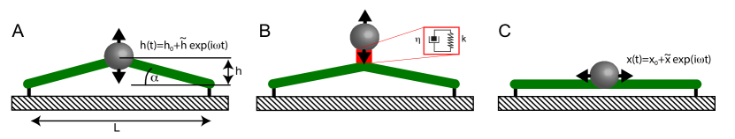
Deformation of an adhered model cell. We will first consider the case of a one-dimensional actively prestressed fibre tethered to a substrate as a minimal model of an adherent cell. Consider the experimental scenario sketched in Fig. 1A where the fibre center is oscillated around a mean height with through a force exerted by an adherent bead. The one-dimensional stress in the fibre has a contribution from active prestress and a deformation-induced viscoelastic contribution Fischer-Friedrich et al. (2016)
| (1) |
where is the prestress- and frequency-dependent complex elastic modulus of the fibre, is the strain with respect to fibre length changes and is the angular frequency of the applied oscillation.
The measured force signal is
| (2) |
where is the angle between the fibre and the substrate (see Fig. 1A) and is a time-dependent geometrical factor. We make the ansatz
| (3) | ||||
| (4) |
where and are average values of the geometrical factor and measured force, respectively, while and characterise emerging oscillatory changes. Here, may take complex values if the respective force oscillations are not in-phase with the imposed oscillations of bead height. We find to first order
| (5) |
Force oscillation amplitudes thus contain a contribution proportional to the active prestress if the oscillation amplitude of the geometrical factor is does not vanish. We will denote this effect as geometrical coupling of active stress into the force signal. In a conventional rheological analysis, that disregards the presence of active prestress, the force amplitude is assumed to scale with the complex elastic modulus of the system for a given strain. Therefore, the effective elastic modulus would be determined to scale as
| (6) |
A short calculation yields (see Supporting Material)
| (7) |
where is the fibre length in a straight conformation (see Fig. 1A).
Thus, the effective shear modulus shows apparent linear stress-stiffening with a slope proportional to and an additional active contribution .
In particular, for sufficiently small values of mean height , the active contribution starts to override the contribution of the actual complex elastic modulus in the measurement and the slope of the apparent stress-stiffening can in principle become arbitrarily large.
So far, the examples of geometrical coupling of active prestress have contributed only a constant contribution to the effective storage modulus of the system because the deformation and the geometrical factor were in phase. However, in general, out-of-phase oscillations of the geometrical factor are possible e.g. in the case of a prestressed material coupled to a viscoelastic connector (see Fig. 1B and Supporting Material).
If an attached bead is deflected horizontally along the fiber (see Fig. 1C), active contributions to the force oscillations are avoided and we have .
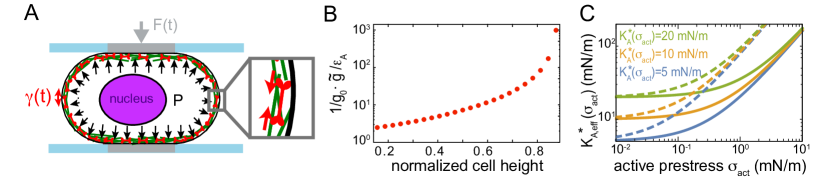
Deformation of a non-adherent model cell. As a second example, we consider a non-adherent cell that is being probed in a parallel plate assay subject to oscillatory height changes (see Fig. 2A). Accordant measurements were for instance performed on mitotic cells in Ref. Fischer-Friedrich et al. (2016). We will regard the cell as a liquid-filled pressurized shell where the shell material is constituted by the plasma membrane and the neighboring actomyosin cortex. To keep our calculations simple, we will assume that the deformation-induced stress in the cortex-membrane layer is a spatially homogeneous, isotropic, in-plane stress governed by an area dilation modulus and an active mechanical prestress . Furthermore, we will assume that viscous contributions of the cytoplasm are negligible Fischer-Friedrich et al. (2016). With these simplifications, Laplace’s law holds at all times for the free cell surface area, where denotes the mean curvature of the cell surface, the two-dimensional mechanical stress in the cortical shell and a balancing pressure excess in the cytoplasm Fischer-Friedrich et al. (2014). Analogous to the previous example, we have
| (8) |
where is again a geometrical factor, is the prestress- and frequency-dependent complex area dilation modulus and is the corresponding surface area shear with amplitude . If the cell is not adhering to the contacting plate, we have a vanishing contact angle and thus according to Laplace’s law where denotes the time-variable contact area of the cell-plate interface. Due to Laplace’s law, the oscillatory variations of the geometrical factor are in phase with surface area oscillations such that is real. We find analogous to Eq. 6
| (9) |
Thus, the effective modulus contains a term linear in active stress giving rise to an (apparent) linear stress-stiffening in addition to possible actual stress-stiffening of the material given by the explicit dependence of on . To illustrate the magnitude of this apparent linear stress-stiffening, we estimated the stiffening-slope for a cell subject to different degrees of confinement as described in Ref. Fischer-Friedrich et al. (2016). It is noteworthy that the stiffening slope is independent of the strain amplitude and may take arbitrarily large values as it diverges for large cell confinement heights (see Fig. 2B).
In conclusion, the two examples presented above show that geometrical coupling may give rise to an apparent linear stress-stiffening during cell-mechanical measurements with stiffening slopes that depend crucially on the chosen measurement setup. Therefore, the associated stiffening slopes are no material parameters.
III Apparent stress-stiffening through shear-induced nematic alignment
So far, we have considered examples of cell-mechanical probing where no shear deformations have been involved. Typically, shear deformations do however play a role and give rise to an additional form of apparent linear stress-stiffening through geometrical effects. Stiffening through fibre-alignment and active prestress has already been discussed in previous papers Stamenović et al. (2002); Ingber et al. (2014). We reformulate it here in the continuum mechanics framework of active gel theory. Furthermore, we incorporate as a new aspect the effect of filament turnover. Nematic alignment of cytoskeletal polymers has been discussed before in the case of viscous flows in active gels Reymann et al. (2016) but has, to our knowledge, not yet been explicitly connected to stress-stiffening in cells.
In the following, we discuss how shear deformations change the nematic order of a polymer network leading to an apparent linear stiffening in the presence of active prestress. For simplicity, we discuss a two-dimensional material. In this case, the nematic order tensor is defined as
| (10) |
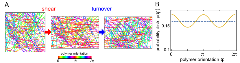
where is the probability distribution of finding a polymer with an orientation of polar angle . We will assume that the network is isotropic in the reference configuration and . Thus, before deformation, . After application of an affine shear deformation to an area element, this orientation distribution will be changed (see Fig. 3A). To illustrate that, consider a small homogeneous shear whose principle axes are, without loss of generality, in x-y-direction. The associated strain tensor reads
| (11) |
In this case, a polymer orientation will on average be realigned by the mapping to first order in , leading to a new probability distribution . In this way, polymer orientations along the x-axis become more likely (see Fig. 3). As a consequence, the active prestress becomes anisotropic. Using Eq. 10, the nematic order tensor is now to first oder in strain . Following active gel theory Prost et al. (2015), we make the ansatz
| (12) |
for active tension in the cytoskeleton, where denotes the difference between the chemical potentials of ATP and its hydrolysis products. If the material is elastic with prestress-dependent shear modulus , the overall traceless stress after a shear deformation is thus
| (13) |
Therefore, in an analysis of the force response that does not compensate for the influence of active prestress, the effective shear modulus would thus be identified as
| (14) |
Since the cytoskeleton turns over through depolymerization and repolymerization processes, induced nematic alignment may be lost over time with the original polymer orientation distribution being reestablished. In the following, we will adapt our above considerations on shear-induced nematic alignment to a viscoelastic material. Adopting the scheme in Reymann et al.Reymann et al. (2016), we will assume that the material turnover takes place on a time scale and that a Maxwell-type dynamics governs the time dependence of the the nematic order tensor such that . In Fourier space, we obtain correspondingly
| (15) |
Thus,
| (16) |
which includes an apparent linear stress-stiffening term in addition to the actual shear modulus. For a homogeneous shear in a three-dimensional material, a corresponding calculation gives
| (17) |
The expected stress-stiffening slope of the storage modulus due to nematic alignment is proportional to . As the parameters and are a priori not known, the magnitude of the associated stress-stiffening slope is not entirely clear. A simple consideration gives an order of magnitude estimate; if polymers of different orientation in the cytoskeletal network do not interact to generate active prestress, active prestress is proportional to the averaged tensor product of filament orientations and we have in 2D ( in 3D). Thus, the associated stress-stiffening slope would be (in 3D: ). It is noteworthy that shear-induced nematic alignment will influence material properties also in the absence of active prestress. However, in this case, effects of nematic alignment and emergent anisotropies on the force response are second order in strain and thus negligible for small deformations Landau and Lifshitz (1986). Only in the presence of active prestress, these effects become first order and non-negligible.
IV Discussion
We have presented theoretical considerations that predict cellular force response to scale linearly with active mechanical prestress even in the absence of actual nonlinear material properties (i.e. if ). The resulting apparent stress-stiffening roots in an increase of measured force amplitudes due to geometrical coupling of active prestress and/or shear-induced nematic alignment of the underlying polymer network during dynamic deformation of the cellular material.
In particular, geometrical coupling may change effective cell moduli in the presence of cytoskeletal contractility (= active prestress) by orders of magnitude depending on the choice of measurement parameters (see Fig. 2). This phenomenon may contribute to the puzzling diversity of cell mechanical moduli reported in the literature Hoffman et al. (2006).
Stress-stiffening through active prestress in cells has been shown before in several experimental studies with mainly linear stiffening trends and slopes between 0.2-45 (see Table 1) Stamenović et al. (2002); Wang et al. (2002); Stamenović et al. (2004); Kollmannsberger et al. (2011); Schlosser et al. (2015); Fischer-Friedrich et al. (2016). An influence of geometrical coupling or shear-induced nematic alignment on previously reported stress-stiffening slopes is conceivable.
| Reference | Method | Approximate stiffening slope | Cells |
|---|---|---|---|
| Wang et al.Wang et al. (2002), Stamenović et al.Stamenović et al. (2002) | magnetic twisting cytometry and traction force microscopy | adhered HASM cells | |
| Stamenović et al.Stamenović et al. (2004) | magnetic twisting cytometry and traction force microscopy | to , for to Hz | adhered HASM cells |
| Kollmansberger et al.Kollmannsberger et al. (2011) | magnetic tweezer | adhered, several cell lines | |
| Fischer-Friedrich et al.Fischer-Friedrich et al. (2016) | AFM cell confinement | 33 and 45 for and Hz | suspended, mitotic HeLa cells |
If the geometrical factor of geometrical coupling is changing in phase with applied strain during the measurement, geometrical coupling gives merely rise to a frequency-independent addition to the effective storage modulus. If the actual storage modulus approaches zero at small frequencies, the active prestress contribution may thus be identified as . If the coupling geometrical factor can be estimated, this offers the chance to measure both the complex elastic modulus and the active prestress jointly from one rheological measurement: To this end, the oscillatory force signal needs to be divided by the time-dependent geometrical factor to obtain a stress signal for rheological analysis (see also Fischer-Friedrich et al. Fischer-Friedrich et al. (2016)). The resulting modulus is then the actual elastic modulus of the material. On the other hand, dividing the baseline force by the time-averaged geometrical factor yields the active prestress. In some ideal measurement setups, geometrical coupling can be entirely avoided (see Fig. 2C). However, such setups are not easily experimentally realised.
Shear-induced nematic alignment generates a frequency-dependent apparent stiffening of actively prestressed material. However, resulting apparent stress-stiffening is independent of the measurement method and thus a true material parameter. Thus, disentangling the active stiffness contribution from the actual passive modulus might not even considered to be necessary. Many measurements report cell rheology which exhibits diverging moduli at high frequencies and short time scales Kollmannsberger and Fabry (2011); Trepat et al. (2008). As active stiffness contributions through shear-induced nematic alignment are bounded by , they are expected to give negligible contributions to an effective elastic modulus at large frequencies.
It is likely that the geometrical stress-stiffening effects described here may also contribute to nonlinear mechanics of cells in the presence of external mechanical prestress. However, exertion of a constant external prestress to viscoelastic cellular material leads in general to i) large deformations (e.g. through a constant strain rate in a rheometer) and ii) large inherent anisotropies of the material. Both effects further complicate a theoretical description considerably. Our results indicate that effective elastic moduli may become entirely determined by active prestress for sufficiently large values of prestress. In the context of external prestress, this could provide an alternative explanation for the recent finding of externally prestressed collagen networks whose shear moduli were found to be collagen concentration independent at sufficiently large mechanical loadings Licup et al. (2015).
In summary, our findings show that geometrical effects may disguise the actual stress-dependence of cellular elastic moduli during cell-mechanical measurements in the presence of active prestress. New approaches in experimental design are necessary to separate apparent stiffening from actual stress-induced stiffening or softening of cells.
Acknowledgements
I thank Benjamin Friedrich, Stefan Münster and Ben Fabry for discussions on the topic and critical reading of the manuscript.
References
- Kollmannsberger and Fabry (2011) Kollmannsberger, P., and B. Fabry, 2011. Linear and Nonlinear Rheology of Living Cells. Ann Rev Mater Res 41:75–97.
- Kollmannsberger and Fabry (2009) Kollmannsberger, P., and B. Fabry, 2009. Active soft glassy rheology of adherent cells. Soft Matter 5:1771–1774.
- Lange et al. (2017) Lange, J. R., C. Metzner, …, B. Fabry, 2017. Unbiased High-Precision Cell Mechanical Measurements with Microconstrictions. Biophys J 112:1472–1480.
- Stricker et al. (2010) Stricker, J., T. Falzone, and M. L. Gardel, 2010. Mechanics of the F-actin cytoskeleton. J Biomech 43:9–14.
- Ingber et al. (2014) Ingber, D. E., N. Wang, and D. Stamenović, 2014. Tensegrity, cellular biophysics, and the mechanics of living systems. Rep Prog Phys 77:046603.
- Wang et al. (2002) Wang, N., I. M. Tolić-Nørrelykke, …, D. Stamenović, 2002. Cell prestress. I. Stiffness and prestress are closely associated in adherent contractile cells. Am J Physiol-Cell Ph 282:C606–C616.
- Stamenović et al. (2004) Stamenović, D., B. Suki, …, J. E. Buy, 2004. Rheology of airway smooth muscle cells is associated with cytoskeletal contractile stress. J Appl Physiol 96:1600–1605.
- Fischer-Friedrich et al. (2016) Fischer-Friedrich, E., Y. Toyoda, …, F. Jülicher, 2016. Rheology of the Active Cell Cortex in Mitosis. Biophys J 111:589–600.
- Fernández et al. (2006) Fernández, P., P. A. Pullarkat, and A. Ott, 2006. A master relation defines the nonlinear viscoelasticity of single fibroblasts. Biophys J 90:3796–3805.
- Jansen et al. (2013) Jansen, K., R. Bacabac, …, G. Koenderink, 2013. Cells Actively Stiffen Fibrin Networks by Generating Contractile Stress. Biophys J 105:2240–2251.
- Chan et al. (2015) Chan, C. J., A. E. Ekpenyong, …, F. Lautenschl ger, 2015. Myosin II activity softens cells in suspension. Biophys J 108:1856–1869.
- Mizuno et al. (2007) Mizuno, D., C. Tardin, …, F. C. MacKintosh, 2007. Nonequilibrium Mechanics of Active Cytoskeletal Networks. Science 315:370–373.
- Bendix et al. (2008) Bendix, P. M., G. H. Koenderink, …, D. A. Weitz, 2008. A Quantitative Analysis of Contractility in Active Cytoskeletal Protein Networks. Biophys J 94:3126–3136.
- Koenderink et al. (2009) Koenderink, G. H., Z. Dogic, …, D. A. Weitz, 2009. An active biopolymer network controlled by molecular motors. PNAS 106:15192–15197.
- Bustamante et al. (1994) Bustamante, C., J. F. Marko, …, S. Smith, 1994. Entropic elasticity of lambda-phage DNA. Science 265:1599–1600.
- Gardel et al. (2004) Gardel, M. L., J. H. Shin, …, D. A. Weitz, 2004. Elastic Behavior of Cross-Linked and Bundled Actin Networks. Science 304:1301–1305.
- Storm et al. (2005) Storm, C., J. J. Pastore, …, P. A. Janmey, 2005. Nonlinear elasticity in biological gels. Nature 435:191–194.
- Broedersz and MacKintosh (2011) Broedersz, C. P., and F. C. MacKintosh, 2011. Molecular motors stiffen non-affine semiflexible polymer networks. Soft Matter 7:3186.
- Yao et al. (2013) Yao, N. Y., C. P. Broedersz, …, D. A. Weitz, 2013. Stress-Enhanced Gelation: A Dynamic Nonlinearity of Elasticity. Phys Rev Lett 110:018103.
- Sheinman et al. (2012) Sheinman, M., C. P. Broedersz, and F. C. MacKintosh, 2012. Actively Stressed Marginal Networks. Phys Rev Lett 109:238101.
- Licup et al. (2015) Licup, A. J., S. Münster, …, F. C. MacKintosh, 2015. Stress controls the mechanics of collagen networks. PNAS 112:9573–9578.
- Volokh et al. (2000) Volokh, K. Y., O. Vilnay, and M. Belsky, 2000. Tensegrity architecture explains linear stiffening and predicts softening of living cells. J Biomech 33:1543–1549.
- Hochmuth (2000) Hochmuth, R. M., 2000. Micropipette aspiration of living cells. Journal of Biomechanics 33:15–22.
- Fischer-Friedrich et al. (2014) Fischer-Friedrich, E., A. A. Hyman, …, J. Helenius, 2014. Quantification of surface tension and internal pressure generated by single mitotic cells. Sci Rep 4:6213.
- Stamenović et al. (2002) Stamenović, D., Z. Liang, …, N. Wang, 2002. Effect of the cytoskeletal prestress on the mechanical impedance of cultured airway smooth muscle cells. J Appl Physiol 92:1443–1450.
- Reymann et al. (2016) Reymann, A.-C., F. Staniscia, …, S. W. Grill, 2016. Cortical flow aligns actin filaments to form a furrow. eLife 5:e17807.
- Prost et al. (2015) Prost, J., F. Jülicher, and J.-F. Joanny, 2015. Active gel physics. Nat Phys 11:111–117.
- Landau and Lifshitz (1986) Landau, L., and E. Lifshitz, 1986. Theory of Elasticity. Elsevier Ltd., 3rd edition.
- Hoffman et al. (2006) Hoffman, B. D., G. Massiera, …, J. C. Crocker, 2006. The consensus mechanics of cultured mammalian cells. PNAS 103:10259–10264.
- Kollmannsberger et al. (2011) Kollmannsberger, P., C. T. Mierke, and B. Fabry, 2011. Nonlinear viscoelasticity of adherent cells is controlled by cytoskeletal tension. Soft Matter 7:3127.
- Schlosser et al. (2015) Schlosser, F., F. Rehfeldt, and C. F. Schmidt, 2015. Force fluctuations in three-dimensional suspended fibroblasts. Phil Trans R Soc B 370:20140028.
- Trepat et al. (2008) Trepat, X., G. Lenormand, and J. J. Fredberg, 2008. Universality in cell mechanics. Soft Matter 4:1750.
Supporting Material
Geometrical coupling with phase-shifted, frequency-dependent active force contributions
In the first and second example in the main text, geometrical coupling of active prestress has contributed only a frequency-independent addition to the measured storage modulus of the system. In the following, we will present an example where geometrical coupling gives rise to a complex-valued, frequency-dependent addition to the effective elastic modulus of the system for the case of a deflected prestressed fibre with a viscoelastic connector between the bead and the fiber (see Fig. 1B, main text). The bead is deflected in a vertical manner. The combined system has a (complex) spring constant
where and are the effective (complex) spring constants of the connector and the fibre with respect to vertical deflection. One finds
where equals the r.h.s of formula (6), main text. Thus, in the stiffness of the combined system , the active term in contributes in general also to the imaginary part of the system and is frequency-dependent.
Determining the geometrical factor for the case of a deflected prestressed cytoskeletal fibre
In the main text, we where discussing the example of geometrical coupling of active stress in a prestressed cytoskeletal fibre tethered at its end points at a distance (see Fig. 1A, main text). Height oscillations are imposed on the fibre center with . Here, we derive the factor of geometrical coupling for this specific example.
We make a perturbation calculation determining the fibre oscillation dynamics up to first order in height amplitude . We make the following expansions of the dynamic fibre length and the dynamic angle between the substrate and the fibre
Using the geometrical relations and , we obtain
| (18) |
We can thus calculate the time variation of the geometrical factor to first order
where we have used Eqn. (18) in the last transformation step. We conclude that and . The strain amplitude of the fibre is . We thus obtain the coupling geometrical factor as