Acoustically driven x-ray emission and matter collapse in lead
Abstract
The action of focused underwater weak shock waves on a lead sample was revealed to be not restricted by a mechanical influence only. A strong unexpected x-ray emission was registered from the lead foil exposed to those shock waves (sound into x-rays) which were extremely adiabatic compared to processes of x-ray generation. The lead foil, exposed to shock waves, lost a part of its area having the shape of a polygonal hole of the size of . The missing polygon of lead foil looks as a delicately removed part with no damage at the hole surroundings as it should be after a mechanical breaking. This points to a non-mechanical mechanism of hole formation. That missing polygonal lead matter seems to be “disappeared” because the total lead volume was reduced by that amount after exposure to acoustic waves (matter collapse). Both paradoxical phenomena cannot be explained by a combination of known effects and a fundamentally new mechanism is required to underlie them. The concept of electron anomalous states, which encouraged the experiments and specified main features of them, is likely that mechanism.
pacs:
01.55.+b, 43.90.+v, 41.50.+hI INTRODUCTION
External perturbations of matter may result in x-ray radiation. It can occur at braking of moving electrons (Bremsstrahlung) BET ; HAU ; LANDAU ; CHE and also under electron transitions to lower energy levels in atoms (characteristic radiation) BON ; SHA ; VAN . Known x-ray emission phenomena MAT ; DUN ; HUA ; PUTT ; HOR result from those basic ones.
Acoustic action on matter can be strong. Shock waves in liquids result in cavitation COL ; VOG ; LAU ; PEC ; BEN . Ultrasonically driven bubbles in liquids may emit bursts of light when they collapse (sonoluminescence) PEC1 ; HIL ; PUT1 ; PUT2 ; BRE . Shock waves in a soft tissue can provide a strong mechanical action LOSKE . In solids acoustic energy may be converted into visible light OST or infrared radiation AHN (acoustoluminescence).
X-ray emission can occur under action on solids of extremely strong shock waves, with the pressure of GIZ . In this case external mechanical forces on solids are comparable with the atomic ones. The intensity of those shock waves is four orders of magnitude larger than in the experiments reported here (weak shock waves). Furthermore, x-ray emission from radiative shock waves in astrophysics can occur due to heating of a matter up to high temperature NYM . In our experiments weak shock waves result in the negligible increase of temperature, .
In this article we study completely different phenomena compared to the ones mentioned above. Acoustic pulses, propagating through lead, resulted in x-ray emission. This phenomenon may seem to be just one more on the list of various types of x-ray emission. This impression is not correct, since the x-ray emission revealed here cannot be explained by a combination of known effects. In addition, it is also accompanied by a paradoxical matter collapse which has not been observed before. It turns out that a fundamentally new mechanism has to underlie those two phenomena. It is likely that the concept of electron anomalous states IVLEV , which encouraged the experiments and specified main features of them, is this mechanism. Here we only introduce the discovered phenomena. A detailed study of them is out of the scope of this article.
We exposed a “sandwich” (see details in Sec. II), containing a lead foil, to underwater shock waves. The passage of the acoustic pulse through a particular point in the lead occurs during approximately . A perturbation with this characteristic time cannot excite electrons up to the energy scale, corresponding to the frequency . The formal probability of one quantum excitation up to the energy is of the type IVLEV1 ; IVLEV2 . In a multiquanta absorption, quanta should be involved resulting in the probability of the type above IVLEV1 ; IVLEV2 . Therefore, a characteristic x-ray emission in the region is impossible. Also there are no conditions in lead for Bremsstrahlung of that energy since conduction electrons adiabatically follow the acoustic wave acquiring its velocity .
Slow varying acoustic perturbation, in principle, can produce a spatial charge separation resulting in an electric field locally increasing an electron energy with a possibility of a subsequent quanta emission. For example, such mechanism resulted in acoustoluminescence in a semiconductor due to a lattice defect separation OST . But in the bulk lead fast electron processes (keeping a fixed Fermi level) with a characteristic time ABR equalize any acoustically caused charge separation. The only possibility for a charge separation is a lead - dielectric border in the sandwich used in our experiments. One can suppose a deformation of surfaces of those solids when they locally loose contact forming a cavity with opposite charges on separated surfaces. This would remind the charge separation at peeling of an adhesive tape when the separated charges resulted in glow discharge and x-ray Bremsstrahlung PUTT . But in our experiment such discharge avalanche cannot be formed due to air which is unavoidable between the two surfaces. The hypothetical cavity would be filled out by air at atmospheric pressure. In this case an accelerating electron would be braked by air molecules which is not sufficient for x-ray Bremsstrahlung.
One can conclude that a conversion of the acoustic energy into x-rays by known mechanisms is impossible. However, we observed a strong x-ray emission when shock waves come from water and reflect off a sample containing the lead foil (sound into x-rays). The energy of x-ray quanta is estimated to be in the range. The source of the observed x-rays was not in the water (including cavitation bubble dynamics), since it was screened by a relatively thick copper film. As argued in this paper, the source was the lead.
The other experimental observation is also unusual. Under shock wave exposure a small area of the lead foil (of size and of polygonal shape) “disappears” because the total lead volume was reduced by that amount after exposure to acoustic waves (matter collapse). Moreover, the missing polygon of lead foil looked as a delicately removed part with no damage at the hole surrounding as it should at a mechanical breaking. This points to a non-mechanical mechanism of hole formation. A resemblance between two phenomena is manifested in our experiments.
II MATERIALS AND METHODS
Shock waves, as an acoustic type of matter perturbation, are widely used in various fields LOSKE . These waves consist of a narrow front and a relatively long release wave BEN . Usual applications of shock waves relate to their mechanical actions on matter BEI ; PHI ; ILO ; KLA . Shock waves in water can result in cavitation arising from nucleation sites and microbubbles PHI ; ILO ; KLA . During an individual bubble collapse, high-speed microjets of water may be generated LAU . These microjets have sufficient energy to puncture thin plastic or metal film after a multiple action.
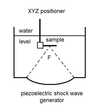
The main element of the setup used was a Piezolith 2501-based shock wave source (Richard Wolf GmbH, Knittlingen, Germany). It consists of approximately 3000 small piezoelectric elements, arranged on a bowl-shaped (radius ) aluminum backing (Fig. 1) with an aperture of . The electric circuit basically consists of capacitor charging unit and a discharge control system. The piezoelectric elements are insulated from water by a flexible membrane. A high voltage discharge across the array of piezoelectric elements generates an abrupt expansion of all elements, producing pressure waves that travel towards the center of the device. The superposition of propagating pressure waves, due to a hydrodynamic nonlinear distortion, produces a shock wave in the vicinity of the focal region (see Fig. 1). In this region, in the shape of a cigar measuring about , aligned along the axis of symmetry, the positive pressure pulse has more
an of the maximum peak positive amplitude.
All samples were exposed to pressure waves generated at a voltage between 2.5 and and a discharge rate of . At this voltage, the pressure waveform, measured at with a PVDF needle hydrophone (Imotec GmbH, Würselen, Germany), had a peak positive amplitude at the pressure interval . Pressure values are specified in Figs. 3 - 11. The temperature inside the water tank was approximately . Special care was taken to avoid air gaps or bubbles at the water-sample interface or between layers inside the sample. The shock wave velocity in water is connected to the pressure in the front by the relation COL ; PEC
| (1) |
where is the sound velocity in water at .
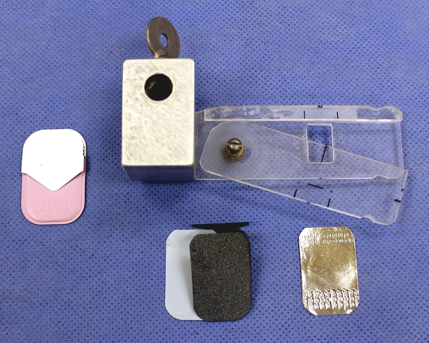
As a sample in Fig. 1 a “sandwich”, based on Carestream Dental-Insight intraoral x-ray film () and manufactured by Carestream Health Inc. (Rochester NY, USA), was used. Each individual film was packaged in a light-tight plastic (vinyl) envelope to protect the film from light. The front side is supposed to face the x-ray tube; the back side has an opening tab to open the film during processing. A black paper wrapper ( thick) protects the film base (sheet of cellulose acetate of thickness) from light and damage. The x-ray sensitive emulsion faces towards the front side of the envelope. A lead foil ( thick) is on the back side.
Each plastic envelope, fixed with the specially designed Lucite holder shown in Fig. 2, was fastened horizontally and centered at the focus . The x-ray sensitive emulsion was facing towards the shock waves coming from the water. Waves propagated through the outer plastic cover, the front black paper, the x-ray film, the black paper, the lead foil, and the back plastic cover. The water level was set above . A small strip of water resistant duck tape was used to seal the opening tab of each x-ray film envelope.
Shock waves, created near the focus were not the only mechanical effect on the sample. Another mechanical effect was due to cavitation in the focal region. This occurs because the water contains nucleation sites and microbubbles PHI ; ILO ; KLA . Cavitation produces secondary shock waves which also impact the sample. In addition, shock wave-induced microjets of water are generated during individual or multiple bubble collapses LAU . These microjets have sufficient energy to puncture thin plastic or metal film after a multiple action.
III SOUND INTO X-RAYS
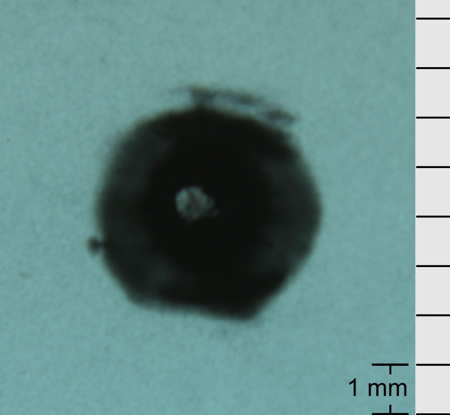
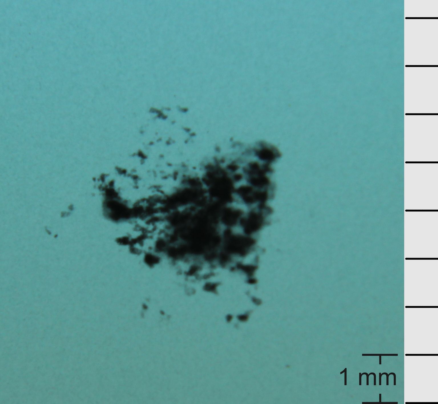
Various types of sandwiches (samples in Fig. 1) were exposed to shock waves in water. We have started the research obtaining the image shown in Fig. 3, where the sandwich used contained a thick cooper foil.
The exposed spot on the x-ray film in Fig. 3 could be interpreted as produced by x-rays emitted from the focal region in the water. The diameter of that region () coincides with the spot size; however, in that case x-rays had to be of at least to overcome the attenuation in the copper foil. This finding also looks strange since the spot in Fig. 3 has the obvious tendency for polygonal formation but the focal region does not have such shape. The definite exclusion of that scenario followed from the absence of an exposed spot (a clean x-ray film after development) after removing the lead foil from the sandwich keeping the rest as it was.
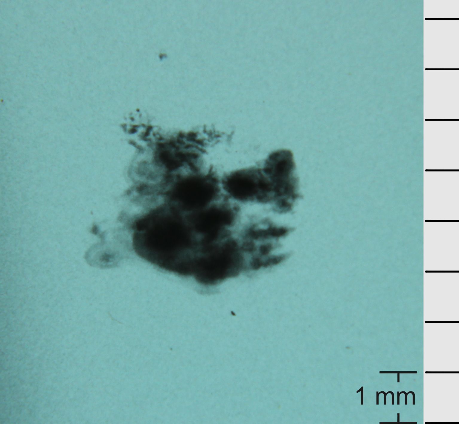
The presence of the copper layer in the sandwich, described in Fig. 3, was not crucial for the phenomenon. In the sandwich, described in Fig. 4, the copper film was substituted by a layer of plastic but the x-ray film was still exposed.
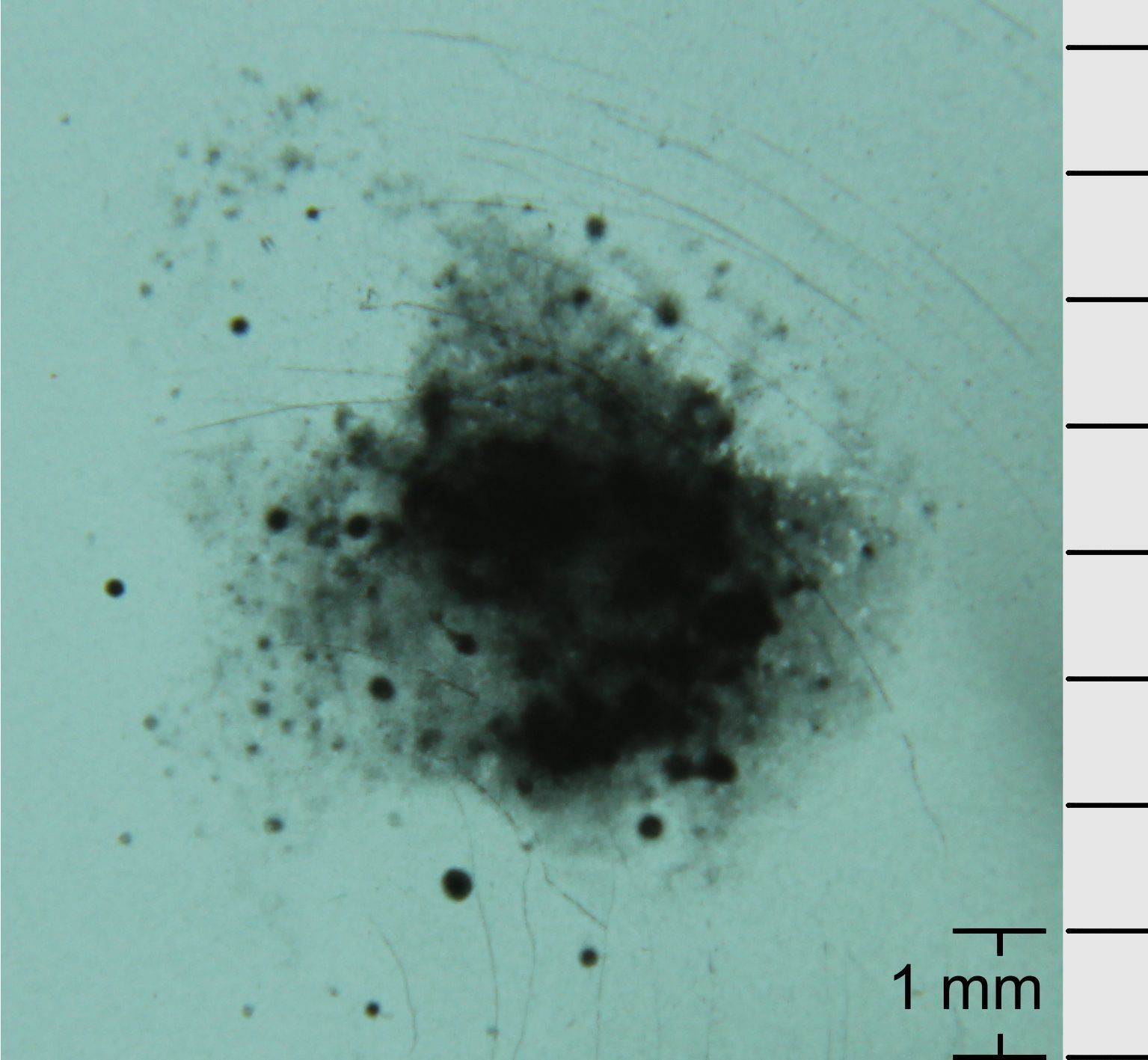
Visible light did not contribute to film exposure. This was proved by using light protecting black papers in the sandwich described in Fig. 5.
In the sandwich described in Fig. 6, the light protected x-ray film was placed behind the lead foil. This arrangement opened the possibility to study a propagation of the emitted quanta in space and to measure their spectrum.
So the source of x-rays, detected as shown in Figs. 3 - 6, was the shock wave-exposed lead foil. The underwater shock wave consists of a narrow wave front having a width and a release wave, , following the front (see Ref. LOSKE and references therein). The shortest characteristic time of acoustic perturbations in lead is determined by the duration of the shock wave front in water. is approximately the ratio of the width of the shock wave front and the velocity of the shock wave
| (2) |
We did not measure the energy of emitted x-ray quanta precisely. However, we know that the x-ray emission reduces approximately twice after penetration through the black paper which is the usual light protection of the x-ray films used. It was also demonstrated that the emitted x-rays hardly penetrate through the wet black paper. Therefore, one can roughly estimate the energy of emitted x-ray quanta to be in region. This corresponds to the quanta frequency .
In Fig. 6 the total time of exposure to x-rays was . This produces the same darkness of the spot as an x-ray ramp from a dental x-ray tube with a duration. So the estimated x-ray flux, during the acoustic pulse action, was approximately times higher than a flux from a dental x-ray tube.
Which is the mechanism producing the observed x-ray emission from lead (sound into x-rays)? The perturbation with the characteristic time (2) cannot excite electrons up to the energy scale, corresponding to the frequency . The formal probability of one quantum excitation up to the energy is of the type IVLEV1 ; IVLEV2 . In the multiquanta absorption, quanta should be absorbed to increase an electron energy up to the scale IVLEV1 ; IVLEV2 . But in our experiment the acoustic perturbation of the lead is not very strong (almost elastic regime) since it relates to a variation of approximately of the lead density at the position of the acoustic peak.
Therefore, the -th order of a perturbation theory, a product of small values, corresponds to the probability mentioned above IVLEV1 ; IVLEV2 . Therefore a characteristic x-ray emission in the region is impossible. Also there are no conditions in lead for Bremsstrahlung of that energy since conduction electrons adiabatically follow the acoustic wave acquiring its velocity .
As discussed in Sec. I, a possible macroscopic effect (a charge separation resulting in local electric fields) of the slow acoustic pulses cannot lead to x-rays. Therefore the observed x-ray emission from shock wave exposed lead is paradoxical since it contradicts the known mechanisms.
IV MATTER COLLAPSE
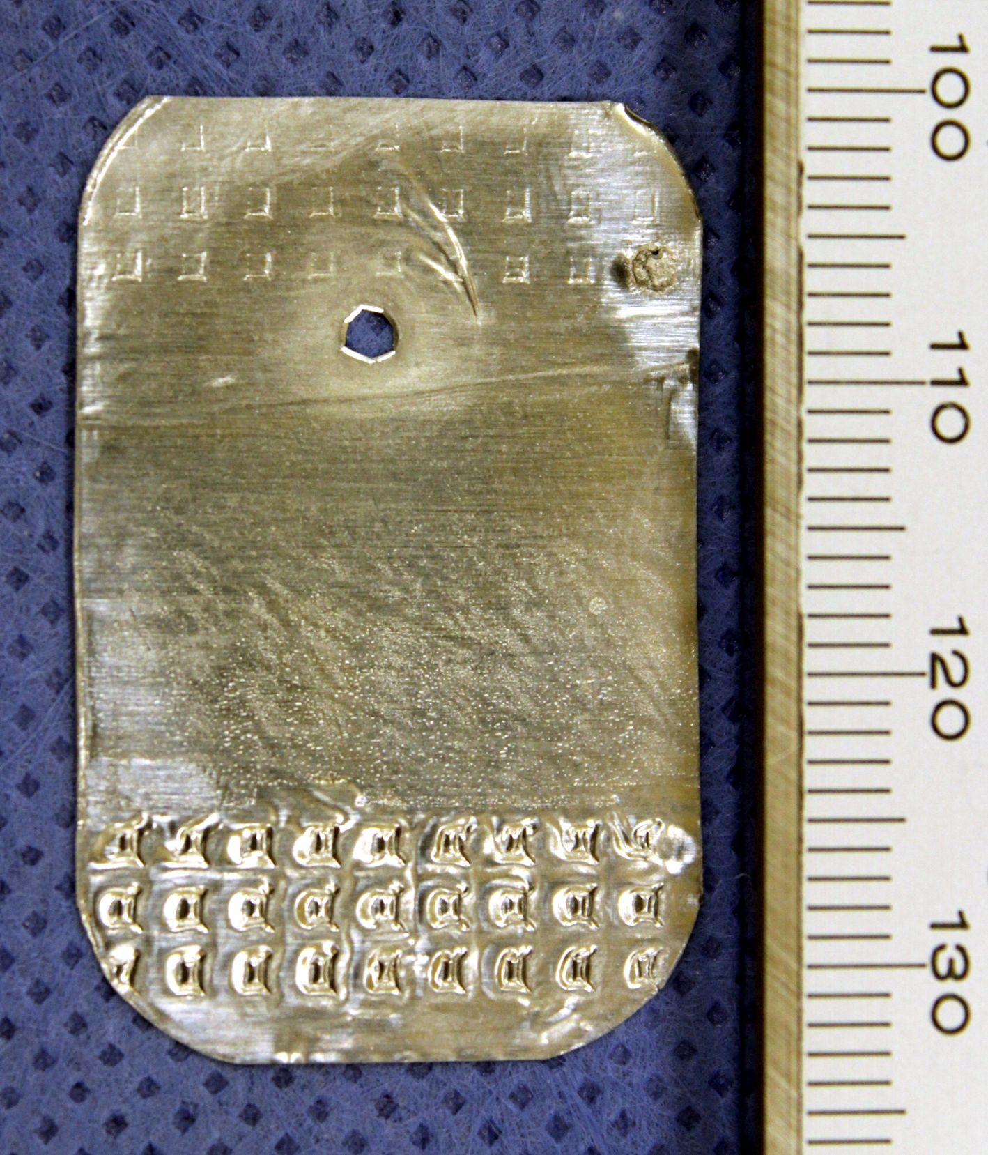
In our experiments we also studied another effect of shock waves, which reflect from a surface of the sandwich containing the lead foil of thickness. Each shock wave generates an acoustic pulse propagating through the metal. At the energy used in our study, the lead foil could not be mechanically perforated by one shock wave. One shock wave only produced a residual plastic deformation of the foil which got slightly bent in the direction (the axis) of acoustic propagation. Further repeated shock waves lead to an accumulation of those deformations. After a
critical number (on the order of hundred) of shock waves the foil became perforated. This was accompanied by a strong deformation of the foil area surrounding the perforated region as in Fig. 7. In this case the foil was in contact with water.
In Figs. 8 - 11 the lead foil was hermetically sealed inside the factory plastic envelope (Fig. 2) and was not directly exposed to underwater shock waves. However, those shocks, that weakly reflected off the water-plastic interface, also resulted in acoustic pulses in the metal.
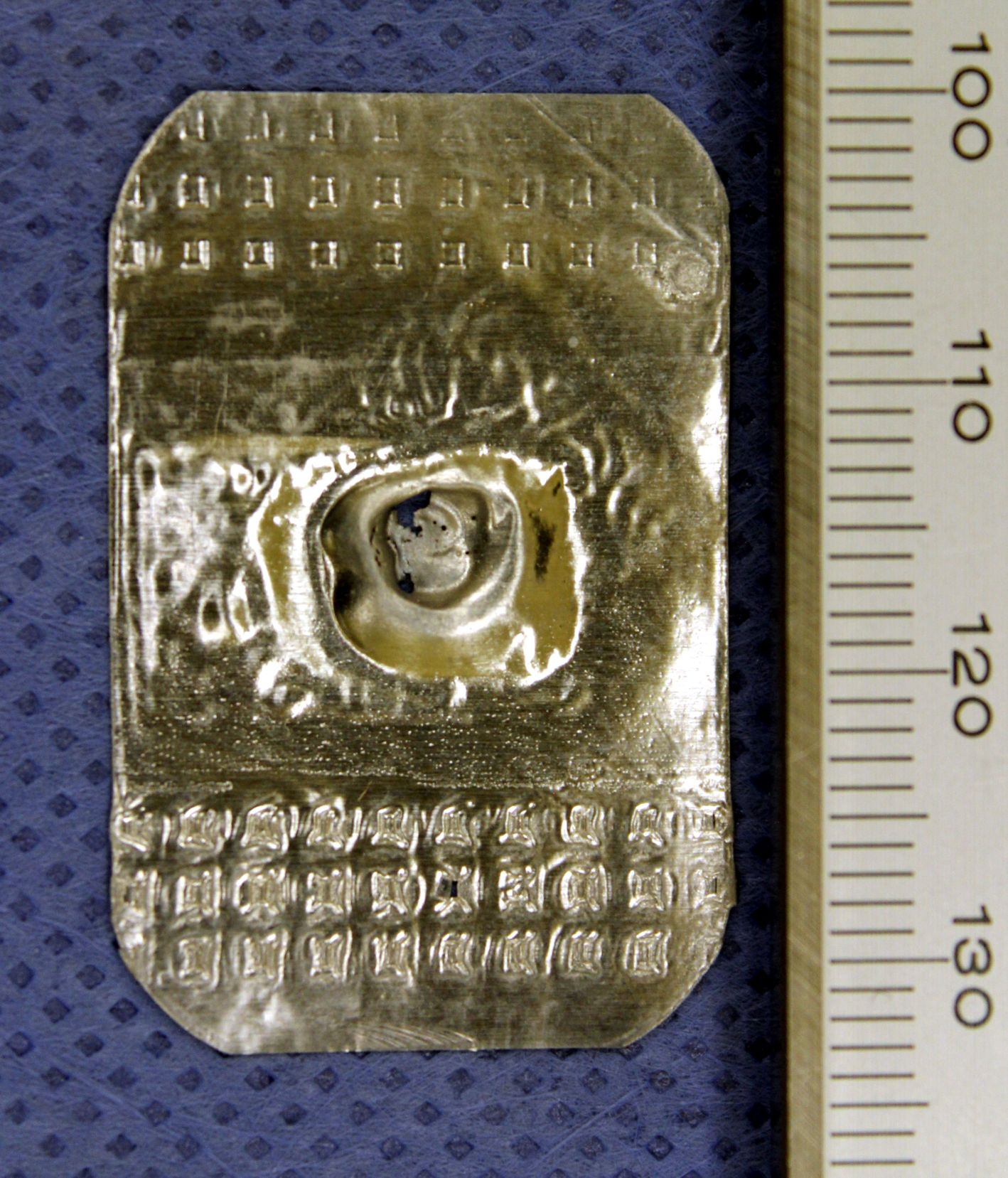
Whereas the foil damages in Fig. 7 are due to mechanical actions of shock waves the damages in Figs. 8 - 11 are substantially different. The scenario of pure mechanical creation of those holes by acoustic pulses would look straightforward. In this case, after accumulation of small deformations, the foil in the center would become thinner and finally get perforated. The lead matter, that initially filled out the hole, would be distributed around it. By the action of the contacting plastic film a rugosity of the foil would be created near the hole.
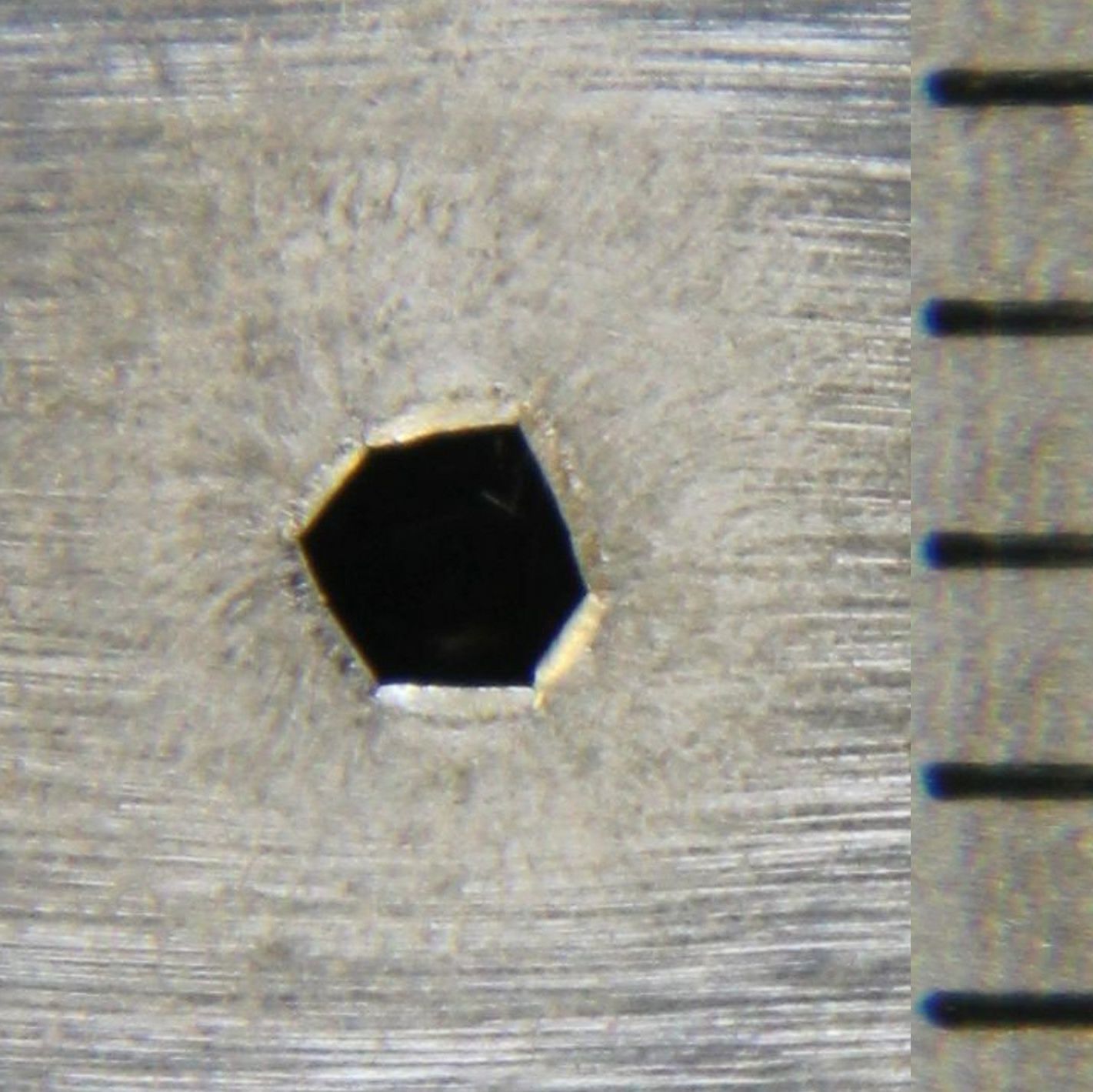
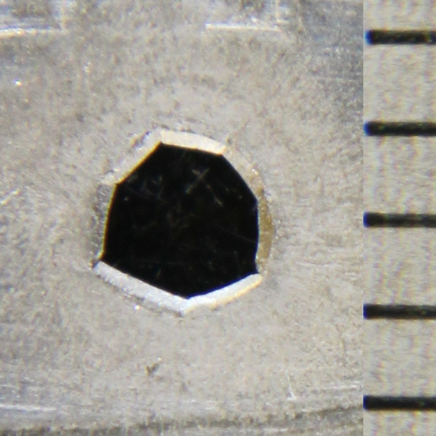
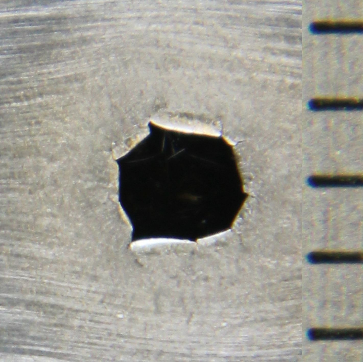
The absence of a rugosity around the holes in Figs. 8 - 11 contradicts the scenario of pure mechanical breaking. The microscope magnifications of the hole, shown in Figs. 12 and 14, are incompatible with a usual mechanical perforation. In Fig. 13 the cross-section of the foil at the hole shows that the foil was bent towards the
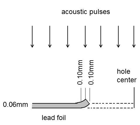
direction of the incoming acoustic pulses. This also stands against a mechanical hole formation. Fig. 13 is schematically based on confocal microscope analysis (not detailed here). According to it, of the hole interior is collected on the hole border. This is also visible in Figs. 12 and 14. Therefore of the hole interior was somehow transferred away from the hole.
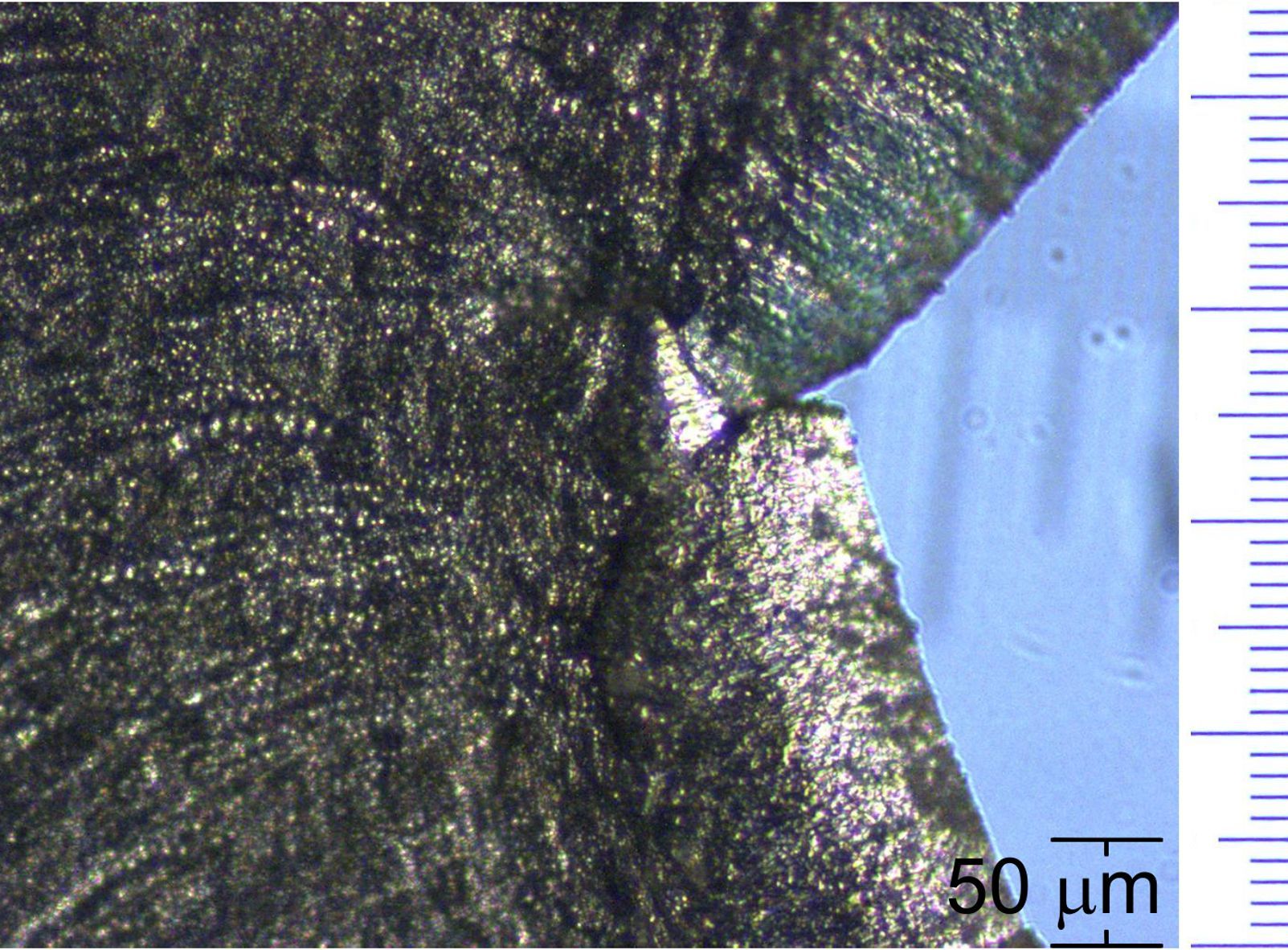
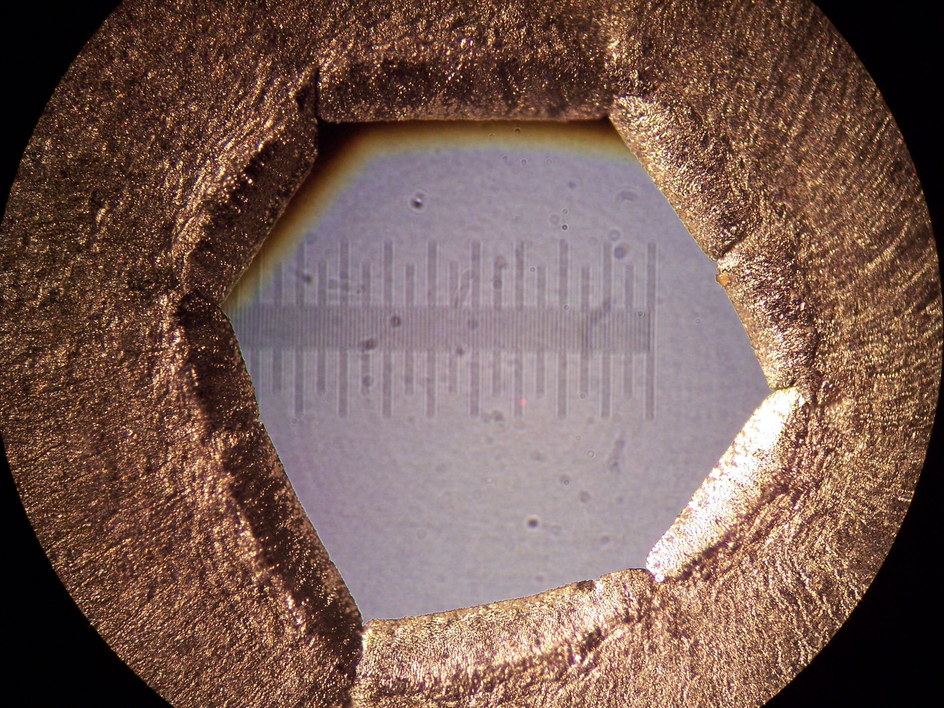
All holes, in Figs. 8 - 11, look as if the missing lead matter would have been delicately removed with no disturbance of the surroundings. Those holes have a polygonal shape. The number of sides of the polygon increases from Fig. 9 (hexagon) to Fig. 11 (octagon) under the enhancement of the total acoustic flux to the foil. As follows from Figs. 12 and 14, micron-sized lead particles are formed on the lead foil surface facing the acoustic pulses. The particles are better seen on the microscope magnification of the part of the hole border shown in Fig. 14. Those particles are absent on the back side (exit side of the acoustic pulses) of the lead foil.
There was another exciting phenomenon. The lead matter, looking as delicately removed from the holes, “disappeared”. This corresponds to the aforementioned of the hole interior. It was not found in the envelope as lost macroscopic fragments. The plastic surfaces, which were in contact with the lead, looked as before shock wave action, i.e., they were not damaged, appeared to be clean, and without traces of lead. Fig. 10 corresponds to “disappearance” of of lead. Lead melting is impossible since a conversion of the entire acoustic energy into heat would increase the temperature of the missing lead part by only.
A conversion of the missing lead matter into the micron-sized particles, shown in Figs. 12 and 14, seems impossible since their total volume is approximately of the “disappeared” hole interior.
One can suppose that the missing of the hole interior was transferred by secondary acoustic pulses propagating along the lead foil away from the hole. In principle, such a pulse can be generated by the incident acoustic pulse, propagating across the lead foil, which has the duration (2). So the hypothetical secondary pulse would have the spatial extension along the lead foil. It is easy to evaluate that each of 250 secondary pulses (referred to the hole in Fig. 9) would carry away from the hole ten times increased lead density localized within . Therefore, the hypothetical secondary pulse would be a strong shock wave propagating along the lead foil. But there is no source for such grandiose effect since the incident pulse, propagating across the lead foil, is weaker than a shock wave (Sec. V). So secondary pulses cannot be generated and therefore do not transfer lead matter away from the hole.
The above phenomenon can be qualified as matter collapse, i.e., a non-conventional matter redistribution. Into what does the missing lead matter go over?
V THE MECHANISM
The velocity of the shock waves in water in the focal plane in Fig. 1 is approximately . It slightly exceeds the speed of sound in water . In the macroscopic description there is a jump of velocity at the shock wave front. In reality this jump is smeared out over approximately . Therefore the characteristic time, related to the shock wave front propagation, is given by Eq. (2).
The metal foil was placed at the focal region of the shock wave source with the size of a few millimeters. Within this area one can consider a plane shock wave which collides the foil normally (in the direction). We are interested on short time processes occurring during the front wave time (2). The density of the metal is larger than the density of water. Therefore the shock front in water reflected almost elastically from the metal border. In this process the momentum per unit area of the water-metal interface was transferred to the metal. See also BEN ; BEI . This momentum, acquired by the metal, leads to the acoustic pulse propagating from its surface. Here is the speed of sound in the metal and is the positive longitudinal displacement localized on . It results in the local variable density in the metal, where is the equilibrium density. The local velocity in the metal is .
Below we estimate the balance of momentum transfer per unit area of the water-metal interface. It can be written as
| (3) |
where and are the lengths of the pulse (along the axis) in metal and water, respectively. Angular brackets mean an average on . Since the average of is zero, one should account the square of this term in the left-hand side of (3). The maximal velocity in the pulse, propagating in the metal, is estimated as
| (4) |
For lead , , and therefore the maximal value of can be estimated as 0.49. This estimate is approximate since we use the limit of small . However it shows that and the pulse, propagating through the lead, is not a shock wave but rather a strong acoustic perturbation.
The two paradoxical phenomena, the x-ray emission and matter collapse, are expected to be consequences of a certain mechanism which underlies both. To understand the basis of these intriguing phenomena let us consider the concept of anomalous states developed in Ref. IVLEV .
In condensed matter, certain macroscopic perturbations can form anomalous electron wells due to a local reduction of zero point electromagnetic energy. These wells are narrow, on the order of the Lamb radius IVLEV , and are localized around an atomic nucleus which is of size. The well depth is . The electron spectrum in the well is continuous and non-decaying IVLEV . The latter is true when the well with electrons is at rest. In a condensed matter, under thermal vibrations resulting in Bremsstrahlung of the captured electrons, states in the anomalous well acquire a small but finite width. That well with captured electrons may be treated as an anomalous atom. The wells can be created by some perturbation which rapidly varies in space, on the scale .
In condensed matter such perturbations may relate to acoustic pulses. In this process the short scale is the length of the standing de Broglie wave of reflected lattice atoms resulting in a spatial variation of charge density. In the acoustic pulse, propagating through the metal, the lattice site acquires the velocity (4). When the pulse continues its motion, the site returns to its initial position with the velocity due to a reflection from other sites. In this process the quantum interference of forth and back motions results in the modulation of the charge density on the scale
| (5) |
where is the mass of the lattice site. For lead and the above estimates give . Therefore the perturbation of lead by the acoustic pulses is effective for creation of anomalous electron states.
The quantum coherence of the incident and reflected de Broglie waves can exist if it is not destroyed by thermal fluctuations. For this reason, the velocity of the macroscopic motion of lattice sites should exceed their velocity of thermal motion. For lead at room temperature () the velocity is approximately .
One should note that in copper the above mentioned acoustic pulses will hardly create anomalous states. The copper density and the mass of the copper atom correspond to which is a too long spatial perturbation compared to . So, heavy metals are preferable for formation of anomalous states. Note that for copper at room temperature () the velocity is approximately .
According to the Thomas-Fermi approach LANDAU1 , in heavy atoms the energy to remove all electron from a neutral atom is . For lead (atomic number ) . Since the Thomas-Fermi method is approximate, in reality one can estimate the ground state energy per electron in lead as minus a few .
After the fast (within the time ) formation of the anomalous well the initial atomic state, in the region, is “unable” to immediately vary and remains the same as before. But now this state is non-stationary corresponding to an electron flow toward the anomalous well to form the anomalous atom. A typical time of this process is . Faster processes are less effective due to strong spatial oscillations (compared to the initial wave function) of new states localized in the anomalous well. This is similar to smallness of a matrix element when one wave function rapidly oscillates. So, according to quantum mechanical uncertainty, the formation of the anomalous well is accompanied by quanta emission in the region. Further quanta emission from the formed well is weak since the states in the well are almost non-decaying. An intensive high energy () emission can be induced by some external sources.
One can see that the deep (of the range) anomalous well, from where the x-ray emission occurs, is formed by adiabatic acoustic perturbations with the typical time . This resolves the paradoxical observation of x-ray emission under the slow varying driving force (Sec. III).
There is another unusual aspect of anomalous atoms. The size of this atom () is three orders of magnitude smaller than the conventional one. If in a part of a solid all atoms undergo the transition to the anomalous state that macroscopic region reduces times in volume. This process can be qualified as matter collapse. The collapsed matter (with times enhancement of density) looks as a dramatically different concept IVLEV .
In our experiments the missing macroscopic part of lead, initially filling the areas of the polygonal holes in Figs. 8 - 11, did not disappear but could be converted into collapsed matter of micron size. Such “speck of dust”, invisible even with a magnifying lens, imitates matter “disappearance”. Another possibility is that the observed micron-sized particles on the lead surface are collapsed (or partly collapsed) matter. Further research is required to establish which scenario is realized. Anyway, it would be extremely unusual to mechanically collect after an experiment that heavy matter artificially created. That heavy matter, up to times enhancement in density, is unknown to be found in nature.
The paradoxical x-ray emission and the lead matter “disappearance” cannot be explained by a combination of known effects. A different concept is required which underlies both intriguing phenomena. The concept of anomalous states satisfies this condition. It explains the common mechanism of both phenomena and also why they are observed in lead but not in copper.
VI DISCUSSION
The main purpose of this paper is to introduce the unexpected paradoxical phenomena providing convincing experimental arguments for their existence. Further quantitative studies, for example spectrum measurements of the emitted x-rays, are to be done. The second purpose is to link those paradoxical phenomena to the mechanism likely responsible for them.
As known, usual applications of shock waves relate to their mechanical actions on matter. We report a completely different type of a shock wave action. Two intriguing phenomena were observed.
1. Sound into x-rays. A strong x-ray emission from the lead under the action of acoustic pulses which are extremely adiabatic, times slower, compared to fast processes of x-ray generation was observed. The estimated x-ray flux, during acoustic pulse action, was approximately times higher than the flux from a dental x-ray tube. Our phenomenon strongly differs from ones when x-ray emission occurs due to known mechanisms initiated by extremely strong shock waves (artificial explosions, processes in supernovae in astrophysics, etc.).
2. Matter collapse. “Disappearance” of a macroscopic part of lead matter exposed to acoustic pulses was noticed. The “disappeared” matter has the shape of a polygonal hole in the lead foil with non-damaged surroundings of the hole. The polygonal shape of spots and holes confirms a resemblance between two phenomena. A direct mechanical effect of the acoustic pulses, as a cause of that hole formation, is excluded. In that case, the acoustic pulses would strongly damage the hole surroundings as was observed in previous experiments.
The two above paradoxical phenomena were discovered in table-top experiments and remind, at first sight, routine acoustic studies. This impression is not correct. Both phenomena cannot be explained by a combination of known effects. It turns out that a fundamentally new mechanism has to underlie them. The concept of electron anomalous states, which encouraged the experiments and specified main features of them, likely is this mechanism.
It relates to the formation in condensed matter of anomalous electron wells, because of a local reduction of zero point electromagnetic energy. The wells are narrow, , and deep, on the order of a few . The well formation is due to short scale () modulations of charge density caused by the interference of de Broglie waves of lattice sites. This interference occurs, on that short scale, between the incident and reflected lattice site participating in shock wave motion with the velocity . The thermal motion of lattice sites, with the velocity , cannot destroy that interference since in lead is substantially smaller than .
Atomic electrons are collected inside the anomalous well forming an anomalous atom and resulting in two effects. First, they emit x-ray quanta by transitions to lower levels. Second, the anomalous atom is three orders of magnitude smaller than a usual one. The lead matter, that initially filled the hole region, transformed into anomalous state reducing its macroscopic volume times. In other words, the macroscopic volume is converted into a heavy particle of micron size. This “speck of dust”, invisible even with a magnifying lens, imitates matter “disappearance”. The origin of micron size lead particles on the lead surface, facing acoustic pulses, may be of the above mentioned nature but this needs further studies.
The theory of anomalous states IVLEV explains (i) the paradoxical x-ray emission caused by extremely adiabatic perturbations, (ii) paradoxical matter collapse, and (iii) the lack of effect in copper in contrast to lead. A detailed study of the phenomena discussed is not the goal of this paper.
Furthermore, there is another aspect related to quanta emission from anomalous matter. All electrons of the single anomalous lead atom, in principle, can emit approximately by transitions
to lower states IVLEV . These processes do not occur automatically since anomalous states are weakly decaying. One can ask the question: what kind of external perturbation triggers off that
avalanche releasing per atom? In this case of lead would release originating from a reduction of zero point electromagnetic energy IVLEV .
VII CONCLUSIONS
Acoustic pulses, propagating through lead, unexpectedly resulted in strong x-ray emission (sound into x-rays). Those pulses are extremely adiabatic compared to atomic processes of x-ray generation which have the formal probability . Bremsstrahlung mechanisms are excluded.
The lead foil, exposed to acoustic pules, misses a part of its area in the shape of a polygonal hole of the size of (matter collapse). The missing polygon of the lead foil looks as a delicately removed part with no damage at the hole surroundings as it should be after a strong mechanical breaking. That missing polygonal lead matter seems “disappeared” because no traces of it were found.
The discovered phenomena of sound into x-rays and matter collapse require a fundamentally new mechanism to underlie them. The concept of electron anomalous states IVLEV is likely that mechanism.
Acknowledgements.
The work was supported by CONACYT through grant 237439.References
- (1) H. A. Bethe and W. Heitler, Proc. Phys. Soc. Lond. 146, 83 (1934).
- (2) E. Haug and W. Nakel, The elementary processes of bremsstrahlung (River Edge NJ: World Scientific, 2004).
- (3) V. B. Berestetskii, E. M. Lifshitz, and L. P. Pitaevskii, Quantum Electrodynamics (Pergamon Press, 1980).
- (4) J. G. Chervanak and A. Liuzzi, Phys. Rev. A 12, 26 (1975).
- (5) Advances in X-ray Spectroscopy, Ed. by C. Bonnelle and C. Mandé (Elsevier, 1982).
- (6) X-ray Spectroscopy, Ed. by S. K. Sharma, (InTech, Croatia, 2012).
- (7) X-ray Absorption and X-ray Emission Spectroscopy: Theory and Applications, Ed. by J. A. Van Bokhoven and C. Lamberti, (John Willey and Sons, 2016).
- (8) D. L. Matthews at al., Phys. Rev. Lett. 54, 110 (1985).
- (9) J. Dunn, A. L. Osterheld, R. Shepherd, W. E. White, V. N. Shlyaptsev, and R. E. Stewart, Phys. Rev. Lett. 80, 2825 (1998).
- (10) Z. Huang and K. J. Kim, Phys. Rev. Special Topics - Accelerators and beams 10, 03481 (2007).
- (11) C. G. Camara, J. V. Escobar, J. R. Hird, and S. J. Putterman, Nature 455, 1089 (2008).
- (12) J. Horvat and R. A. Lewis, Optics Lett. 34, 2195 (2009).
- (13) R. H. Cole, Underwater Explosions (Dover Publications Inc., New York, 1965).
- (14) A. Vogel and S. Bush, Annu. J. Acoust. Soc. Am. 100, 148 (1996).
- (15) W. Lauterborn and C. D. Ohl, Appl. Sci. Res. 58, 63 (1998).
- (16) R. Pecha and B. Gompf, Phys. Rev. Lett. 84, 1328 (2000).
- (17) G. Ben-Dor, T. Elperin, O. Igra, and A. Lifschitz, Handbook of Shock Waves (Academic, San Diego, San Francisco, New York, Boston, 2001).
- (18) Z. Q. Wang, R. Pecha, B. Gompf, and W. Eisenmenger, Phys. Rev. E 59, 1777 (1999).
- (19) S. Hilgenfeldt, S. Grossmann, and D. Lohse, Nature 398, 402 (1999).
- (20) S. J. Putterman and K. R. Weninger, Annu. Rev. Fluid Mech. 32, 445 (2000).
- (21) C. G. Camara, S. D. Hopkins, K. S. Suslick, and S. J. Putterman, Phys. Rev. Lett. 98, 064301 (2007).
- (22) M. P. Brenner, Rev. Mod. Phys. 74, 425 (2002).
- (23) A. M. Loske, Medical and Biomedical Applications of Shock Waves (Springer Int. Publ., Switzerland, 2017).
- (24) I. V. Ostrovskii, A. K. Rozhko, and V. N. Lysenko, Sov. Tech. Phys. Rev. Lett. 5, 377 (1979).
- (25) K. J. Ahn, F. Milde, and A. Knorr, Phys. Rev. Lett. 98, 027401 (2007).
- (26) B. A. Gizhevskii, V. R. Galakhov, D. A. Zatsepin, L. V. Elokhina, T. A. Belykh, E. A. Kozlov, S. V. Naumov, V. L. Arbuzov, K. V. Shalnov, and M. Neumann, Phys. of Solid State 44, 1380 (2002).
- (27) T. K. Nymark, C. Fransson, and C. Kozma, Astronomy and Astrophys. 449, 171 (2006).
- (28) B. I. Ivlev, arXiv:1701.00520
- (29) B. Ivlev, Phys. Rev. A 70, 032110 (2004).
- (30) J. P. Palomares-Báez, B. Ivlev, and J. L. Rodríduez-Lopez, Phys. Rev. A 76, 052103 (2007).
- (31) A. A. Abrikosov, Fundamentals of the Theory of Metals (North-Holland, 1988).
- (32) K. Beissner, Acustica 62, 255 (1987).
- (33) A. Philipp, M. Delius, C. Scheffczyk, A. Vogel, and W. Lauterborn, J. Acoust. Soc. Am. 93, 2496 (1993).
- (34) J. I. Iloreta, Y. Zhou, G. N. Sankin, P. Zhong, and A. J. Szeri, Phys. Fluids 19, 86103 (2007).
- (35) E. Klaseboer, S. W. Fong, C. K. Turangan, B. C. Khoo, A. J. Szeri, M. L. Calvisi, G. N. Sankin, P. Zhong, J. Fluid Mech. 593, 33 (2007).
- (36) L. D. Landau and E. M. Lifshitz, Quantum Mechanics (Pergamon Press, 1977).