Spectroscopic study of hyperfine properties in 171Yb3+:Y2SiO5
Abstract
Rare-earth ion doped crystals are promising systems for quantum communication and quantum information processing. In particular, paramagnetic rare-earth centres can be utilized to realize quantum coherent interfaces simultaneously for optical and microwave photons. In this article, we study hyperfine and magnetic properties of a Y2SiO5 crystal doped with 171Yb3+ ions. This isotope is particularly interesting since it is the only rare–earth ion having electronic spin and nuclear spin , which results in the simplest possible hyperfine level structure. In this work we determine the hyperfine tensors for the ground and excited states on the optical 2FF5/2(0) transition by combining spectral holeburning and optically detected magnetic resonance techniques. The resulting spin Hamiltonians correctly predict the magnetic-field dependence of all observed optical-hyperfine transitions, from zero applied field up to fields where the Zeeman interaction is dominating the hyperfine interaction. Using the optical absorption spectrum we can also determine the order of the hyperfine levels in both states. These results pave the way for realizing solid-state optical and microwave quantum memories based on a 171Yb3+:Y2SiO5 crystal.
I INTRODUCTION
Rare-earth ion-doped crystals (REIC) are of particular interest in domains of quantum information processing and quantum communication Tittel et al. (2010); Thiel et al. (2011); Bussières et al. (2013); de Riedmatten and Afzelius (2015); Goldner et al. (2015). Thanks to their long optical and spin coherence times at low temperatures Macfarlane (2002); Böttger et al. (2009) and wide optical inhomogeneous linewidths (in the GHz range) they have been actively studied to realize optical quantum memories Tittel et al. (2010); Bussières et al. (2013) and quantum processors Longdell and Sellars (2004); Ahlefeldt et al. (2013). In this context the optically addressable hyperfine transitions of Eu3+:Y2SiO5 showed coherence lifetimes up to six hours Arcangeli et al. (2014); Zhong et al. (2015). Recent progress of optical memory experiments includes spin-wave storage Jobez et al. (2015); Gündoğan et al. (2015); Seri et al. (2017), as well as memories with high efficiency Sabooni et al. (2013); Jobez et al. (2014), and long storage time Heinze et al. (2013). Quantum applications of REIC involve photonic entanglement storage Saglamyurek et al. (2011); Clausen et al. (2011) of different types Tiranov et al. (2015, 2016) and light-matter teleportation at a telecom wavelength Bussières et al. (2014). The interface between single RE ions have also been demonstrated Kolesov et al. (2012); Utikal et al. (2014), which opens the way to quantum processing using these systems.
RE ions with an odd number of electrons such as Nd3+, Er3+ and Yb3+ are known to form paramagnetic centers (Kramers doublets) when doped into low-symmetry crystal sites Macfarlane and Shelby (1987), with magnetic moments of the order of the electronic Bohr magneton GHz/T. Due to this fact they can be interfaced with microwave photons through a superconducting resonator Probst et al. (2013); Wolfowicz et al. (2015); Chen et al. (2016). This approach gives additional tools for hybrid quantum technologies, since the nuclear hyperfine transitions for some istotopes can also provide long coherence time, as for instance more than one second in 167Er3+:Y2SiO5 Rančić et al. (2018). The transfer of coherence between the electronic and nuclear spins with high-fidelity was achieved for 145Nd3+:Y2SiO5 with nuclear coherence times up to 9 ms Wolfowicz et al. (2015).
Ytterbium has a number of advantages comparing with other rare-earths. The [Xe]4f13 electronic configuration of Yb3+ results in a simple energy level structure consisting of only two electronic multiplets: 2F7/2 and 2F5/2 for ground and excited states, respectively. The optical line between the lowest energy levels of the ground and excited multiplets (around 980 nm) is easily accessible by standard diode lasers, while commercial single photon sources based on InGaAs quantum dots are also covering these optical energies Loredo et al. (2016). The favorable optical branching ratio connecting lowest crystal field levels, in comparison to other REIC systems, gives additional advantages for the single ion detection using optical cavity enhancement Ding et al. (2016); Miyazono et al. (2016). Another advantage comes from the specific isotope 171Yb, which has the lowest non–zero nuclear spin (). This fact greatly simplifies the energy level structure facilitating the spectral tailoring and the spectroscopic study using standard methods, as for instance it limits the number of different transition lines observed in absorption spectra, making them easier to identify.
The basic spectroscopic properties of a naturally doped Yb3+:Y2SiO5 crystal were presented in Ref. Welinski et al. (2016). These included absorption coefficients as a function of polarization, characterization of radiative lifetimes and branching ratios, as well as magnetic properties of the ground and excited state. In this work we present a detailed spectroscopic study of the particular isotope 171Yb3+ in the same Y2SiO5 host, using spectral hole burning (SHB) and optically detected magnetic resonance (ODMR) techniques. We find that the 171Yb3+ hyperfine tensor, deduced from standard electron paramagnetic resonance (EPR) measurements in Ref. Welinski et al. (2016), does not correctly predict the zero-field ODMR resonances, nor their magnetic field dependence in the intermediate non-linear field regime where the hyperfine and Zeeman interactions have similar strength. We attribute this to the problem of properly determining the hyperfine tensor from EPR data when the tensor is anisotropic and the doping site has low symmetry ( in this case).
For the same reason, the spin Hamiltonian of 167Er3+:Y2SiO5 crystal was recently refined by combining the EPR measurements at low and high fields Chen et al. (2018). Using our SHB and ODMR data together with previous EPR measurements Welinski et al. (2016) allows us to determine the spin Hamiltonian for both the ground and excited states of 171Yb3+:Y2SiO5. The predictions of the new spin Hamiltonians were compared with experimental data in a large range of magnetic fields, yielding an excellent agreement. Using the absorption profile measurements and selective ODMR we also determine the order of the energy levels on the optical 2FF5/2(0) transition.
We note that throughout this article we call the low-field regime where the hyperfine energy is larger than the hyperfine interaction, the intermediate regime where they have similar energy, and the high-field regime where the Zeeman energy is larger than the hyperfine energy. In any case we do not consider the regime where the Zeeman interaction is non-linear due to perturbations from higher-lying crystal field levels Mehta et al. (2000).
We additionally observe peculiar transformations of holes to antiholes in the SHB spectra, and vice versa, as the field intensity is varied across a nonlinear field regime. We interpret this as a change in spin cross-relaxation rates due to a transformation of the wavefunctions in the studied magnetic field region.
The article is organized as follows. In Section II we briefly describe the spin Hamiltonian utilized for spin systems and our 171Yb3+:Y2SiO5 crystal. In Section III we describe the setup and methods of SHB and ODMR. Section IV shows the main results: the measurement of ground state and excited state hyperfine splittings as a function of the external magnetic field and the refined spin Hamiltonian parameters. In Section V we discuss the observed exchange of wavefunctions under an avoided crossing condition. We finally discuss the implications of our findings and give an outlook in Section VI.
II BASIC PROPERTIES OF 171Yb3+:Y2SiO5
II.1 Spin Hamiltonian
Let us consider a system with both electronic and nuclear spin 1/2 ( and ). In this case, the electronic spin is coupled with its nuclear spin through the hyperfine interaction tensor , and the effective spin Hamiltonian involving the interaction with an external magnetic field can be written as Abragam and Bleaney (1970)
| (1) |
Here, and are the coupling tensors of the electronic and nuclear Zeeman interactions, respectively, while and are the electronic and nuclear magnetons. The quadrupolar interaction term , present only for spin number systems, does not appear for the of 171Yb. This strongly simplifies the spectroscopic analysis of crystalline systems doped with this isotope if compared to, for instance, 167Er3+:Y2SiO5, which has . The nuclear Zeeman interaction is considered to be isotropic with . It is included for completeness, but it gives too small energy shifts to be detected for the range of magnetic fields used in this work.
If there is no applied magnetic field (), then the spin Hamiltonian can be diagonalized analytically, resulting in four states with energies
| (2) |
where , and are the eigenvalues of the tensor. In the Y2SiO5 crystal, 171Yb3+ ions substitute Y3+ ions in sites with point symmetry (see Sec. II.2). For this low symmetry the tensor has three independent eigenvalues , such that the zero-field hyperfine structure consists of four non-degenerate energy levels. If these are known, then the tensor eigenvalues can be calculated analytically using Eq. (2). In Section IV we use ODMR and SHB measurements to determine the zero-field energy level splittings, from which the tensor elements are calculated.
In point symmetry, the orientation of the hyperfine tensor with respect to the crystal axes is not given by the symmetry. Hence, to fully characterize the hyperfine tensor one also needs to determine three orientation angles. This can be done by applying a magnetic field (), and studying the energy levels as a function of the field vector (by varying its angle and/or strength). In this work we were also aided by the fact that the electronic Zeeman tensors were fully known Welinski et al. (2016), both for the ground and excited states. Therefore only the three orientation angles of the were free parameters when fitting the field measurement data to the spin Hamiltonian, as we discuss in detail in Section IV.
In conventional EPR spectroscopy, all six independent elements of the tensor (for site symmetry) are deduced from measurements with applied magnetic fields. When using EPR at the common 9.7 GHz X band, the energy splittings are mostly given by the electronic Zeeman interaction part in Eq. (1), while the hyperfine interaction often has much less impact in this energy range. There is thus a question if X-band EPR data is sufficient to accurately fit all six elements in a symmetry. It should be emphasized that a hyperfine tensor fitted using EPR data will generally be accurate in the range of fields where it was measured, as was the case in Ref. Welinski et al. (2016). However, as shown in this work, the accuracy of those hyperfine elements can be insufficient in order to predict the energy levels outside this measurement region. Particularly in the low-field regime, where the hyperfine interaction dominates, or in the intermediate field region where both the Zeeman and hyperfine interactions have similar strengths.

II.2 Crystal properties and optical spectra
Our crystal is a Y2SiO5 host doped with 10 ppm 171Yb3+ with 95 % isotope purity. It was grown via the Czochralski method and cut along the , and polarization extinction axes Li et al. (1992). The sides parallel to these axes have lengths , and , and the faces corresponding to the plane were polished to reduce light scattering. Y2SiO5 is a common crystal in the rare-earth community Equall et al. (1994); Fraval et al. (2004); Böttger et al. (2006); Zhong et al. (2015), as it allows RE dopant ions to reach long coherence times due to the low nuclear spin density of the crystal. It has a monoclinic structure and it belongs to the space group.
Yb3+ ions can replace Y3+ in the host crystal in two different sites of point symmetry, usually called site I and site II Kurkin and Chernov (1980). Furthermore, each crystallographic site consists of two magnetic sub-sites which are related to each other via the symmetry axis (this axis coincide with the axis).
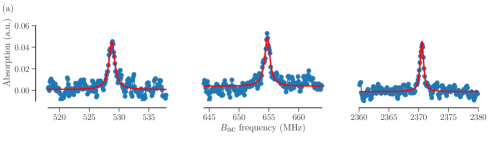
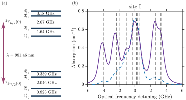
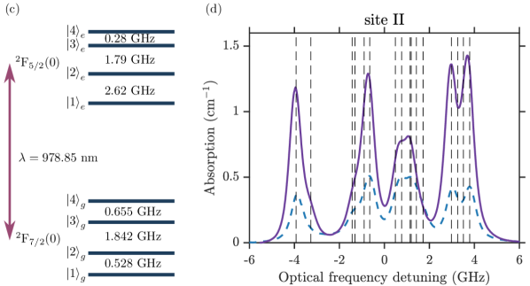
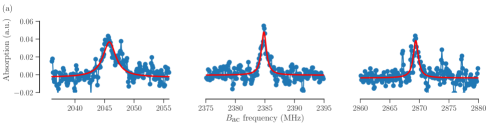
The optical transition 2FF5/2(0) under study here couples the lowest crystal field levels of the two electronic multiplets, with a vacuum wavelength of nm for site I and nm for site II Welinski et al. (2016). The optical absorption spectra of this transition are shown in Figure 1, which were recorded at zero magnetic field. Since both the ground and excited states have four non-degenerate energy levels, each spectrum consists of 16 optical-hyperfine transitions that are inhomogeneously broadened. The inhomogeneous profile was measured to be Lorentzian, with a full-width at half-maximum (FWHM) of 800 MHz for site I and 560 MHz for site II. Although the spectra are partly resolved, one cannot accurately measure the hyperfine splittings using the inhomogeneous absorption spectra. For this purpose we employ the SHB measurement technique.
III EXPERIMENTAL METHODS
III.1 Spectral Hole Burning
The SHB technique is commonly used to investigate transitions in systems with strong inhomogeneous broadening Macfarlane and Shelby (1981). In solid state crystals, inhomogeneous broadening is often due to uneven strains in different positions of the crystal Stoneham (1969), so that each ion can have a different detuning compared to the centre of the frequency distribution of a certain transition.
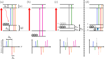
In this context, SHB consists in irradiating the sample with laser light at a specific frequency inside the broadened spectrum (the burn pulse), so to pump atoms away into other energy levels via an excited state. A scan of a weaker probe around the pump frequency will then reveal a series of holes and antiholes corresponding to transitions from states with increased or decreased population, respectively (Figure 2).
If the inhomogeneous broadening is much larger than the ground and excited state splittings, then a burn pulse at a fixed frequency can resonantly excite atoms in different classes, where for each class the burn pulse excites a different optical-hyperfine transition Macfarlane and Shelby (1981); Hastings-Simon et al. (2008a). Each class will produce a pattern of holes and anti-holes, see Figure 2, and the resulting spectrum can be hard to interpret. Since for this system the inhomogeneous broadening is smaller than some of the hyperfine splittings, the number of contributing classes will depend on the exact burn frequency. It also causes an asymmetric hole/anti-hole pattern, with respect to the burn frequency. The hole/antihole structures can be studied as a function of the time between the pump and the probe pulses to reveal population lifetimes Hastings-Simon et al. (2008a), and as a function of other external parameters such as magnetic field and temperature Cruzeiro et al. (2017).
In this paper, we focus on the dependence of the position of spectral holes/antiholes on an external magnetic field, which induces shifts in frequency of the holes/antiholes as the energy level splittings are varied by Zeeman interactionRiesen and Szabo (2010).
As will be explained in Section IV, we can also observe holeantihole transformations as the magnetic field is varied across a critical value (Figure 2c). This phenomenon marks the passage from a low field regime with highly nonlinear energy shifts due mostly to the hyperfine interaction, to a high field, linear regime dominated by Zeeman interactions. Hole/antihole transformations have been observed in different contexts including modification of the cross-relaxation Riesen et al. (2007) or temperature dependant phononic relaxation Hastings-Simon et al. (2008b), or as a result of superhyperfine resonances between a thulium dopant and aluminium ions in a Tm3+:YAG crystal Ahlefeldt et al. (2015).
III.2 Optically Detected Magnetic Resonance
Magnetic resonance techniques can be combined with SHB to study transitions in domains different than the optical one Laplane et al. (2016); Zhong et al. (2015); Fernandez-Gonzalvo et al. (2015); Macfarlane et al. (2014). Hyperfine and Zeeman interactions often induce splittings in the microwave range in atomic ensembles, and these splittings can be varied by using an external magnetic field in a broad range between MHz and GHz frequencies, requiring a broadband investigation technique. We thus rely on ODMR to study transitions between spin states for various conditions of external magnetic field. The technique consists in burning a spectral hole and observing its variation as a microwave transition is addressed at the same time as an optical probe beam is on at a constant frequency (Figure 2d) Macfarlane and Shelby (1987). When the microwave radiation is in resonance with the spin transition, the latter will be detected as a population change, which in turn reduces the amplitude of the spectral hole.
To find the spin resonance and obtain information such as its central frequency and linewidth, we measure the hole amplitude while scanning the microwave frequency. The measurements can be repeated at various external magnetic field values so to obtain information about its influence on the transition.
III.3 Experimental setup
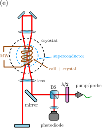
Our setup is shown schematically in Figure 2e. The crystal is placed inside a cryostat in vacuum and at temperature of about . A copper coil surrounds the crystal with its longitudinal axis coincident with the crystal axis, and it is used in the ODMR technique to address the spin transitions by generating a microwave field (up to in frequency, fed with power up to ). A superconductor coil surrounds the crystal and copper coil, and allows us to apply static fields up to in directions orthogonal to . In the experiments presented here, we use a pulsed laser beam (through an acousto-optic modulator, not shown), generated by a tunable external cavity diode laser and injected into a single mode polarization maintaining fibre. A half-waveplate right after the fibre output allows the adjustment of polarization according to the orientation of the crystal extinction axis of maximal absorption ( axis). The light is then focused on the sample in a spot with a diameter of and traverses it in four passes to further increase the amount of light absorbed. Finally, a beam splitter redirects the output light on a silicon photodiode.
IV Experimental results
In principle the SHB measurement can provide all required information about energy splittings for the ground and excited states. However, the interpretation of the SHB data is difficult as most anti-holes depend on both the ground and excited state splittings. Moreover, as explained in Figure 2c, one can observe so-called pseudoholes, where one would expect to see an anti-hole, further complicating the interpretation.
To facilitate the analysis we first detect the hyperfine splittings of the ground state at zero magnetic field, using the ODMR technique explained previously (see Section III.2). For this purpose the laser producing the burn and probe pulses was tuned to the middle of the absorption structure (Figure 1) in order to address a maximum number of classes and to produce a population difference for a high number of spin transitions.
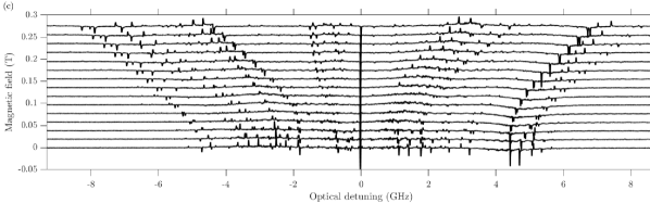
For optical site I we detected four ODMR lines at MHz (Figure 3a), while for site II five ODMR lines were found at MHz (see Appendix, Figure S6). The ODMR linewidths varied between 0.5 and 1.2 MHz. These were limited by the inhomogeneous linewidths and possibly by additional power broadening.
Using the detected ODMR resonances and Eq. (2), one can easily calculate the eigenvalues of the ground-state hyperfine tensor. These are listed in Table 1 and the corresponding energy level diagrams are reported in Figure 3a and Figure 1. These eigenvalues of the ground-state hyperfine tensor differ considerably from the ones obtained from high field EPR measurements Welinski et al. (2016). This clearly demonstrates the problem of standard X-band EPR for characterizing a highly anisotropic magnetic interaction in the case of low site symmetry. The strength of using zero-field ODMR is that we can determine the eigenvalues of the hyperfine tensors, independently of their orientation with respect to the crystal axes and without using any non-linear fitting algorithm. In EPR measurements the eigenvalues and orientation angles (six parameters) are fitted simultaneously to the experimental data, which can introduce errors.
The ODMR technique can also be used to measure hyperfine splittings of the excited state. However, due to the limited excited state lifetime (order of 1 ms for both sites Welinski et al. (2016)), excited-state ODMR resonances were not observed in these experiments. The SHB technique was thus used for this purpose. To clearly identify the SHB features stemming from the zero-field hyperfine splittings in the excited state, we found it necessary to also study the SHB spectra as a function of applied magnetic field, as explained in the following.
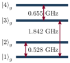
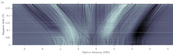
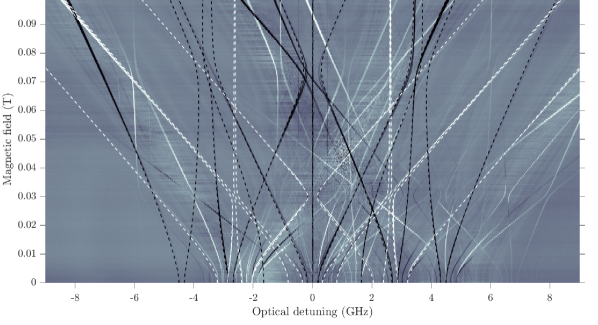
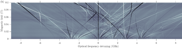
The SHB pattern appearing inside the absorption profile was recorded at different external magnetic fields in various directions. For each measurement, the normalization signal, that is the absorption profile without hole burning, was subtracted from the acquired trace so to leave only the hole/antihole structure visible (Figure 3b) (also see Appendix, Figure S6). An example of such a measurement, for site I and magnetic field parallel to axis, is shown in Figure 3b. It clearly shows a series of lines which shift linearly at high magnetic fields and go into a nonlinear regime at lower magnetic fields. The holes and antiholes appear as darker and brighter lines, respectively. By following these lines, one can precisely characterize the nonlinear behavior for various magnetic field regimes. The widths of the observed spectral holes were of a few MHz, which were mainly limited by the laser linewidth.
In conventional SHB methods the side holes usually correspond to the structure of the excited state (see Figure 2). Therefore, one expects to observe 6 holes on each side of the main hole, corresponding to the 6 possible spin transitions of the excited state. However, in our case we detect a much higher number of side holes, with some of them corresponding to the ground state energy levels. This happens due to the relatively fast relaxation rate on the spin transitions, which is comparable to the burning time used in our experiment (hundreds of milliseconds). In this situation, the optical pumping process effectively empties not only the addressed level but also the ones which are connected to it by fast relaxation processes. This leads to the emergence of pseudo holes at the positions expected for the antiholes, as also explained in Figure 2c, and it complicates the interpretation of the spectra, in particular the identification of the zero-field hyperfine resonances in the excited state.
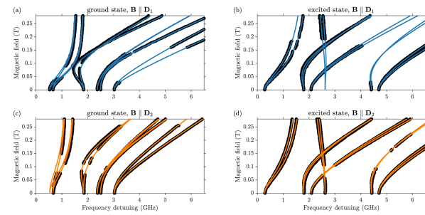
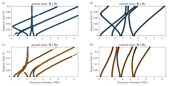
In a first step we use the zero-field ground state structure determined from the ODMR data to identify, without ambiguity, several SHB resonances that only depend on the ground state splitting. We use this information to extract the field dependence of the corresponding transitions in a large range of magnetic field amplitudes from the recorded SHB spectra (Figure 3). The extracted data for site I is shown in Figure 4.
In a second step we determine the excited state structure, which requires distinguishing between pseudo-holes and holes. To this end we use the fact that we generally observe two distinct regions of non-linear Zeeman effects in the SHB data. For the particular case of site I and parallel to the axis, these appear at around 30 and 80 mT, see Figure 3b. The one at 30 mT is due to a non-linear Zeeman effect in the ground state, as seen in the already identified transition energies of the ground state shown in Figure 4a. The one at 80 mT is then due to the excited state Zeeman effect, which is consistent with the fact that the effective -factor is twice as low in the excited state Welinski et al. (2016). Hence, SHB holes that display non-linear behaviour in both the 30 and 80 mT region are pseudo-holes that can be disregarded from the excited state analysis.
In a third step we identify strong SHB side holes, particularly at higher fields, which only display the non-linearity at around 80 mT, as shown in Figure 4b. To further confirm that these are indeed true holes depending on the excited state hyperfine splits, we use the complete set of resonances from both ground and excited states to calculate the position of all expected anti-holes. Many of these are seen in the SHB spectra (see Appendix Figure S9), and most importantly none of these predicted lines are in contradiction with the observed SHB spectra. The same approach was also used to analyse the SHB spectra recorded with parallel to the axis for site I, see Figure 4c-d. We also fully measured and analyzed the SHB spectra along the and axes for site II (see Appendix Figure S7).
The analysis presented above allows us to clearly identify excited-state hyperfine resonances at zero magnetic field, from which the eigenvalues of the excited state hyperfine tensor are calculated. These are listed in Table 1 and the corresponding energy level diagrams are reported in Figure 1, both for site I and II.
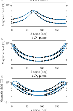
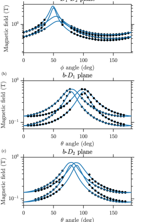
It remains to determine the orientation of the hyperfine tensors with respect to the crystal axes. To this end we use the fact that accurate Zeeman tensors were already determined Welinski et al. (2016) for Yb3+ ions having no hyperfine states (I = 0). The hyperfine tensors are thus fitted with respect to the tensors, both for the ground and excited states. In the fit we use the hyperfine splittings as a function of magnetic field, measured using the SHB technique. In addition, for the ground state we can use the X-band EPR data from Ref. Welinski et al. (2016) (see Appendix, Figure S8), which was recorded at higher fields in between 100 mT and 1 T. The fitting procedure and the definition of all rotation matrices are given in the Appendix. The fitted rotation angles for all tensors are listed in Table 1.
| Site I | Site II | |||
|---|---|---|---|---|
| ground | excited | ground | excited | |
| (GHz) | 0.481 | 1.44 | -0.1259 | 2.34 |
| (GHz) | 1.159 | 1.82 | 1.1835 | 2.90 |
| (GHz) | 5.251 | 7.20 | 4.8668 | 6.49 |
| (∘) | 72.25 | 73.88 | 45.86 | 51.07 |
| (∘) | 92.11 | 84.76 | 11.13 | 14.11 |
| (∘) | 63.92 | 90.13 | 2.97 | -0.67 |
| 0.31 | 0.8 | 0.13 | 1.0 | |
| 1.60 | 1.0 | 1.50 | 1.4 | |
| 6.53 | 3.4 | 6.06 | 3.3 | |
| (∘) | 72.8 | 77 | 59.10 | 54 |
| (∘) | 88.7 | 84 | 11.8 | 23 |
| (∘) | 66.2 | -7 | -12.6 | -10 |
To determine the order of the energy splittings we fit the absorption profiles measured at zero magnetic field (Figure 1). Only the central frequency offset is used as a free parameter while all the differences between absorption lines are fixed using a certain order of the energy splittings. From the four different possibilities we find that only one gives a spectrum in a good agreement with the experiment. The order found for both sites is depicted in Figure 1. This result is further confirmed using separate ODMR measurements by selective optical excitation at the outermost absorption lines. We note that the order is given by the relative signs between the tensor eigenvalues, while the simultaneous sign change of two elements does not alter the order. The signs combination corresponding to the determined order is given in Table 1 for sites I and II.
V Discussion
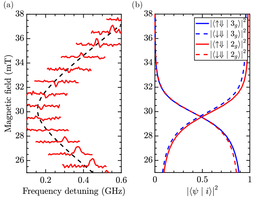
A close look at the spectral hole map on Figure 3 (especially in the nonlinear regime around 30 mT) reveals many positions of the antiholes with peculiar behaviour. As an example in Figure 5a we show the variation in hole amplitude of the 2046 MHz transition (at zero magnetic field). As seen it features the expected anti-hole behaviour below and above 30 mT, while at around 30 mT it features a pseudo hole. Similar behaviour can be seen also for other lines corresponding to various optical classes (Figure 3).
We believe this is due to the strong admixing of the corresponding wavefunctions which appears for this magnetic field region. Indeed, from the decomposition of the eigenstates involved in this spin transition (Figure 5b) one can see the strong non-linear dependence and flip of the electronic wavefunctions associated with the avoided crossing. This can lead to an enhanced relaxation process on this transition, which leads to the observed transformation of the antihole into a pseudo hole. We note that a similar hole-antihole transformation was reported in ruby Riesen et al. (2007), and the given explanation was also based on the modified cross-relaxation under external magnetic fields.
The phenomenon of pseduo holes is also observed at zero magnetic field, where the antiholes corresponding to and transitions (823 MHz and 339 MHz, respectively) appear as pseudo holes. To explain this we assume that the relaxation process at these transitions is faster than for other spin transitions, which makes the optical pumping to be efficient for both of the connected levels (see Figure 2c). The flip-flop relaxation process between different Yb3+ ions could be dominant for low magnetic field in this material , as observed in Nd3+ doped Y2SiO5 Cruzeiro et al. (2017), however, further studies of the relaxation rates are required to conclude about the nature of the observed phenomena.
The same behavior is also present for other antiholes on this map, whose energy splittings involve the excited state spin transition, and can be found in many parts of the spectrum. Although this effect complicates the reading of the holeburning spectra, as discussed above, it potentially can also give additional information about the relative relaxation dynamics between particular sets of spin transitions.
Finally we briefly discuss the relative orientations of the and tensors, and their anisotropy in the ground and excited states. By evaluating the values for hyperfine tensors from Table 1 one can see that the orientations of the and tensors for each state are almost parallel. For site I the orientation of the maximum -factor is close to , while for site II it is close to . The ratio between the tensor elements is nearly constant for each component. This indicates that each state on the optical 2FF5/2(0) transition is close to a pure multiplet Welinski et al. (2016).
VI CONCLUSION and OUTLOOK
In conclusion, we have characterized the hyperfine interaction of the 171Yb3+:Y2SiO5 crystal on the optical 2FF5/2(0) transition. We refined the hyperfine tensor of the ground state and determined its parameters for the excited state. The largest principal values of these tensors are oriented along similar directions for the ground and excited states, which are close to axis for site I and close to axis for site II. The hyperfine tensors are also similarily oriented as the corresponding Zeeman tensors Welinski et al. (2016). This simplifies the description of the spin Hamiltonians of 171Yb3+:Y2SiO5 crystal.
Our characterization of 171Yb3+:Y2SiO5 is in good agreement with previously obtained results for the Zeeman tensors of both the ground and excited states. However, for the hyperfine tensors of the ground states, the eigenvalues are very different as compared to those obtained through conventional EPR measurements in the high-field regime Welinski et al. (2016). While both hyperfine tensors accurately predict the high-field regime (within experimental errors), only the hyperfine tensors given here work well at low fields. We believe this to be due to the difficulty to accurately determining all eigenvalues in the high-field regime, where the hyperfine interaction is a perturbation to the Zeeman interaction. This problem was also recently encountered in a 167Er3+:Y2SiO5 crystal Chen et al. (2018), hence it appears to be a general problem that has not yet been studied in EPR studies.
We also observed pseudo-holes in the SHB spectra, at positions where anti-holes were expected. We attribute this to cross-relaxation processes that efficiently couples certain hyperfine states. Further studies of the hole lifetimes at different magnetic fields, particularly for the highly non-linear regime, can give the information about relaxation rates and can help to attribute the observed effects to the particular relaxation process.
The spin Hamiltonian presented here allows the calculation of transition frequencies in the spin and optical domain under arbitrarily orientated external magnetic fields, over a wide range of magnetic fields. We note, however, that the effective spin Hamiltonian will break down for very high magnetic fields (for fields significantly higher than 1 Tesla), where perturbations from the nearest crystal-field level cannot be neglected. In particular, the Hamiltonian can be used to accurately predict so-called ZEFOZ (ZEro First-Order Zeeman) points, where the sensitivity of transitions to magnetic field perturbations are highly suppressed Fraval et al. (2004). In a parallel work the Hamiltonian was used to predict ZEFOZ and near ZEFOZ points in 171Yb3+:Y2SiO5, at zero and low external magnetic fields, respectively Ortu et al. (2018). Furthermore, it was experimentally demonstrated that a strong enhancement of both optical and spin coherence times appears in these points, with optical coherence times reaching up to 200 µs and spin coherence times reaching up to 4 ms Ortu et al. (2018). Based on these results we believe the 171Yb3+:Y2SiO5 crystal is very promising for optical quantum memories Tittel et al. (2010); Bussières et al. (2013), microwave-to-optical transducers Fernandez-Gonzalvo et al. (2015) and coupling to superconducting qubits in the microwave range Probst et al. (2013).
ACKNOWLEDGEMENTS
The authors thank F. Fröwis and E. Z. Cruzeiro for useful discussions, as well as Claudio Barreiro for technical support. We acknowledge funding from EUs H2020 programme under the Marie Skłodowska-Curie project QCALL (GA 675662), ANR under grant agreement no. 145-CE26-0037-01 (DISCRYS), Nano’K project RECTUS and the IMTO Cancer AVIESAN (Cancer Plan, C16027HS, MALT).
References
- Tittel et al. (2010) W. Tittel, M. Afzelius, T. Chanelière, R. Cone, S. Kröll, S. Moiseev, and M. Sellars, Laser & Photonics Reviews 4, 244 (2010).
- Thiel et al. (2011) C. Thiel, T. Böttger, and R. Cone, Journal of Luminescence 131, 353 (2011).
- Bussières et al. (2013) F. Bussières, N. Sangouard, M. Afzelius, H. de Riedmatten, C. Simon, and W. Tittel, Journal of Modern Optics 60, 1519 (2013).
- de Riedmatten and Afzelius (2015) H. de Riedmatten and M. Afzelius, “Quantum light storage in solid state atomic ensembles,” in Engineering the Atom-Photon Interaction: Controlling Fundamental Processes with Photons, Atoms and Solids, Nano-Optics and Nanophotonics, edited by A. Predojević and M. W. Mitchell (Springer International Publishing, 2015) Chap. 9, pp. 241–268.
- Goldner et al. (2015) P. Goldner, A. Ferrier, and O. Guillot-Nöel, “Rare earth-doped crystals for quantum information processing,” in Handbook on the Physics and Chemistry of Rare Earths, Vol. 46, edited by J.-C. G. Bünzli and V. K. Pecharsky (Elsevier Science Publishers, North Holland, Amsterdam, 2015) Chap. 267, pp. 1–78.
- Macfarlane (2002) R. M. Macfarlane, J. Lumin. 100, 1 (2002).
- Böttger et al. (2009) T. Böttger, C. W. Thiel, R. L. Cone, and Y. Sun, Phys. Rev. B 79, 115104 (2009).
- Longdell and Sellars (2004) J. J. Longdell and M. J. Sellars, Phys. Rev. A 69, 032307 (2004).
- Ahlefeldt et al. (2013) R. L. Ahlefeldt, D. L. McAuslan, J. J. Longdell, N. B. Manson, and M. J. Sellars, Phys. Rev. Lett. 111, 240501 (2013).
- Arcangeli et al. (2014) A. Arcangeli, M. Lovrić, B. Tumino, A. Ferrier, and P. Goldner, Phys. Rev. B 89, 184305 (2014).
- Zhong et al. (2015) M. Zhong, M. P. Hedges, R. L. Ahlefeldt, J. G. Bartholomew, S. E. Beavan, S. M. Wittig, J. J. Longdell, and M. J. Sellars, Nature 517, 177 (2015).
- Jobez et al. (2015) P. Jobez, C. Laplane, N. Timoney, N. Gisin, A. Ferrier, P. Goldner, and M. Afzelius, Phys. Rev. Lett. 114, 230502 (2015).
- Gündoğan et al. (2015) M. Gündoğan, P. M. Ledingham, K. Kutluer, M. Mazzera, and H. de Riedmatten, Phys. Rev. Lett. 114, 230501 (2015).
- Seri et al. (2017) A. Seri, A. Lenhard, D. Rieländer, M. Gündoğan, P. M. Ledingham, M. Mazzera, and H. de Riedmatten, Phys. Rev. X 7, 021028 (2017).
- Sabooni et al. (2013) M. Sabooni, Q. Li, S. Kröll, S., and L. Rippe, Phys. Rev. Lett. 110, 133604 (2013).
- Jobez et al. (2014) P. Jobez, I. Usmani, N. Timoney, C. Laplane, N. Gisin, and M. Afzelius, New Journal of Physics 16, 083005 (2014).
- Heinze et al. (2013) G. Heinze, C. Hubrich, and T. Halfmann, Phys. Rev. Lett. 111, 033601 (2013).
- Saglamyurek et al. (2011) E. Saglamyurek, N. Sinclair, J. Jin, J. A. Slater, D. Oblak, F. Bussières, M. George, R. Ricken, W. Sohler, and W. Tittel, Nature 469, 512 (2011).
- Clausen et al. (2011) C. Clausen, I. Usmani, F. Bussieres, N. Sangouard, M. Afzelius, H. de Riedmatten, and N. Gisin, Nature 469, 508 (2011).
- Tiranov et al. (2015) A. Tiranov, J. Lavoie, A. Ferrier, P. Goldner, V. B. Verma, S. W. Nam, R. P. Mirin, A. E. Lita, F. Marsili, H. Herrmann, C. Silberhorn, N. Gisin, M. Afzelius, and F. Bussières, Optica 2, 279 (2015).
- Tiranov et al. (2016) A. Tiranov, P. C. Strassmann, J. Lavoie, N. Brunner, M. Huber, V. B. Verma, S. W. Nam, R. P. Mirin, A. E. Lita, F. Marsili, M. Afzelius, F. Bussières, and N. Gisin, Physical Review Letters 117, 240506 (2016).
- Bussières et al. (2014) F. Bussières, C. Clausen, A. Tiranov, B. Korzh, V. B. Verma, S. W. Nam, F. Marsili, A. Ferrier, P. Goldner, H. Herrmann, C. Silberhorn, W. Sohler, M. Afzelius, and N. Gisin, Nature Photonics 8, 775 (2014).
- Kolesov et al. (2012) R. Kolesov, K. Xia, R. Reuter, R. Stöhr, A. Zappe, J. Meijer, P. Hemmer, and J. Wrachtrup, Nat Commun 3, 1029 (2012).
- Utikal et al. (2014) T. Utikal, E. Eichhammer, L. Petersen, A. Renn, S. Götzinger, and V. Sandoghdar, Nat Commun 5, (2014).
- Macfarlane and Shelby (1987) R. Macfarlane and R. Shelby, in Spectroscopy of Crystals Containing Rare Earth Ions, Modern Problems in Condensed Matter Sciences, Vol. 21, edited by A. Kaplyankii and R. Macfarlane (Elsevier Science Publishers, North Holland, Amsterdam, 1987) Chap. 4, pp. 51–184.
- Probst et al. (2013) S. Probst, H. Rotzinger, S. Wünsch, P. Jung, M. Jerger, M. Siegel, A. V. Ustinov, and P. A. Bushev, Phys. Rev. Lett. 110, 157001 (2013).
- Wolfowicz et al. (2015) G. Wolfowicz, H. Maier-Flaig, R. Marino, A. Ferrier, H. Vezin, J. J. L. Morton, and P. Goldner, Phys. Rev. Lett. 114, 170503 (2015).
- Chen et al. (2016) Y.-H. Chen, X. Fernandez-Gonzalvo, and J. J. Longdell, Phys. Rev. B 94, 075117 (2016).
- Rančić et al. (2018) M. Rančić, M. P. Hedges, R. L. Ahlefeldt, and M. J. Sellars, Nature Physics 14, 50 (2018).
- Loredo et al. (2016) J. C. Loredo, N. A. Zakaria, N. Somaschi, C. Anton, L. de Santis, V. Giesz, T. Grange, M. A. Broome, O. Gazzano, G. Coppola, I. Sagnes, A. Lemaitre, A. Auffeves, P. Senellart, M. P. Almeida, and A. G. White, Optica 3, 433 (2016).
- Ding et al. (2016) D. Ding, L. M. C. Pereira, J. F. Bauters, M. J. R. Heck, G. Welker, A. Vantomme, J. E. Bowers, de Dood Michiel J. A., and D. Bouwmeester, Nature Photonics 8, 385 (2016).
- Miyazono et al. (2016) E. Miyazono, T. Zhong, I. Craiciu, J. M. Kindem, and A. Faraon, Applied Physics Letters 108, 011111 (2016).
- Welinski et al. (2016) S. Welinski, A. Ferrier, M. Afzelius, and P. Goldner, Phys. Rev. B 94, 155116 (2016).
- Chen et al. (2018) Y.-H. Chen, X. Fernandez-Gonzalvo, S. P. Horvath, J. V. Rakonjac, and J. J. Longdell, Phys. Rev. B 97, 024419 (2018).
- Mehta et al. (2000) V. Mehta, O. Guillot-Noel, D. Gourier, Z. Ichalalene, M. Castonguay, and S. Jandl, Journal of Physics: Condensed Matter 12, 7149 (2000).
- Abragam and Bleaney (1970) A. Abragam and B. Bleaney, Electronic Paramagnetic Resonance of Transition Ions (Clarendon Press, Oxford UK, 1970).
- Li et al. (1992) C. Li, C. Wyon, and R. Moncorge, IEEE Journal of Quantum Electronics 28, 1209 (1992).
- Equall et al. (1994) R. W. Equall, Y. Sun, R. L. Cone, and R. M. Macfarlane, Phys. Rev. Lett. 72, 2179 (1994).
- Fraval et al. (2004) E. Fraval, M. J. Sellars, and J. J. Longdell, Phys. Rev. Lett. 92, 077601 (2004).
- Böttger et al. (2006) T. Böttger, C. W. Thiel, Y. Sun, and R. L. Cone, Phys. Rev. B 73, 075101 (2006).
- Kurkin and Chernov (1980) I. Kurkin and K. Chernov, Physica B+C 101, 233 (1980).
- Macfarlane and Shelby (1981) R. M. Macfarlane and R. M. Shelby, Opt. Lett. 6, 96 (1981).
- Stoneham (1969) A. M. Stoneham, Rev. Mod. Phys. 41, 82 (1969).
- Hastings-Simon et al. (2008a) S. R. Hastings-Simon, M. Afzelius, J. Minář, M. U. Staudt, B. Lauritzen, H. de Riedmatten, N. Gisin, A. Amari, A. Walther, S. Kröll, E. Cavalli, and M. Bettinelli, Phys. Rev. B 77, 125111 (2008a).
- Cruzeiro et al. (2017) E. Z. Cruzeiro, A. Tiranov, I. Usmani, C. Laplane, J. Lavoie, A. Ferrier, P. Goldner, N. Gisin, and M. Afzelius, Phys. Rev. B 95, 205119 (2017).
- Riesen and Szabo (2010) H. Riesen and A. Szabo, Physics Procedia 3, 1577 (2010).
- Riesen et al. (2007) H. Riesen, B. F. Hayward, and A. Szabo, Journal of Luminescence 127, 655 (2007).
- Hastings-Simon et al. (2008b) S. R. Hastings-Simon, B. Lauritzen, M. U. Staudt, J. L. M. van Mechelen, C. Simon, H. de Riedmatten, M. Afzelius, and N. Gisin, Phys. Rev. B 78, 085410 (2008b).
- Ahlefeldt et al. (2015) R. L. Ahlefeldt, M. F. Pascual-Winter, A. Louchet-Chauvet, T. Chanelière, and J.-L. Le Gouët, Phys. Rev. B 92, 094305 (2015).
- Laplane et al. (2016) C. Laplane, E. Z. Cruzeiro, F. Fröwis, P. Goldner, and M. Afzelius, Phys. Rev. Lett. 117, 037203 (2016).
- Fernandez-Gonzalvo et al. (2015) X. Fernandez-Gonzalvo, Y.-H. Chen, C. Yin, S. Rogge, and J. J. Longdell, Phys. Rev. A 92, 062313 (2015).
- Macfarlane et al. (2014) R. M. Macfarlane, A. Arcangeli, A. Ferrier, and P. Goldner, Phys. Rev. Lett. 113, 157603 (2014).
- Ortu et al. (2018) A. Ortu, A. Tiranov, S. Welinski, F. Fröwis, N. Gisin, A. Ferrier, P. Goldner, and M. Afzelius, Nature Materials 17, 671 (2018).
Appendix A Hamiltonian definition
The tensors and for the ground and excited states can be diagonalized in their respective principle axis systems. To express them in the crystal frame we define a rotation with the usual Euler angle convention:
| (3) |
| (4) |
where is the rotation matrix with Euler angles for convention
where
| (5) |
| (6) |
The interaction tensors for the second magnetic subsite are defined using an additional -rotation around the symmetry axis of the crystal and given by
| (7) | |||
| (8) |
Additional rotation is applied to go from the crystal frame to the laboratory frame and was found to contain the angles lower than 5∘. The orientation of the and axes was checked using the polarization dependent absorption measurement. The expected error in their determination could reach 10∘. The The interaction tensors in crystal frame for the ground and excited state are found to be (in GHz)
The corresponding -tensors used for the fitting are