Seeding Dispersal Modeling For Systems of Planar Microbial Biofilms
Abstract
We propose a modeling approach to study how mature biofilms spread and colonize new surfaces by predicting the formation and growth of satellite colonies generated by dispersing biofilms. This model provides the basis for better understanding the fate and behavior of dispersal cells, phenomenon that cannot, as yet, be predicted from knowledge of the genome. The model results were promising as supported by the experimental results. The proposed approach allows for further improvements through more detailed sub-models for front propagation, seeding, availability and depletion of resources. The present study was a successful proof-of-concept in answering the following questions: Can we predict the colonization of new sites following biofilm dispersal? Can we generate patterns in space and time to shed light on seeding dispersal? That are fundamental issues for developing novel approaches to manipulate biofilm formation in industrial, environmental and medical applications.
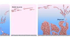
It is now well accepted that microorganisms lead social lives and engage in complex behavior in response to other organisms and the extracellular environment. By adopting coordinated chemical and physical interactions, microorganisms establish complex communities attached to a surface and embedded in a self-produced extracellular polymeric matrix, enabling cells to develop efficient survival strategies Nadell et al. (2016). This sessile lifestyle is called biofilm, and it represents the dominant mode of microbial life in many natural, medical and engineered systems Hall-Stoodley et al. (2004); Mattei et al. (2017). Cells in biofilms undergo developmental programs resulting in an ordered and predictable transition through a series of stages, each based on stage-specific expression of genes Flemming et al. (2016). The biofilm developmental program culminates with the release of free-living cells that can colonize new habitats, possibly richer in resources McDougald et al. (2011), as seen Fig. 1. While detachment is a passive process of cell loss resulting from sloughing of cells and erosion from the biofilm, active or seeding dispersal is coordinated via regulatory systems in response to a number of cues (e.g., alteration in the availability of nutrients, oxygen depletion, levels of iron) and signals (e.g., acyl-homoserine lactones, diffusible fatty acids, cell-cell autoinducing peptides) Guilhen et al. (2017). Thus, seeding dispersal can occur in the complete absence of flowing conditions, and does not depend upon shear forces that removes cells from the biofilm. Another interesting feature of seeding dispersal is that cells appear to have a distinct phenotypes different from those of biofilm and planktonic cells, increasing cell ability to colonize a greater range of habitats important for niche expansion Chua et al. (2014); D’Acunto et al. (2015). Thus, dispersal represents an important adaptive strategy with profound impacts on the survival and fitness of microorganisms. It allows biofilm populations to spread and colonize new surfaces, avoiding overcrowding, depletion of resources and competition among cells in the local environment, and promoting the rejuvenation of biofilms Barraud et al. (2015). Furthermore, dispersal is linked to the generation or maintenance of genetic variation, with significant outcomes for the success of those bacteria in the environment Purevdorj-Gage et al. (2005); Chua et al. (2014); D’Acunto et al. (2015). Although dispersal is advantageous from the microbial standpoint, it may negatively affect some industrial and medical processes. For instance, through dispersed cells, biofilm can spark new infections within the host and result in the transmission of bacteria between different hosts Koo et al. (2017). Furthermore, dispersal may promote, for example, the spread of parasitism phenomena in animals and plants Villa et al. (2017), biodeterioration of historical and artistic objects Cappitelli et al. (2011); Villa et al. (2016) and fouling in food-processing equipment (Cappitelli et al., 2014). The existence of a programmed generation of dispersed cells appears increasingly clear, but the challenge now is to provide the mechanistic understanding of biofilm dispersal. Thus, the principal questions that motivate this work are: Can we predict the colonization of new sites following biofilm dispersal? Can we generate patterns in space and time to shed light on seeding dispersal? We propose a modeling approach to study the growth of mono-layer microbial biofilm on inert surfaces by focusing on the biofilm spread induced by dispersal, predicting the formation and growth of satellite colonies generated by dispersing biofilms. The importance of this work relies on the fact that the fate and behavior of dispersal cells cannot, as yet, be predicted from knowledge of the genome. Thus, a mathematical modelling of biofilm dispersion is urgently needed. The planar geometry we focus on is proper of biofilm growth in oligotrophic environments (e.g., reverse osmosis membranes, stone monuments, surgical gauze, contact lenses, water supply pipes), where nutrient constraints limit microbial growth to thin mono-layered biofilms. The growth of this biofilm is characterized by two main phenomena: the biomass expansion due to the growth of primary existing colonies, and the formation of new colonies due to the attachment of dispersed cells released by the primary ones, i.e., seeding dispersal.
In analogy with an approach originally introduced for turbulent premixed combustion Pagnini and Bonomi (2011) and wild-land fire propagation Pagnini and Mentrelli (2014); Kaur et al. (2016), the proposed mechanistic model is built up as follows. Biofilm colony growth is modeled by using the Level Set Method Sethian and Smereka (2003), while seeding dispersal is simulated through the Probability Density Function (PDF) corresponding to the diffusive process that governs the bacteria dispersal behavior. The seeds attachment depends on their concentration and environmental resources availability, with the latter characterized by its initial spatial distribution, and by the depletion effect due to the presence of mature biofilm colonies. The initial configuration of environmental resource availability can be modeled by setting a specific scenario or by using a random distribution.
The surface of mature biofilm colony is generally composed by an ensemble of biofilms spots with where the total number depends on time because of merging and birth of colonies. Let be a function defined on the domain of interest such that the iso-line describes the evolution the boundaries of , i.e., the evolution of the colonies fronts. Then the motion of the fronts of biofilm colonies is determined by the Level Set Equation:
| (1) |
In the following, the outward normal velocity is assumed constant, i.e., .
Let the mature colonies be able to release a sufficient large number of cells whose dispersion is characterised by a random motion. Let be the -realization of the trajectory of a dispersed cell with an average position and initially located in , such that . Cell trajectories are described by the one-particle density function , where is the Dirac -function. Moreover, let the regions occupied by the colonies be conveniently marked by an indicator function . Then, an effective indicator , , of the region surrounded by a random front is obtained by using the sifting property of the -function and by averaging the indicator function:
| (2) | |||||
where is the PDF of the seeding bacteria. In this work, is assumed to be Gaussian.
Function provides the probability that dispersed bacteria cells arrive in a point from different sources . However, to relate this probability of arrival to a successful formation of a new biofilm colony spot, a criterion associated with a reversible/irreversible attachment due to environmental conditions and biological time scales is needed. With this aim, we introduce the integral field
| (3) |
that stores the signals received from the active biofilm domain in the temporal interval . We denote by the timescale of signal storing and it is determined by
| (4) |
where represents the environmental distribution of resources in absence of biofilm and accounts for the resource depletion performed by the biofilm.
The feedback mechanism between and is given by the procedure
| (5) |
that is: when into a considered spot a certain amount of dispersed cells have established and endured a certain amount of time (that accounts for the environmental availability of resources) then a new colony is generated. Hence, the indicator function results to be
| (6) |
Equation (4) shows an interplay between the availability of resources offered from the surrounding environment and the resource depletion performed by the growth of the biofilm colonies. This simple formulation of the timescale for the waiting times of free cells seeding is able to generate a plethora of patterns of biological relevance. In the following, the term is assumed constant in time, because it represents the availability of resources before the action of biofilm, and this changes slower than the biofilm evolution. The term is modeled by the following Poisson problem
| (7a) | |||
| (7b) |
where is the bacterial density inside the colonies and an absorption kinetic coefficient. In our case, the bacterial density inside the colony is constant, and the latter equation becomes
| (8) |
where corresponds to in the rescaled setting and differs for the physical dimensions. The dynamic governed by (8) depends only on and, in spite of its simplicity, it manages to represent availability of biofilm resources, determining the temporal dynamics of seeding dispersal.
In order to prove the potentiality of the proposed approach, an experimental test case has been designed and realized. Pseudomonas aeruginosa strain PAO1 (MH873) was used in this study as a model system of bacterial biofilms. In fact, the metabolically versatile P. aeruginosa PAO1 is an opportunistic pathogen of plants, animals, and humans and is ubiquitously distributed in soil and aquatic habitats. Furthermore, the bacterium is genetically characterized and amenable to mutagenesis and ”omics” based approaches Stover et al. (2000); Walker et al. (2004). The microorganism was maintained at -80∘C in suspensions containing 20% glycerol and 2% peptone, and was grown aerobically in Tryptic Soy Broth (TSB medium) for 15h at 30∘C. Dispersion experiments were conducted by using the colony-biofilm culturing system. Briefly, 2 sterile black polycarbonate filter membranes (0.22 pore size and 25mm diameter) were placed in each Petri dish containing Tryptic Soy Agar (TSA medium), at a distance of 2 mm from each other. Bacterial cells are trapped completely by the membrane filters having a pore size smaller than the bacterial size, while nutrients and metabolites diffuse across membranes easily. Fifty of cell suspension containing cells were used to inoculate the central filter membrane. The plates were incubated at 30∘C for 72h. Every 24h the Petri dishes were observed, and the dispersal phenomenon was documented by capturing imagines with both a camera and a stereomicroscope (magnification 12X).
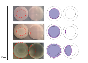
Numerical solution of model (1)–(8) has been computed by setting the physical parameters as follows: , inside the membranes and zero outside, and the diffusion coefficient of the Gaussian PDF equal to . The numerical set-up is based on a 2D mesh with grid step . The numerical test concerns the two membranes: the inoculated one and seeding target (the two external dashed lines in Fig. 2). These circular membranes have radius in grid step units and center in (the inoculated membrane) and (the target membrane). At the initial instant, a mature biofilm colony is assumed to be present in the inoculated membrane with circular profile centered in the center of the membrane and radius . Furthermore, the availability of the environmental food needs to be set and it is represented by in (4). In particular, is assumed to be constant in time and ranging through a linear interpolation procedure from , when is inside the inner disk with radius , to , when is outside the membranes (see the dashed circles in the right side of Fig. 2). This assumption corresponds to a very large timescale for generating a new colony outside the membranes, which corresponds to unfavorable conditions.
The computation was done by using the facilities of BCAM by running an OpenMp-parallel finite difference C/Fortran code. Its routines rely on a general-purpose library written in Fortran2008/OpenMP, LSMLib (http://ktchu.serendipityresearch.org/software/lsmlib/). The latter provides robust and efficient tools for studying the evolution of co-dimensional fronts moving in one-, two- and three-dimensional domains. ENO algorithms are used for the sake of computing accurate space derivatives, while for the advancement in time a second order Runge–Kutta scheme was implemented.
Figure 2 shows the growth of a primary colony in the inoculated membrane and its colonization of the target membrane by a seeding dispersal mechanism both for the experimental data (the pair of membranes on the left) and the proposed modeling approach (the pair on the right). The Level Set Method describes the growth of the colony: first the primary one that is living in the inoculated membrane (left side membrane) and later the secondary one in the target membrane. The seeding and the attachment mechanism, which are responsible for the colonization of the target membrane, are well reproduced by the model. In spite of the fact that the present comparison is qualitative, it shows that the present approach is able through its modular structure to model the growth of the biomass colony and to take into account the different processes that simultaneously occur. In particular, the present approach provides a method to link a sharp interface model for the growth of biofilm colonies and a statistical treatment for biofilm seeding. The modular structure allows for a detailed front propagation through a more detailed expression for the normal velocity of the colony front and a more detailed bacterial migration through a new statical characterization. The comparison between the experimental pictures and some frame of evolution of the model is promising and, thanks to the modular structure, the present approach emerges as a novel and useful method for understanding the complex dynamics displayed by microbial biofilm.
To conclude, we remark that one of the main motivations for studying biofilm dispersal is to provide a mechanistic model to predict how cells attach and proliferate to seed new biofilms. An increased understanding of the fate of dispersed cells will offer a broad conceptual framework for developing novel approaches to manipulate biofilm formation (either discouraging or promoting biofilm development) in industrial, environmental and medical applications. Thus, the ability to unravel the mechanisms of dispersal would have a great socio-economical significance, with profound implications for global health, as well as for the management of environmental microorganisms in biogeochemical cycling processes and biotechnological applications of biofilms. Another argument supporting the significance of this model is that could be potentially applied to eukaryotes that show ”biphasic” life cycles characterized by a dispersive phase and a sessile phase (e.g., corals, bryozoans, cancer cells).
GP acknowledges the support by the Basque Government through the BERC 2014-2017 program and by Spanish Ministry of Economy and Competitiveness MINECO through BCAM Severo Ochoa excellence accreditation SEV-2013-0323 and through project MTM2016-76016-R ”MIP”. AT is supported by the PhD Grant ”La Caixa” 2014. LF acknowledges Progetto Giovani GNFM 2016 ”Comportamenti emergenti ed auto-organizzazione in sistemi iperbolici di reazione-diffusione in ambito biologico ed ecologico.” FV has received funding from the European Union Seventh Framework Programme (FP7-PEOPLE-2012-IOF) under grant agreement No. 328215.
References
- Nadell et al. (2016) C. D. Nadell, K. Drescher, and K. R. Foster, Nat. Rev. Microbiol. 14, 589 (2016).
- Hall-Stoodley et al. (2004) L. Hall-Stoodley, J. W. Costerton, and P. Stoodley, Nat. Rev. Microbiol. 2, 95 (2004).
- Mattei et al. (2017) M. R. Mattei, L. Frunzo, B. D’Acunto, Y. Pechaud, F. Pirozzi, and G. Esposito, J. Math. Biol. (2017), doi 10.1007/s00285-017-1165-y.
- Flemming et al. (2016) H. C. Flemming, J. Wingender, U. Szewzyk, P. Steinberg, S. A. Rice, and S. Kjelleberg, Nat. Rev. Microbiol. 14, 563 (2016).
- McDougald et al. (2011) D. McDougald, S. A. Rice, N. Barraud, P. D. Steinberg, and S. Kjelleberg, Nat. Rev. Microbiol. 10, 39 (2011).
- Guilhen et al. (2017) C. Guilhen, C. Forestier, and D. Balestrino, Mol. Microbiol. 105, 188 (2017).
- Chua et al. (2014) S. L. Chua, Y. Liu, J. K. Yam, Y. Chen, R. M. Vejborg, B. G. Tan, S. Kjelleberg, T. Tolker-Nielsen, M. Givskov, and L. Yang, Nat. Commun. 5, 4462 (2014).
- D’Acunto et al. (2015) B. D’Acunto, L. Frunzo, I. Klapper, and M. R. Mattei, Math. Biosci. 259, 20 (2015).
- Barraud et al. (2015) N. Barraud, S. Kjelleberg, and S. A. Rice, Microbiol. Spectr. 3 (2015), doi:10.1128/microbiolspec.MB-0015-2014.
- Purevdorj-Gage et al. (2005) B. Purevdorj-Gage, W. J. Costerton, and P. Stoodley, Microbiology 151, 1569 (2005).
- Koo et al. (2017) H. Koo, R. N. Allan, R. P. Howlin, P. Stoodley, and L. Hall-Stoodley, Nat. Rev. Microbiol. 15, 740 (2017).
- Villa et al. (2017) F. Villa, F. Cappitelli, P. Cortesi, and A. Kunova, Front. Microbiol. 8, 654 (2017).
- Cappitelli et al. (2011) F. Cappitelli, O. Salvadori, D. Albanese, F. Villa, and C. Sorlini, Biofouling 28, 257 (2011).
- Villa et al. (2016) F. Villa, P. Stewart, I. Klapper, J. Jacob, and F. Cappitelli, BioScience 66, 285 (2016).
- Cappitelli et al. (2014) F. Cappitelli, A. Polo, and F. Villa, Food Eng. Rev. 6, 29 (2014).
- Pagnini and Bonomi (2011) G. Pagnini and E. Bonomi, Phys. Rev. Lett. 107, 044503 (2011).
- Pagnini and Mentrelli (2014) G. Pagnini and A. Mentrelli, Nat. Hazards Earth Syst. Sci. 14, 2249 (2014).
- Kaur et al. (2016) I. Kaur, A. Mentrelli, F. Bosseur, J.-B. Filippi, and G. Pagnini, Commun. Nonlinear Sci. Numer. Simul. 39, 300 (2016).
- Sethian and Smereka (2003) J. A. Sethian and P. Smereka, Ann. Rev. Fluid Mech. 35, 341 (2003).
- Stover et al. (2000) C. K. Stover, X. Q. Pham, A. L. Erwin, S. D. Mizoguchi, P. Warrener, M. J. Hickey, F. S. Brinkman, W. O. Hufnagle, D. J. Kowalik, M. Lagrou, R. L. Garber, L. Goltry, E. Tolentino, S. Westbrock-Wadman, Y. Yuan, L. L. Brody, S. N. Coulter, K. R. Folger, A. Kas, K. Larbig, R. Lim, K. Smith, D. Spencer, G. K. Wong, Z. Wu, I. T. Paulsen, J. Reizer, M. H. Saier, R. E. Hancock, S. Lory, and M. V. Olson, Nature 406, 959 (2000).
- Walker et al. (2004) T. S. Walker, H. P. Bais, E. Deziel, H. P. Schweizer, L. G. Rahme, R. Fall, and J. M. Vivanco, Plant Physiol. 134, 320 (2004).