Frequency and wavenumber selective excitation of spin waves through
coherent energy transfer from elastic waves
Abstract
Using spin-wave tomography (SWaT), we have investigated the excitation and the propagation dynamics of optically-excited magnetoelastic waves, i.e. hybridized modes of spin waves and elastic waves, in a garnet film. By using time-resolved SWaT, we reveal the excitation dynamics of magnetoelastic waves through coherent-energy transfer between optically-excited pure-elastic waves and spin waves via magnetoelastic coupling. This process realizes frequency and wavenumber selective excitation of spin waves at the crossing of the dispersion relations of spin waves and elastic waves. Finally, we demonstrate that the excitation mechanism of the optically-excited pure-elastic waves, which are the source of the observed magnetoelastic waves, is dissipative in nature.
pacs:
63.20.kk, 75.30.Ds, 75.40.Gb, 75.78.JpThe development of spintronics is attracting a lot of attention due to the scaling limits of silicon based electronics. One of the concepts for future spintronic devices relies on the transfer of data via collective oscillations of spins Khitun et al. (2001, 2008); Schneider et al. (2008); Serga et al. (2010); Lenk et al. (2011); Chumak et al. (2014, 2015), so-called spin waves or magnons. This approach is expected to provide novel functionalities such as multi-bit parallel processing Khitun et al. (2008), low-energy consumption Chumak et al. (2015), and quantum computation Khitun et al. (2001). In this framework, femtosecond laser pulses have already demonstrated great potential, given their ability in the generation, manipulation and observation of the precessional motion of electron spins, even with femtosecond period and nanometer wavelength Kirilyuk et al. (2010); Bossini et al. (2016); Walowski and Münzenberg (2016). Moreover, an all-optical scheme allows real-time imaging of the photo-induced spatially propagating spin waves Satoh et al. (2012); Au et al. (2013); Yoshimine et al. (2014); Iihama et al. (2016); Yoshimine et al. (2017); Hashimoto et al. (2017); Kamimaki et al. (2017); Savochkin et al. (2017) and the reconstruction of spin wave dispersions Hashimoto et al. (2017).
In magnetic media, spin waves and lattice vibrations (phonons or elastic waves) are hybridized due to the magnetoelastic coupling Chikazumi (1997). In particular, when an elastic wave and a spin wave have the same frequency and wavenumber, one can observe hybridization behavior, so-called magnetoelastic waves. The concept of a magnetoelastic wave was first suggested by C. Kittel Kittel (1958) and then extensively investigated theoretically Schlömann (1960); Kobayashi et al. (1971); Weiler et al. (2011); Dreher et al. (2012); Ruckriegel et al. (2014); Shen and Bauer (2015) and experimentally Eshbach (1962, 1963); Strauss (1965); Comstock (1965); Lemanov et al. (1971); Weiler et al. (2011, 2012); Afanasiev et al. (2014); Janusonis et al. (2016); Kikkawa et al. (2016).
It has recently been discovered that magnetoelastic waves can be generated by femtosecond optical excitation via the magnetoelastic coupling Ogawa et al. (2015). The optical generation of magnetoelastic waves allowed the manipulation of spin textures, such as magnetic bubbles and domain walls Ogawa et al. (2015). Although in this study the excitation of the magnetoelastic waves was attributed to impulsive stimulated Raman scattering (ISRS), this interpretation is controversial since the reported excitation fluence dependence exhibited threshold behavior, which has never been observed in any previous ISRS experiment Kirilyuk et al. (2010); Bossini and Rasing (2017).
In this study, we investigate the excitation mechanism of the optically-generated magnetoelastic waves in a ferrimagnetic garnet film by spin-wave tomography (SWaT). By using time-resolved SWaT, we found that the magnetoelastic waves are excited by a coherent energy transfer from the optically-excited elastic waves to the hybridized magnetoelastic waves, due to the magnetoelastic coupling, as schematically shown in Fig. 1. Moreover, we demonstrate the manipulation of frequency and wavenumber of these magnetoelastic waves by applying an external magnetic field.
For our investigation, we chose a 4 m thick bismuth-doped iron-garnet of Lu2.3Bi0.7Fe4.2Ga0.8O12, which is known to give a strong magneto-optical response Helseth et al. (2001, 2002); Hansteen et al. (2004) and has a strong magnetoelastic coupling Comstock (1965). The sample was grown on a [001] oriented gadolinium gallium garnet substrate by liquid phase epitaxy.
The propagation of optically-excited magnetoelastic waves was observed with a time-resolved magneto-optical imaging system based on a pump-and-probe technique and a rotation analyzer method Hashimoto et al. (2014). The experimental configuration is schematically shown in Fig. 2(a). We used an 1 kHz amplified laser system generating 100 fs pulses with a central wavelength of 800 nm, which were divided into two beams: pump and probe. Some measurements were performed tuning the wavelength of the pump beam to 400 nm, via frequency-doubling of the fundamental wavelength with a BBO crystal. Employing an optical parametric amplifier, the wavelength of the probe beam was tuned to 630 nm, because in this spectral range the sample shows a large saturation Faraday rotation angle of (5.2 0.3) degrees and high transmissivity (41 ) Helseth et al. (2001, 2002); Hansteen et al. (2004). The pump beam was circularly polarized while all the observed waveforms shown in this study were independent of the polarization of the pump beam, including linear and circular polarizations. The pump beam was focused to a several-m spot in diameter on the sample surface with a fluence of 1.2 J cm-2. The probe beam was linearly polarized and weakly focused on the sample surface with a diameter of roughly 1 mm. The probe fluence was 0.2 mJ cm-2. The transmitted probe beam was detected with a CCD camera. The spatial resolution of the obtained images was 1 m. The absolute angle of the polarization plane of the probe beam was measured by using the rotation analyzer method with an accuracy of a few millidegrees Hashimoto et al. (2014). The obtained images of light polarization show elastic waves through a photoelasticity Yamazaki et al. (2004) and spin waves through a magneto-optical effect Zvezdin and Kotov (1997), respectively. All the waveforms discussed in this study show strong magnetic field dependences and are thus attributed to spin waves. The obtained images of the spin waves were independent of the orientation of the polarization plane of the probe beam Zvezdin and Kotov (1997), implying that the spin waves were observed through the Faraday effect. An in-plane external magnetic field was applied along the [100] axis to control the orientation of the magnetization and to keep the sample in a single domain structure. All the experiments discussed here were performed at room temperature. We define in Fig. 2(a) the coordinates () and , which is the angle between the magnetization () and the wavevector of the magnetoelastic wave (k).
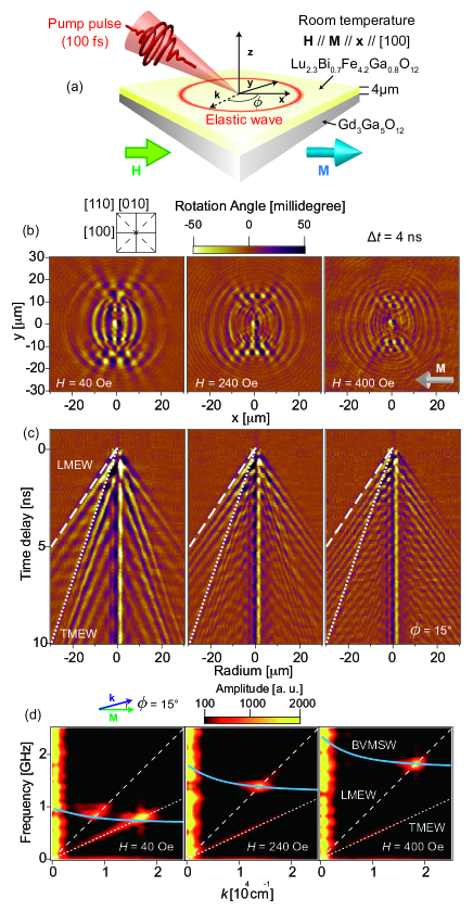
Figure 2(b) shows the magneto-optical images observed under the magnetic fields of 40 Oe, 240 Oe, and 400 Oe. The time delay between the pump and probe pulses () was set to 4 ns. We observed spin waves propagating with complicated waveforms, showing radial and concentric structures. Their magnetic nature was demonstrated by their strong magnetic field dependences, shown in Fig. 2(b). Moreover, their magnetoelastic nature was indicated by their propagation speeds of 2.9 km/s (dotted line in Fig. 2(c)) and 6.2 km/s (dashed line in Fig. 2(c)), which show good agreement with the propagation speeds of the transverse and the longitudinal elastic waves Spencer and Lecraw (1958), respectively. The convincing proof of their magnetoelastic nature is obtained by the SWaT spectra, which characterizes all propagating waves by their frequency and wavenumber Hashimoto et al. (2017). Figure 2(d) shows the SWaT spectra obtained from the data shown in Figs. 2(b) and 2(c). We found that the dominant contribution to the signal appears around the crossing of the dispersion relations of elastic waves and spin waves, which is a clear indication of magnetoelastic waves. At = 15 degree, pure-spin waves have negative group velocity so that the phase and the energy of the spin wave propagate in opposite directions. This is inconsistent with the feature of the modes discussed in this study, hence ruling out the possibility to interpret our results in a purely spin-wave picture.
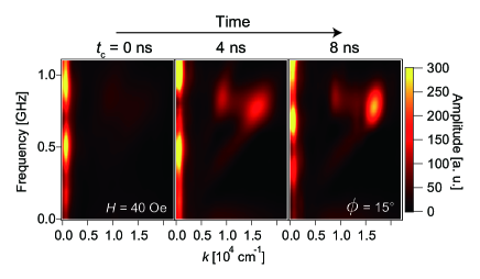
The temporal change in the amplitudes of the magnetoelastic waves is seen in time-resolved SWaT spectra obtained by applying a Gaussian time window for the calculation of the time-Fourier transform (Fig. 3(a)) Hashimoto et al. (2017). We see that the amplitudes of the magnetoelastic waves gradually increase in time. This trend rules out impulsive stimulated Raman scattering (ISRS) as a possible excitation mechanism, since the impulsive nature of the ISRS process entails that the maximum amplitude of the magnetic oscillations occurs right after the photo-excitation Kirilyuk et al. (2010); Bossini and Rasing (2017). Instead, we attribute the excitation of the magnetoelastic waves to the following process. First, the optical excitation of the sample generates -elastic waves due to the absorption of the pump pulse. Then, the -elastic waves subsequently excite magnetoelastic waves due to the magnetoelastic coupling.
In the SWaT spectra, magnetoelastic waves are observed as sharp peaks. This feature results from the excitation mechanism of magnetoelastic waves, which is a coherent energy transfer between optically-generated elastic waves and spin waves due to the magnetoelastic coupling. Spin waves excited by magnetoelastic coupling have a phase determined by the phase of their sources, i.e. the propagating elastic waves. Thus, spin waves generated at different times and different positions interfere. This interference is constructive only at the crossing of the dispersion curves of spin waves and elastic waves but destructive in other regions Hashimoto et al. (2017). This process, caused by the coherence of the magnetoelastic waves, results in the selective excitation of magnetoelastic waves at a specific frequency and wavenumber.
Consequently, the identification of the excitation mechanism of the magnetoelastic waves shifts to the potential pathways of the laser-excitation of the -elastic waves.
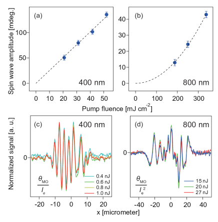
The optical excitation of the pure-elastic waves is attributed to the absorption of light by the sample. This conclusion is drawn by comparing two experiments performed with two different central pump wavelength (): 400 nm and 800 nm. The LuIG film is almost transparent at 800 nm but shows large absorption around 400 nm, attributed to a charge transfer transition (CTT) Helseth et al. (2001, 2002); Hansteen et al. (2004). Magnetoelastic waves generated by the pump pulses with different wavelengths show similar propagation but different excitation fluence dependences of their amplitudes (Fig. 4). The amplitude of the magnetoelastic waves increases linearly with the excitation fluence for = 400 nm (Fig. 4(a)) but quadratically for = 800 nm (Fig. 4(b)). These excitation fluence dependences are confirmed by comparing the cross sections of spin waves observed with different excitation fluence at the different pump wavelength. By using the data obtained with hundreds of pixels of the CCD camera, the accuracy of the interpretation is significantly improved. Figures 4(c) and 4(d) show the cross sections of the spin waves generated by pump beams with = 400 nm and = 800 nm, respectively. The data were normalized with the corresponding excitation fluence dependences shown in Figs. 4(a) and 4(b). The good agreements of the normalized data shown in each figure confirm the excitation fluence dependences of the spin waves shown in Figs. 4(a) and 4(b). We ascribe these behaviors to the excitation fluence dependences of -elastic waves, which are generated by the absorption of the pump beam through linear absorption for = 400 nm but through two-photon absorption for = 800 nm Boyd (2003). The -elastic waves excite magnetoelastic waves due to the magnetoelastic coupling, with a strength proportional to the amplitude of the elastic waves. It is well established that the absorption of light can trigger lattice excitations Thomsen et al. (1986); Zeiger et al. (1992). Disentangling the microscopic process responsible for the generation of elastic waves is beyond the scope of the present work. The main statement demonstrated here is that the absorption of light plays a crucial role in the excitation of the magnetoelastic waves, unlike the non-dissipative ISRS picture invoked in earlier work Ogawa et al. (2015). This difference may be due to the different wavelengths of the pump beams. In the study of Ref. Ogawa et al. (2015), the central wavelength of the pump beam was set to 1300 nm: in this spectral range the garnet film is fully transparent so that both one- and two-photon absorption processes cannot take place, thus leaving the ISPS process as the dominant mechanism.
Elastic waves are widely used in a number of devices such as surface acoustic wave touch screens and radio frequency filters. Thus, the mechanism demonstrated in this study, i.e. the coherent energy transfer between elastic waves and spin waves, has great potential for applications, like a frequency and wavelength selective elastic wave absorber and a spin wave transmitter. The spectral features of these devices could be controlled by applying a magnetic field. This mechanism works in ferro- and ferrimagnetic materials for any elastic waves, longitudinal, transverse, and even surface acoustic waves Dreher et al. (2012). Elastic waves do not have to be optically excited, but can be induced by other stimuli as well Dreher et al. (2012).
Our work may change the perspective concerning the role of the lattice in optically-induced spin dynamics. So far, the majority of the experiments displayed an unavoidable and undesired scattering effect of phonons on spin waves, causing damping and relaxation of the magnetic excitation. Our work provides a different scenario: the lattice vibration is now one of the ideal sources of spin waves, with frequency and wavevector selectivity.
In summary, the excitation and propagation dynamics of magnetoelastic waves in a garnet film has been investigated with a time-resolved magneto-optical imaging system Hashimoto et al. (2014). The dominant role played by the magnetoelastic coupling in the data has been demonstrated. Combining our experimental investigations with the recently developed SWaT analysis, we could identify the pathways of the excitation of spins through the coherent energy transfer from the photo-induced elastic waves to the magnetoelastic waves due to the magnetoelastic coupling. Moreover, we reveal that the excitation mechanism of the optically-generated elastic waves is dissipative in nature, being due to the absorption of light. Finally, we discussed potential applications using the coherent energy transfer between elastic waves and spin waves, investigated in this study.
Acknowledgements.
We thank Dr. T. Satoh, Dr. L. Dreher, and Prof. B. Hillebrands for fruitful discussions and Dr. S. Semin, A. van Roij and A. Toonen for their technical support. This work was financially supported by de Nederlandse Organisatie voor Wetenschappelijk Onderzoek (NWO), de Stichting voor Fundamenteel Onderzoek der Materie (FOM), the EU-FP7 project FemtoSpin (grant no. 281043), and ERC grant agreement No 339813 (EXCHANGE). Also, this work was financially supported by JST-ERATO Grant Number JPMJER1402, and World Premier International Research Center Initiative (WPI), all from MEXT, Japan.References
- Khitun et al. (2001) A. Khitun, R. Ostroumov, and K. L. Wang, Physical Review A 64, 062304 (2001).
- Khitun et al. (2008) A. Khitun, M. Bao, and K. L. Wang, IEEE Transactions on Magnetics 44, 2141 (2008).
- Schneider et al. (2008) T. Schneider, A. A. Serga, B. Leven, B. Hillebrands, R. L. Stamps, and M. P. Kostylev, Applied Physics Letters 92, 022505 (2008).
- Serga et al. (2010) A. A. Serga, A. V. Chumak, and B. Hillebrands, Journal of Physics D-Applied Physics 43, 264002 (2010).
- Lenk et al. (2011) B. Lenk, H. Ulrichs, F. Garbs, and M. Münzenberg, Physics Reports 507, 107 (2011).
- Chumak et al. (2014) A. V. Chumak, A. A. Serga, and B. Hillebrands, Nature Communications 5, 4700 (2014).
- Chumak et al. (2015) A. V. Chumak, V. I. Vasyuchka, A. A. Serga, and B. Hillebrands, Nature Physics 11, 453 (2015).
- Kirilyuk et al. (2010) A. Kirilyuk, A. V. Kimel, and T. Rasing, Reviews of Modern Physics 82, 2731 (2010).
- Bossini et al. (2016) D. Bossini, S. Dal Conte, Y. Hashimoto, A. Secchi, R. V. Pisarev, T. Rasing, G. Cerullo, and A. V. Kimel, Nature Communications 7, 10645 (2016).
- Walowski and Münzenberg (2016) J. Walowski and M. Münzenberg, Journal of Applied Physics 120, 140901 (2016).
- Satoh et al. (2012) T. Satoh, Y. Terui, R. Moriya, B. A. Ivanov, K. Ando, E. Saitoh, T. Shimura, and K. Kuroda, Nature Photonics 6, 662 (2012).
- Au et al. (2013) Y. Au, M. Dvornik, T. Davison, E. Ahmad, P. S. Keatley, A. Vansteenkiste, B. Van Waeyenberge, and V. V. Kruglyak, Physical Review Letters 110, 097201 (2013).
- Yoshimine et al. (2014) I. Yoshimine, T. Satoh, R. Iida, A. Stupakiewicz, A. Maziewski, and T. Shimura, Journal of Applied Physics 116, 043907 (2014).
- Iihama et al. (2016) S. Iihama, Y. Sasaki, A. Sugihara, A. Kamimaki, Y. Ando, and S. Mizukami, Physical Review B 94, 020401(R) (2016).
- Yoshimine et al. (2017) I. Yoshimine, Y. Y. Tanaka, T. Shimura, and T. Satoh, EPL 117, 67001 (2017).
- Hashimoto et al. (2017) Y. Hashimoto, S. Daimon, R. Iguchi, Y. Oikawa, K. Shen, K. Sato, D. Bossini, Y. Tabuchi, T. Satoh, B. Hillebrands, G. E. W. Bauer, T. H. Johansen, A. Kirilyuk, T. Rasing, and E. Saitoh, Nature Communications 8, 15859 (2017).
- Kamimaki et al. (2017) A. Kamimaki, S. Iihama, Y. Sasaki, Y. Ando, and S. Mizukami, Physical Review B 96, 014438 (2017).
- Savochkin et al. (2017) I. V. Savochkin, M. Jäckl, V. I. Belotelov, I. A. Akimov, M. A. Kozhaev, D. A. Sylgacheva, A. I. Chernov, A. N. Shaposhnikov, A. R. Prokopov, V. N. Berzhansky, D. R. Yakovlev, A. K. Zvezdin, and M. Bayer, Scientific Reports 7, 391002 (2017).
- Chikazumi (1997) S. Chikazumi, Physics of Ferromagnetism (Oxford University Press, 1997).
- Kittel (1958) C. Kittel, Physical Review 110, 836 (1958).
- Schlömann (1960) E. Schlömann, Journal of Applied Physics 31, 1647 (1960).
- Kobayashi et al. (1971) T. Kobayashi, R. C. Barker, and A. Yelon, IEEE Transactions on Magnetics 7, 755 (1971).
- Weiler et al. (2011) M. Weiler, L. Dreher, C. Heeg, H. Huebl, R. Gross, M. S. Brandt, and S. T. B. Goennenwein, Physical Review Letters 106, 117601 (2011).
- Dreher et al. (2012) L. Dreher, M. Weiler, M. Pernpeintner, H. Huebl, R. Gross, M. S. Brandt, and S. T. B. Goennenwein, Physical Review B 86, 134415 (2012).
- Ruckriegel et al. (2014) A. Ruckriegel, P. Kopietz, D. A. Bozhko, A. A. Serga, and B. Hillebrands, Physical Review B 89, 184413 (2014).
- Shen and Bauer (2015) K. Shen and G. E. W. Bauer, Physical Review Letters 115, 197201 (2015).
- Eshbach (1962) J. R. Eshbach, Physical Review Letters 8, 357 (1962).
- Eshbach (1963) J. R. Eshbach, Journal of Applied Physics 34, 1298 (1963).
- Strauss (1965) W. Strauss, Proceedings of the Institute of Electrical and Electronics Engineers 53, 1485 (1965).
- Comstock (1965) R. L. Comstock, Proceedings of the IEEE 53, 1508 (1965).
- Lemanov et al. (1971) V. V. Lemanov, A. V. Pavlenko, and A. N. Grishmanovskii, Soviet Physics Jetp-Ussr 32, 389 (1971).
- Weiler et al. (2012) M. Weiler, H. Huebl, F. S. Goerg, F. D. Czeschka, R. Gross, and S. T. B. Goennenwein, Physical Review Letters 108, 176601 (2012).
- Afanasiev et al. (2014) D. Afanasiev, I. Razdolski, K. M. Skibinsky, D. Bolotin, S. V. Yagupov, M. B. Strugatsky, A. Kirilyuk, T. Rasing, and A. V. Kimel, Physical Review Letters 112, 147403 (2014).
- Janusonis et al. (2016) J. Janusonis, C. L. Chang, T. Jansma, A. Gatilova, V. S. Vlasov, A. M. Lomonosov, V. V. Temnov, and R. I. Tobey, Physical Review B 94, 024415 (2016).
- Kikkawa et al. (2016) T. Kikkawa, K. Shen, B. Flebus, R. A. Duine, K.-i. Uchida, Z. Qiu, G. E. W. Bauer, and E. Saitoh, Physical Review Letters 117, 207203 (2016).
- Ogawa et al. (2015) N. Ogawa, W. Koshibae, A. J. Beekman, N. Nagaosa, M. Kubota, M. Kawasaki, and Y. Tokura, Proceedings of the National Academy of Sciences of the United States of America 112, 8977 (2015).
- Bossini and Rasing (2017) D. Bossini and T. Rasing, Physica Scripta 92, 024002 (2017).
- Hashimoto et al. (2014) Y. Hashimoto, A. R. Khorsand, M. Savoini, B. Koene, D. Bossini, A. Tsukamoto, A. Itoh, Y. Ohtsuka, K. Aoshima, A. V. Kimel, A. Kirilyuk, and T. Rasing, Review of Scientific Instruments 85, 063702 (2014).
- Helseth et al. (2001) L. E. Helseth, R. W. Hansen, E. I. Il’yashenko, M. Baziljevich, and T. H. Johansen, Physical Review B 64, 174406 (2001).
- Helseth et al. (2002) L. E. Helseth, A. G. Solovyev, R. W. Hansen, E. I. Il’yashenko, M. Baziljevich, and T. H. Johansen, Physical Review B 66, 064405 (2002).
- Hansteen et al. (2004) F. Hansteen, L. E. Helseth, T. H. Johansen, O. Hunderi, A. Kirilyuk, and T. Rasing, Thin Solid Films 455-456, 429 (2004).
- Yamazaki et al. (2004) H. Yamazaki, O. Matsuda, and O. B. Wright, physica status solidi (c) 1, 2991 (2004).
- Zvezdin and Kotov (1997) A. K. Zvezdin and V. A. Kotov, Modern Magnetooptics and Magnetooptical Materials (CRC Press, 1997).
- Spencer and Lecraw (1958) E. G. Spencer and R. C. Lecraw, Physical Review Letters 1, 241 (1958).
- Boyd (2003) R. W. Boyd, Nonlinear Optics, 2nd ed. (ACADEMIC PRESS, 2003).
- Thomsen et al. (1986) C. Thomsen, H. T. Grahn, H. J. Maris, and J. Tauc, Physical Review B 34, 4129 (1986).
- Zeiger et al. (1992) H. J. Zeiger, J. Vidal, T. K. Cheng, E. P. Ippen, G. Dresselhaus, and M. S. Dresselhaus, Physical Review B 45, 768 (1992).