On EM Reconstruction of a Multi Channel Shielded Applicator for Cervical Cancer Brachytherapy: A Feasibility Study.
Abstract
Electromagnetic tracking (EMT) is a promising technology for automated catheter and applicator reconstructions in brachytherapy. In this work, a proof-of-concept is presented for reconstruction of the individual channels of a shielded tandem applicator dedicated to intensity modulated brachytherapy. All six channels of a straight prototype was reconstructed and the distance between two opposite channels was measured. A study was also conducted on the influence of the shield on the data fluctuation of the EMT system. The differences with the CAD specified dimensions are under 2 mm. The pair of channels which has one of it more distant from the generator have higher inter-channel distance with higher variability. In the first 110 cm reconstruction, all inter-channel distances are within the geometrical tolerances. According to a paired Student t-test, the data given by the EM system with and without the shield applicator tip are not significantly different. This study shows that the reconstruction of channel path within the mechanical accuracy of the applicator is possible.
Corresponding author: Luc Beaulieu
email: Luc.Beaulieu@phy.ulaval.ca
1 Introduction
The emergence of electromagnetic tracking (EMT) technologies is currently paving the way for a variety of novel applications in the medical field. It is used in surgical procedures [1] and gives accurate measurements [2] [3]. The Aurora system is composed of a field generator which produces a known EM field within a known volume inside of which, a small sensor is placed. The system uses mutual induction to compute the position of the sensor. Brachytherapy is used to treat numerous body sites including prostate, breast and gynecology. Many different applications of minimally invasive image-guided brachytherapy has been demonstrated like magnetic resonance tomography, computed tomography or ultrasound imaging data. All of those reconstruction imaging method have their limitations in term of accuracy [4] [5] [6].
It is possible to use the EMT to perform reconstructions [7]. At the same time, shielded applicators for brachytherapy treatment are making their way to improve dosimetric results [8]. According to the literature, EMT tracking accuracy is sensible to conductive metal which causes distorsion in the magnetic field [9][10].
In this technical note, a proof-of-concept is presented for reconstruction of the individual channels of a prototype shielded tandem applicator. The technique has the potential to considerably speed up planning and quality assessment tasks in the clinic, particularly in this context when imaging of such applicator might be impossible due to artifacts. The purpose of this work is to show the feasibility of EMT for automated multi-channel shielded applicator reconstruction.
2 Materials and Methods
2.1 Straight shielded applicator
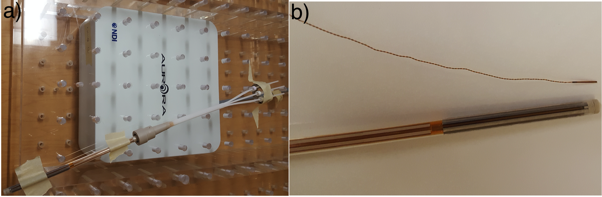
The applicator used in this work corresponds to the straight, intra-uterine portion of a shielded multi-channel tandem prototype previously described by Han et al. (Figure 1a) [11]. All 6 channels were reconstructed (Figure 1b). The inter-channel distances of each opposite channel pair, measured along the complete 14 cm length, was used as a metric for validation of EMT reconstruction i.e. absence of geometric distortion. Figure 1c shows that this nominal distance is 4 mm. The shielded part is composed of a MR-compatible non-ferromagnetic alloy made of 95tungsten, 3.5 nickel, and 1.5 copper) as described in Han et al. [11].

2.2 EM tracking system
The shielded tandem applicator was reconstructed using the Aurora® V3 EMT system (NDI, Waterloo, Ontario, Canada) using model 610090 5 degree-of-freedom (DOF) sensor (Figure 2). The sensor is a cylinder of 0.8 mm diameter and 11 mm long (Figure 2).
2.3 Effect of shield on data fluctuation
Repeated measurements of EMT sensor positions within the detection volume, with and without the shielded applicator present, was used to study the potential perturbation effect of the shield on the sensor reading (Figure 2). The effective detection volume is a cube of 50 cm side [3]. Nine different positions (one near each corner of the sensible cube volume and the center) were taken each time with the sensor directly on the applicator and without the applicator. Each measurement last 10 s with a frequency of 40 measurements/s. On Figure 2, the shielded end of the applicator is place next to the sensor.
3 Results and discussion
3.1 Straight shielded applicator
The distance between the center of 2 opposite channels is nominally 4 mm from center-to-center (Figure 1c). Table 1 reports the experimental distances computed from the EMT reconstructed channel. The average distance from each pair met the nominal distance within one standard deviation. It is also important to note that each hole is 1.4 mm in depth (or 0.7 mm from its center).
| Pair | Average | Standard deviation |
|---|---|---|
| number | (mm) | (mm) |
| 3-6 | 4.33 | 0.40 |
| 2-5 | 3.88 | 0.26 |
| 1-4 | 4.14 | 0.35 |
The distance were also computed along the reconstruction path.
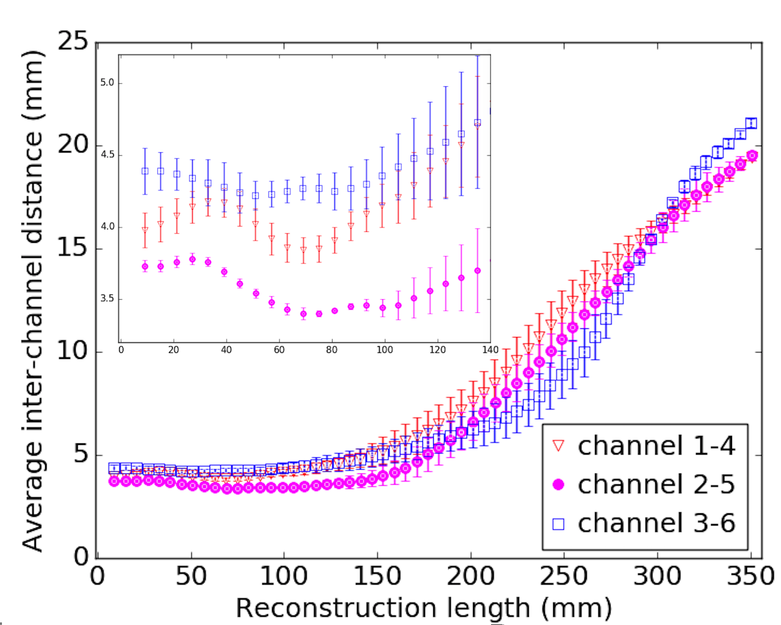
On Figure 3, one can see that most of the shielded part has its inter-channel distance within the geometrical tolerance of the setup. The channel which were on the side during the reconstruction have their inter-channel distance always below the nominal value. Meaning that during the reconstruction, the sensor tend to be on the side of the channel nearer to the middle of the applicator.
3.2 Stability of the reading with a shield present
With the nine different positions, a paired t-test was made. According to it, the data given by the EMT system with and without the shielded applicator tip are not significantly different (Table 2). A p-value < 0.05 was considered statistically significant. The absolute maximum difference is 0.05 mm which was for the Z axis. The standard deviation of all the differences between the acquired position with and without the shield near are sub-millimetric which is comparable with the standard deviation of the computed position.
| Position | p | |(mm) | (mm) |
|---|---|---|---|
| X | 0.09 | 0.02 | 0.04 |
| Y | 0.72 | 0.01 | 0.05 |
| Z | 0.38 | 0.05 | 0.03 |
3.3 Limitations
The off-the-shelf sensor used in this work is smaller than the channel diameter of the applicator. This could explain part of the fluctuation seen in the interchannel distance measurements. Furthermore, the applicator geometrical tolerance, corresponding to the channels radius, has to be taken into account. As such, a maximum of 0.7 mm difference has been considered in the analysis, represented by the dashed lines in Figure 3. After the first 110 cm of reconstruction, the measured interchannel-distances increases. As we approach the end of the shielded portion of the applicator, channels start to move away from each other and the sensor with it. Also, since the sensor is smaller than the channel radius, the effect of gravity on the sensor position within the channels cannot be ruled out.
While this is a shielded applicator, each channel is not completely closed (Figure 1c) and material is non-ferromagnetic. As such the results obtained here might not be generalizable to all shielded geometries, in particular those that would fully enclose the sensor, acting as a Faraday cage.
4 Conclusion
It appears feasible to use EMT technology to reconstruct shielded applicators, such as the one presented here. The observed deviations shows no statistically significant effect on the presence of the shielding material in the detection volume (static configuration). This study demonstrates that it is possible to reconstruct the channel path within the mechanical accuracy of the applicators.
Acknowledgments
This work was supported by the National Sciences and Engineering Research Council of Canada (NSERC) via the NSERC-Elekta Industrial Research Chair. Daline Tho acknowledges support from the Medical Physics Training Network CREATE NSERC grant # 432290.
References
- [1] C. Nafis, V. Jensen, L. Beauregard, and P. Anderson. Method for estimating dynamic EM tracking accuracy of surgical navigation tools. 6141:152–167, March 2006.
- [2] Elodie Lugez, Hossein Sadjadi, David R. Pichora, Randy E. Ellis, Selim G. Akl, and Gabor Fichtinger. Electromagnetic tracking in surgical and interventional environments: usability study. International Journal of Computer Assisted Radiology and Surgery, 10(3):253–262, 2015.
- [3] Northern Digital Inc.
- [4] Jose Richart, Antonio Otal, Silvia Rodriguez, Ana Isabel Nicolás, Marina DePiaggio, Manuel Santos, Javier Vijande, Facundo Ballester, and Jose Perez-Calatayud. A practical mri-based reconstruction method for a new endocavitary and interstitial gynaecological template. Journal of Contemporary Brachytherapy, 7(5):407–414, 10 2015.
- [5] Ryan L. Smith, Annette Haworth, Vanessa Panettieri, Jeremy L. Millar, and Rick D. Franich. 3d catheter reconstruction in hdr prostate brachytherapy for pre-treatment verification using a flat panel detector. Physica Medica: European Journal of Medical Physics, 2017/06/28.
- [6] Amy Limbacher, Evelyn Sebastian, Di Yan, and Jun Zhou. Discrepancies of catheter reconstruction in ultrasound imageguided prostate high-dose-rate brachytherapy and their effects on dosimetry. Brachytherapy, pages S114–S115, 2017/06/28.
- [7] Antonio L. Damato, Akila N. Viswanathan, Sarah M. Don, Jorgen L. Hansen, and Robert A. Cormack. A system to use electromagnetic tracking for the quality assurance of brachytherapy catheter digitization. Medical Physics, 41(10):n/a–n/a, 2014.
- [8] Matthew J. Webster, Slobodan Devic, Te Vuong, Dae Yup Han, Dan Scanderbeg, Dongju Choi, Bongyong Song, and William Y. Song. HDR Brachytherapy of rectal cancer using a novel grooved-shielding applicator design. Medical Physics, 40(9): n/a–n/a,2013.
- [9] S. Boutaleb, E. Racine, O. Fillion, A. Bonillas, G. Hautvast, D. Binnekamp, and L. Beaulieu. Performance and suitability assessment of a real-time 3d electromagnetic needle tracking system for insterstial brachytherapy. Contemporary Brachytherapy, 7(4):n/a–n/a, 2014.
- [10] Jun Zhou, Evelyn Sebastian, Victor Mangona, and Di Yan. Real-time catheter tracking for high-dose-rate prostate brachytherapy using an electromagnetic 3d-guidance device: A preliminary performance study. Medical Physics, 40(2):021716–n/a, 2013. 021716.
- [11] Dae Yup Han, Matthew J. Webster, Daniel J. Scanderbeg, Catheryn Yashar, Dongju Choi, Bongyong Song, Slobodan Devic, Ananth Ravi, and William Y. Song. Direction-modulated brachytherapy for high-dose-rate treatment of cervical cancer. i: Theoretical design. International Journal of Radiation Oncology*Biology*Physics, 89(3):666 – 673, 2014.