Ultrafast Carrier Dynamics in VO2 across the Pressure-Induced Insulator-to-Metal Transition
Abstract
We utilize near-infrared pump – mid-infrared probe spectroscopy to investigate the ultrafast electronic response of pressurized VO2. Distinct pump–probe signals and a pumping threshold behavior are observed even in the pressure-induced metallic state showing a noticeable amount of localized electronic states. Our results are consistent with a scenario of a bandwidth-controlled Mott-Hubbard transition.
pacs:
Vanadium dioxide (VO2) exhibits a sharp insulator-to-metal transition (IMT) accompanied by a transformation from the monoclinic to rutile crystal structure above a critical temperature K Morin (1959). The strong coupling between electronic and lattice subsystems during the phase transition has attracted continuing interest for more than a half century Goodenough (1971); Imada et al. (1998); Basov et al. (2011). The dimerized vanadium chains of the monoclinic phase suggests that a Peierls distortion underlies the insulating state, but electronic correlation leading to the carrier localization typical of a Mott insulator is also observed Biermann et al. (2005); Qazilbash et al. (2007); Weber et al. (2012); Huffman et al. (2017). The IMT in VO2 under the influence of temperature Koethe et al. (2006); Kim et al. (2006); Qazilbash et al. (2007); Budai et al. (2014), strain Gray et al. (2016); Park et al. (2013); Aetukuri et al. (2013) and chemical substitution Marezio et al. (1972); Pouget et al. (1974) has been extensively studied. Some of these studies show that the electronic and structural phase transitions are separable within certain temperature ranges, pointing to a primary role for electron correlation as the driving mechanism for the IMT Kim et al. (2006); Gray et al. (2016).
Application of external pressure offers an attractive way to distinguish the influence of the structural instability from the correlation effects on the IMT in VO2. At sufficiently high pressures, VO2 becomes metallic while keeping the dimerized monoclinic structure Arcangeletti et al. (2007); Marini et al. (2010, 2014); Baldini et al. (2016); Bai et al. (2015); Chen et al. (2017). External pressure induces only an isostructural transformation of the lattice at room temperature Mitrano et al. (2012); Bai et al. (2015); Chen et al. (2017). Thus, in this case the pressure-driven IMT should be dominated by changes in the electronic band structure which is necessarily different from that of the temperature-induced IMT. Therefore, it is highly desirable to unravel how the band structure of the monoclinic metallic phase VO2 changes under high pressure. Unfortunately, in a high-pressure diamond anvil cell (DAC), conventional photoemission spectroscopy cannot be used, and X-ray absorption spectroscopy suffers from limited energy resolution Marini et al. (2014).
Information about the electron and lattice dynamics in VO2 can be obtained using time-resolved techniques that probe the evolution of the non-thermal IMT Cavalleri et al. (2001, 2004, 2005); Hilton et al. (2007); Kim et al. (2006); Kübler et al. (2007); Pashkin et al. (2011); Cocker et al. (2012); Wall et al. (2012); Wegkamp et al. (2014); Morrison et al. (2014); O’Callahan et al. (2015); Huber et al. (2016). In particular, time-resolved photoemission spectroscopy Wegkamp et al. (2014) and ultrafast electron diffraction Morrison et al. (2014) have shown that a transient monoclinic but metallic phase can be induced via photoexcitation. A transient monoclinic metallic phase has also been reported for pressurized VO2 Hsieh et al. (2014) using time-resolved reflectivity.
Here we combine an ultrafast near-infrared pump and mid-infrared spectroscopy in a high-pressure DAC to investigate the non-equilibrium dynamics of the pressure-induced IMT in VO2. Our results provide evidence that near-infrared pumping induces additional long-lived charge carriers – even in the pressure-induced metallic phase. The utilized method of the non-degenerate nonlinear spectroscopy enables us to trace the evolution of localized, weakly localized and fully delocalized electronic states in VO2 across the pressure-driven IMT, and to draw conclusions about the appropriate correlated band structure.
We use VO2 single crystals grown by thermal decomposition of V2O5 Budai et al. (2014); sup . The samples were polished to thicknesses of 20–30 µm and cut into pieces of around 100 µm in diameter. A single piece of VO2 was mounted in an opposing-plate DAC. CsI powder was used as a pressure transmitting medium in order to ensure a direct contact between the sample surface and the front diamond anvil. The pressure inside the DAC was monitored via a standard ruby fluorescence method Mao et al. (1986).
Our pump–probe setup is based on a Ti:sapphire laser amplifier system, providing 55 fs long pulses centered at nm (1.55 eV) with a repetition rate of 250 kHz. A portion of the beam was used for the pumping branch, whereas the remaining part was utilized to generate probe pulses at µm (0.12 eV or 30 THz) using difference frequency mixing between signal and idler pulses from a parametric amplifier. The pump and probe beams were focused non-collinearly on the sample inside the DAC down to a spot size of 30-50 µm (FWHM). We then measure the change in reflectivity of photo-excited VO2 with respect to the reflectivity in the unexcited state. The small photon energy of the probe beam (well below the band gap energy eV of VO2 at ambient conditions) ensures that the pump–probe signal is dominated by the response of free charge carriers Kübler et al. (2007); Pashkin et al. (2011).
Figure 1(a) shows the change of the pump–probe signal of VO2 at 2.1 GPa measured for different pump fluences. All of the curves show a quasi-instantaneous increase of the reflectivity – limited only by the durations of the pump and the probe pulses. The onset is followed by a fast relaxation with a time constant of approximately 0.2 ps sup . At low fluence (green trace), the pump–probe signal vanishes after about 1 ps indicating a return to complete localization of the photo-excited charge carriers. At pump fluences above a threshold a persistant enhanced reflectivity reveals the creation of a metastable metallic phase [see the orange and red trace in Fig. 1(a)]. This state survives for hundreds of picoseconds in agreement with previous studies Becker et al. (1994); Hilton et al. (2007); Brady et al. (2016). All of these observations are fully consistent with the results for ambient conditions published previously Kübler et al. (2007). The threshold fluence corresponds to the photoinjection of a critical density of free charge carriers that screen the Coulomb interaction and thus induce the collapse of the energy gap, thus leading to the metastable metallic state Kim et al. (2006); Kübler et al. (2007); Wegkamp et al. (2014).
The volume of the metastable metallic phase and correspondingly the amplitude of the persistent pump–probe signal grows, when the incident pump fluence is increased above the threshold . This is related to the fact that the penetration depth of the mid-infrared probe and the sample thickness are much larger than the absorption length of the near-infrared pump; the photoexcitation switches only a relatively thin surface layer of the VO2 crystal.
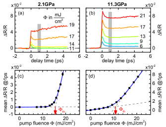
Remarkably, in contrast to the low-pressure regime, at elevated pressures the long-lived excited state is already observed at pump fluences below the threshold as shown in Fig. 1(b). This becomes more obvious when we plot pump–probe signals averaged around 1 ps (where the relaxation process is already completed) as a function of the pump fluence. Figures 1(c) and 1(d) show such plots for the same pressures as for Fig. 1(a) and 1(b), respectively. The threshold behavior is clearly seen in both cases. In contrast to previous works Kübler et al. (2007); Pashkin et al. (2011); Cocker et al. (2012), the threshold is no longer well-defined by the crossing of the high-fluence asymptote with the x-axis, since finite signals are observed down to the lowest pump fluences. Therefore, we define as a crossing point (marked by red arrows) of the two asymptotes (dashed gray lines) illustrated in Fig. 1(c) and (d). Details of the bi-asymptotic fit function are given in sup .
The analysis of pump–probe traces at different pump fluences and various pressures reveals two main parameters that exhibit anomalous pressure behavior: (i) threshold fluence and (ii) slope of the low-fluence asymptote denoted as sup . Figure 2 shows the pressure dependence of these key parameters together with the linear (unpumped) transmissivity and reflectivity at the probe photon energy.
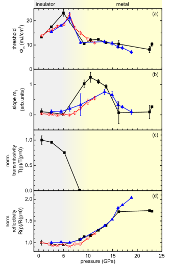
Starting from ambient pressure, the threshold fluence monotonically increases up to a critical pressure of 6-8 GPa as shown in Fig. 2(a). A higher threshold fluence means that more free carriers have to be photoinjected to overcome the correlations and to induce the metastable metallic state. This trend contrasts sharply with the behavior of the thermally driven IMT where a noticeable decrease of the threshold fluence on approaching has been reported Pashkin et al. (2011); Cocker et al. (2012). Most probably this difference is related to the stiffening of the dimerized lattice structure under pressure; that makes the vanadium dimers even more stable and thus raises the energy barrier to the metallic rutile phase. A stiffening of phonons observed in the pressure-dependent Raman measurements Arcangeletti et al. (2007); Bai et al. (2015) as well as enhanced dimerization of the vanadium sublattice seen in X-ray scattering Baldini et al. (2016) confirm this interpretation. Furthermore, the observed increase of with pressure is consistent with the recently reported growth of in VO2 under pressure and the estimations of the corresponding increase in the latent heat of the IMT Chen et al. (2017).
Around the critical pressure we observe an anomalous drop of . Remarkably, it coincides with the vanishing of the linear transmissivity [Fig. 2(c)], the start of the increase in the reflectivity [Fig. 2(d)] as well as the onset of a finite slope of the low-fluence asymptote [Fig. 2(b)]. This effect is highly reproducible and has been observed in independent measurements on three different VO2 samples as presented in Fig. 2. We interpret the observed anomaly as a pressure-driven IMT in VO2. Our results for the linear mid-infrared response agree with the work of Arcangeletti et al. Arcangeletti et al. (2007) where this phenomenon was reported for the first time. The sharper drop in our transmission-versus-pressure data can be explained by the much larger thickness of our sample. The reflectivity starts to increase for due to the presence of delocalized charge carriers and continues to grow as their density and correspondingly the plasma frequency become larger. In agreement with previous reports Arcangeletti et al. (2007); Marini et al. (2010); Mitrano et al. (2012); Bai et al. (2015), our complementary high-pressure Raman measurements confirm that VO2 samples also preserve the monoclinic crystal structure far above sup .
Let us now discuss the nonlinear response of VO2 in the pressure-induced metallic state. The similar shape of the pump-probe response beyond the critical pressure [see Fig. 1(b)] indicates that the majority of vanadium -electrons remain localized even in the pressure-driven metallic phase and a photoexcitation is still able to induce a phase with a higher conductivity. Moreover, the finite values of the threshold fluence [see Fig. 2(a)] suggest that the photo-induced metallization for is governed by the same mechanism as for pressures below . The observed drop of above the IMT can be related to the finite pressure-induced density of free charge carriers that should lead to a partial screening of the Coulomb correlation. As a result, fewer photoinjected carriers are required to achieve the critical concentration that closes the band gap.
A further increase of pressure up to 23 GPa causes gradual lowering of indicating that the density of free charge carriers grows with pressure. Nevertheless, it remains below the critical concentration necessary for complete suppression of the carrier localization. This is consistent with the behavior of the pressure-dependent reflectivity that monotonically increases up to the highest pressures – suggesting that the plasma frequency of free charge carriers just slightly exceeds the frequency of our mid-infrared probe. Assuming an electron mass Qazilbash et al. (2007), we estimate the density of free electrons to be roughly cm-3 – still a factor five lower than the critical concentration of cm-3 needed for the photo-induced phase transition at ambient pressure Pashkin et al. (2011). At high pressures, the critical concentration may be even higher. Thus, the equilibrium density of free carriers for is still well below the critical limit, that could be the reason for the quite moderate decrease of the threshold beyond .
The pressure-induced IMT also leads to the onset of a non-vanishing slope of the low-fluence asymptote [see Fig. 2(b)]. This means that for even pumping well below a threshold fluence can induce a metastable metallic phase with enhanced reflectivity, as illustrated in Fig. 1(b). In other words, a certain amount of long-lived free charge carriers directly proportional to the number of pump photons can be added without reaching the critical concentration for a band gap collapse. Thus, the appearance of a finite is expected to be a non-cooperative phenomenon related to the photoexcitation of weakly localized states (WLS) located near the Fermi level.
The observed behavior of the pump–probe response across the pressure-driven IMT can be understood on the basis of the tentative band diagrams depicted in Fig. 3(a)-(c). Figure 3(a) shows the band structure of VO2 at ambient conditions established in previous studies Goodenough (1971); Biermann et al. (2005); Koethe et al. (2006). The vanadium orbitals overlapping along the axis Pashkin et al. (2011) form two relatively narrow bands usually denoted as . The low- and the high-energy bands correspond to bonding and antibonding combinations of electronic orbitals on a vanadium dimer, respectively. Due to the on-site Coulomb repulsion of electrons, the lower and upper Hubbard bands are formed on the lower and upper energy ends of the bands, respectively Biermann et al. (2005). In our qualitative band diagrams, we present both bands as somewhat broader bands. The band gap is formed between edges of the lower band and the band. Photoexcitation promotes the localized electrons from the band into the high-energy delocalized states [red arrow in Fig. 3(a)] leading to the transient increase of reflectivity.
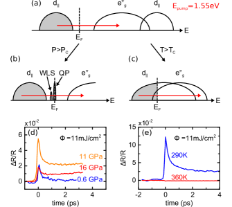
In our measurements, we observe photo-induced free charge carriers typical of the insulating VO2 phase well above the pressure-induced IMT – in stark contrast to the temperature-induced transition. This difference becomes clear by comparing the pump–probe signals measured on our VO2 using the same excitation fluence for different pressures [Fig. 3(d)] and for different temperatures [Fig. 3(e)]. For temperatures above , the metallic VO2 phase possesses the rutile crystal structure without the dimerization. Thus, bands become degenerate and cross the Fermi level together with other bands [see Fig. 3(c)] resulting in a complete delocalization of all electrons of vanadium ions. Therefore, the photoexcitation increases the electronic temperature, but it does not lead to an increase in the total number of free charge carriers. Correspondingly we observe a very weak negative change in for as shown by the red trace in Fig. 3(e). In contrast to this, the pressure-induced metallic state of VO2 is monoclinic sup suggesting that the bands remain split. The drop of the threshold fluence leads to an enhanced photosusceptibility sup , and the sizable pump-induced for signals shown in Fig. 3(d) indicate that a large part of the electrons are localized, i.e., they occupy states in a band which does not cross the Fermi level. This assumption is supported by the X-ray absorption measurements by Marini et al. Marini et al. (2014) which show that the spectral weight transfer needed to achieve the pressure-induced metallic monoclinic phase is much smaller than for the temperature-driven transition to the rutile phase.
In addition, as discussed above, the appearance of finite values suggests the existence of weakly localized states WLS for . These conclusions result in the tentative band diagram shown in Fig. 3(b). It assumes that the band splitting is also preserved above the pressure-induced IMT, and the metallic conductivity originates from a spectral weight transfer to a narrow intragap quasiparticle peak (QP) at the Fermi level. Its low-energy satellite represents the WLS. At elevated pressures well above , the QP gains more spectral weight leading to higher metallic conductivity in line with the observed increase in reflectivity [Fig. 2(d)]. At the same time, the pump–probe signal becomes smaller since the photo-induced relative change in the density of free charge carriers decreases for a given pump fluence [see the decrease from orange to red trace in Fig. 3(d)]. Finally, vanishes at high pressures [Fig. 2(b)] indicating that the WLS merges with the QP.
We now discuss the scenario of the pressure-induced IMT in VO2 via the suggested band diagram. Our data convincingly demonstrate that the dimerized monoclinic structure is preserved across the IMT and even becomes more stable under an initial pressure increase. Thus, the intimate coupling between the electronic and lattice subsystems characteristic of the temperature-driven IMT does not take place for the pressure-induced IMT which should have a predominantly electronic origin. The simplest scenario in this case is an IMT in the nondegenerate Hubbard model. Single-site dynamical mean field theory (DMFT) calculations for such model show that an increasing portion of spectral weight is transferred from the lower and upper Hubbard bands to a quasipartcle peak at the Fermi level when the effective correlation drops below a critical value, while the Hubbard bands persist Zhang et al. (1993); Khomskii (2014). The onset of the QP and the redistribution of spectral weight are governed by the ratio of the Coulomb repulsion and the hopping bandwidth Khomskii (2014).
Application of pressure improves the overlap between the orbitals leading to increased bandwidth and reduced effective correlation that eventually results in a bandwidth-controlled Mott-Hubbard insulator-to-metal transition. Thus, this simple model is capable of explaining the existence of the localized states in the lower-energy band even above the IMT as suggested by our present study [Fig. 3(b)]. The only discrepancy is related to the existence of the WLS that are absent for the bandwidth-controlled IMT in the standard nondegenerate Hubbard model in single-site DMFT Zhang et al. (1993); Khomskii (2014).
We expect two possible reasons to be responsible for the appearance of WLS: (i) The WLS may be caused by lattice defects in the VO2 crystal. Possibly their density increases notably due to the high strain imposed by external pressure O’Callahan et al. (2015). Initially delocalized electrons in the narrow QP are known to possess a high effective mass due to strong correlation effects Qazilbash et al. (2007); Khomskii (2014) and, thus, may be localized and bound to lattice defects. With further increasing pressure and weaker correlation, the effective mass should strongly decrease such that the binding energy of the localized states may drop below the energy of thermal fluctuations at room temperature. As a result, the bound states will be ionized and the WLS peak will merge with the QP. (ii) The WLS may be an intrinsic feature of a realistic Hubbard model which goes beyond the single-site approximation and includes all relevant bands in VO2 and must be solved using contemporary calculation techniques Biermann et al. (2005); He and Millis (2016); Brito et al. (2016). Unfortunately, to the best of our knowledge, no such modeling has yet been performed for a pressure-induced metallic phase of VO2.
In conclusion, we have observed a pressure-induced metallic monoclinic phase of VO2 above a critical pressure of 6-8 GPa by using nonlinear pump–probe spectroscopy. The photo-induced response of VO2 above the pressure-induced IMT is remarkably different as compared to the temperature-driven transition. This behavior agrees well with the scenario of a bandwidth-controlled Mott-Hubbard transition, where the strongly correlated metallic phase appears due to a spectral weight transfer from the Hubbard bands to delocalized states at the Fermi level. Thus, the application of external pressure provides a structural stability of VO2 and reveales the purely electronic character of the insulator-to-metal phase transition.
Acknowledgements.
We thank A. Leitenstorfer and P.M. Oppeneer for fruitful discussions and support. This work was financially supported by the DFG (project 2113-1/1: A.P., J.M.B.). Research at the Oak Ridge National Laboratory for one author (L.A.B.) was supported by the U.S. Department of Energy, Office of Science, Basic Energy Sciences, Materials Sciences and Engineering Division. REM and RFH gratefully acknowledge funding from the National Science Foundation (DMR-1207507).References
- Morin (1959) F. J. Morin, Phys. Rev. Lett. 3, 34 (1959).
- Goodenough (1971) J. B. Goodenough, J. Solid State Chem. 3, 490 (1971).
- Imada et al. (1998) M. Imada, A. Fujimori, and Y. Tokura, Rev. Mod. Phys. 70, 1039 (1998).
- Basov et al. (2011) D. N. Basov, R. D. Averitt, D. van der Marel, M. Dressel, and K. Haule, Rev. Mod. Phys. 83, 471 (2011).
- Biermann et al. (2005) S. Biermann, A. Poteryaev, A. I. Lichtenstein, and A. Georges, Phys. Rev. Lett. 94, 026404 (2005).
- Qazilbash et al. (2007) M. M. Qazilbash, M. Brehm, B.-G. Chae, P.-C. Ho, G. O. Andreev, B.-J. Kim, S. J. Yun, A. V. Balatsky, M. B. Maple, F. Keilmann, H.-T. Kim, and D. N. Basov, Science 318, 1750 (2007).
- Weber et al. (2012) C. Weber, D. D. O’Regan, N. D. M. Hine, M. C. Payne, G. Kotliar, and P. B. Littlewood, Phys. Rev. Lett. 108, 256402 (2012).
- Huffman et al. (2017) T. J. Huffman, C. Hendriks, E. J. Walter, J. Yoon, H. Ju, R. Smith, G. L. Carr, H. Krakauer, and M. M. Qazilbash, Phys. Rev. B 95, 075125 (2017).
- Koethe et al. (2006) T. C. Koethe, Z. Hu, M. W. Haverkort, C. Schüßler-Langeheine, F. Venturini, N. B. Brookes, O. Tjernberg, W. Reichelt, H. H. Hsieh, H.-J. Lin, C. T. Chen, and L. H. Tjeng, Phys. Rev. Lett. 97, 116402 (2006).
- Kim et al. (2006) H.-T. Kim, Y. W. Lee, B.-J. Kim, B.-G. Chae, S. J. Yun, K.-Y. Kang, K.-J. Han, K.-J. Yee, and Y.-S. Lim, Phys. Rev. Lett. 97, 266401 (2006).
- Budai et al. (2014) J. D. Budai, J. Hong, M. E. Manley, E. D. Specht, C. W. Li, J. Z. Tischler, D. L. Abernathy, A. H. Said, B. M. Leu, L. A. Boatner, R. J. McQueeney, and O. Delaire, Nature 515, 535 (2014).
- Gray et al. (2016) A. X. Gray, J. Jeong, N. P. Aetukuri, P. Granitzka, Z. Chen, R. Kukreja, D. Higley, T. Chase, A. H. Reid, H. Ohldag, M. A. Marcus, A. Scholl, A. T. Young, A. Doran, C. A. Jenkins, P. Shafer, E. Arenholz, M. G. Samant, S. S. P. Parkin, and H. A. Dürr, Phys. Rev. Lett. 116, 116403 (2016).
- Park et al. (2013) J. H. Park, J. M. Coy, T. S. Kasirga, C. Huang, Z. Fei, S. Hunter, and D. H. Cobden, Nature 500, 431 (2013).
- Aetukuri et al. (2013) N. B. Aetukuri, A. X. Gray, M. Drouard, M. Cossale, L. Gao, A. H. Reid, R. Kukreja, H. Ohldag, C. A. Jenkins, E. Arenholz, K. P. Roche, H. A. Dürr, M. G. Samant, and S. S. P. Parkin, Nat. Phys. 9, 661 (2013).
- Marezio et al. (1972) M. Marezio, D. B. McWhan, J. P. Remeika, and P. D. Dernier, Phys. Rev. B 5, 2541 (1972).
- Pouget et al. (1974) J. P. Pouget, H. Launois, T. M. Rice, P. Dernier, A. Gossard, G. Villeneuve, and P. Hagenmuller, Phys. Rev. B 10, 1801 (1974).
- Arcangeletti et al. (2007) E. Arcangeletti, L. Baldassarre, D. Di Castro, S. Lupi, L. Malavasi, C. Marini, A. Perucchi, and P. Postorino, Phys. Rev. Lett. 98, 196406 (2007).
- Marini et al. (2010) C. Marini, L. Baldassarre, M. Baldini, A. Perucchi, D. Di Castro, L. Malavasi, S. Lupi, and P. Postorino, High Press. Res. 30, 55 (2010).
- Marini et al. (2014) C. Marini, M. Bendele, B. Joseph, I. Kantor, M. Mitrano, O. Mathon, M. Baldini, L. Malavasi, S. Pascarelli, and P. Postorino, Europhys. Lett. 108, 36003 (2014).
- Baldini et al. (2016) M. Baldini, P. Postorino, L. Malavasi, C. Marini, K. W. Chapman, and H.-k. Mao, Phys. Rev. B 93, 245137 (2016).
- Bai et al. (2015) L. Bai, Q. Li, S. A. Corr, Y. Meng, C. Park, S. V. Sinogeikin, C. Ko, J. Wu, and G. Shen, Phys. Rev. B 91, 104110 (2015).
- Chen et al. (2017) Y. Chen, S. Zhang, F. Ke, C. Ko, S. Lee, K. Liu, B. Chen, J. W. Ager, R. Jeanloz, V. Eyert, and J. Wu, Nano Lett. 17, 2512 (2017).
- Mitrano et al. (2012) M. Mitrano, B. Maroni, C. Marini, M. Hanfland, B. Joseph, P. Postorino, and L. Malavasi, Phys. Rev. B 85, 184108 (2012).
- Cavalleri et al. (2001) A. Cavalleri, C. Tóth, C. W. Siders, J. A. Squier, F. Ráksi, P. Forget, and J. C. Kieffer, Phys. Rev. Lett. 87, 237401 (2001).
- Cavalleri et al. (2004) A. Cavalleri, T. Dekorsy, H. H. W. Chong, J. C. Kieffer, and R. W. Schoenlein, Phys. Rev. B 70, 161102 (2004).
- Cavalleri et al. (2005) A. Cavalleri, M. Rini, H. H. W. Chong, S. Fourmaux, T. E. Glover, P. A. Heimann, J. C. Kieffer, and R. W. Schoenlein, Phys. Rev. Lett. 95, 067405 (2005).
- Hilton et al. (2007) D. J. Hilton, R. P. Prasankumar, S. Fourmaux, A. Cavalleri, D. Brassard, M. A. El Khakani, J. C. Kieffer, A. J. Taylor, and R. D. Averitt, Phys. Rev. Lett. 99, 226401 (2007).
- Kübler et al. (2007) C. Kübler, H. Ehrke, R. Huber, R. Lopez, A. Halabica, R. F. Haglund, and A. Leitenstorfer, Phys. Rev. Lett. 99, 116401 (2007).
- Pashkin et al. (2011) A. Pashkin, C. Kübler, H. Ehrke, R. Lopez, A. Halabica, R. F. Haglund, R. Huber, and A. Leitenstorfer, Phys. Rev. B 83, 195120 (2011).
- Cocker et al. (2012) T. L. Cocker, L. V. Titova, S. Fourmaux, G. Holloway, H.-C. Bandulet, D. Brassard, J.-C. Kieffer, M. A. El Khakani, and F. A. Hegmann, Phys. Rev. B 85, 155120 (2012).
- Wall et al. (2012) S. Wall, D. Wegkamp, L. Foglia, K. Appavoo, J. Nag, R. Haglund, J. Stähler, and M. Wolf, Nat. Commun. 3, 721 (2012).
- Wegkamp et al. (2014) D. Wegkamp, M. Herzog, L. Xian, M. Gatti, P. Cudazzo, C. L. McGahan, R. E. Marvel, R. F. Haglund, A. Rubio, M. Wolf, and J. Stähler, Phys. Rev. Lett. 113, 216401 (2014).
- Morrison et al. (2014) V. R. Morrison, R. P. Chatelain, K. L. Tiwari, A. Hendaoui, A. Bruhacs, M. Chaker, and B. J. Siwick, Science 346, 445 (2014).
- O’Callahan et al. (2015) B. T. O’Callahan, A. C. Jones, J. Hyung Park, D. H. Cobden, J. M. Atkin, and M. B. Raschke, Nat. Commun. 6, 6849 (2015).
- Huber et al. (2016) M. A. Huber, M. Plankl, M. Eisele, R. E. Marvel, F. Sandner, T. Korn, C. Schüller, R. F. Haglund, R. Huber, and T. L. Cocker, Nano Lett. 16, 1421 (2016).
- Hsieh et al. (2014) W.-P. Hsieh, M. Trigo, D. A. Reis, G. A. Artioli, L. Malavasi, and W. L. Mao, Appl. Phys. Lett. 104, 021917 (2014).
- (37) See Supplemental Material below for detailed information about the growth of the VO2 single crystals, the bi-asymptotic fit describing the pump–probe amplitude as function of pump fluence [see Fig. 1(c) and (d)], our Raman measurements on a pressurized VO2 crystal and discussion of special features observed in the measurement data.
- Mao et al. (1986) H. K. Mao, J. Xu, and P. M. Bell, J. Geophys. Res. 91, 4673 (1986).
- Becker et al. (1994) M. F. Becker, A. B. Buckman, R. M. Walser, T. Lépine, P. Georges, and A. Brun, Appl. Phys. Lett. 65, 1507 (1994).
- Brady et al. (2016) N. F. Brady, K. Appavoo, M. Seo, J. Nag, R. P. Prasankumar, R. F. Haglund, and D. J. Hilton, J. Phys. Condens. Matter 28, 125603 (2016).
- Zhang et al. (1993) X. Y. Zhang, M. J. Rozenberg, and G. Kotliar, Phys. Rev. Lett. 70, 1666 (1993).
- Khomskii (2014) D. I. Khomskii, Transition Metal Compounds (Cambridge University Press, Cambridge, 2014).
- He and Millis (2016) Z. He and A. J. Millis, Phys. Rev. B 93, 115126 (2016).
- Brito et al. (2016) W. H. Brito, M. C. O. Aguiar, K. Haule, and G. Kotliar, Phys. Rev. Lett. 117, 056402 (2016).