Disentangling magnetic order on nanostructured surfaces
Abstract
We present a synchrotron-based X-ray scattering technique which allows disentangling magnetic properties of heterogeneous systems with nanopatterned surfaces. This technique combines the nm-range spatial resolution of surface morphology features provided by Grazing Incidence Small Angle X-ray Scattering and the high sensitivity of Nuclear Resonant Scattering to magnetic order. A single experiment thus allows attributing magnetic properties to structural features of the sample; chemical and structural properties may be correlated analogously. We demonstrate how this technique shows the correlation between structural growth and evolution of magnetic properties for the case of a remarkable magnetization reversal in a structurally and magnetically nanopatterned sample system.
pacs:
Knowledge of the relations between morphology and physical properties of nano-scaled objects is key to engineer the functionalities of devices in nanotechnology. Prominent examples are the size- and shape-dependent magnetic properties of nanoparticles Mehdaoui et al. (2011); Reddy et al. (2012), which are highly relevant for medical diagnostics and therapy, of ferromagnetic components in magnonic devices Topp et al. (2010) and magnetoplasmonic systems Armelles et al. (2013), or the size- and composition-dependent reactivity and morphological changes of catalytically active nanoparticles during chemical reaction Wettergren et al. (2014); Hejral et al. (2016). Thus, methods yielding the nanostructure morphology with high spatial resolution and enabling comprehensive chemical characterization or delivering precise information on magnetic properties are indispensable tools in nanoscience: Physicochemical characterization is routinely accomplished by methods such as high-resolution transmission electron microscopy Li et al. (2012), grazing incidence small angle X-ray scattering Rauscher et al. (1999), mass spectroscopy Park et al. (2005), dynamic light scattering Pecora (2000), surface plasmon resonance Juvé et al. (2013), or spectroscopic methods using radiation from infrared to X-ray wavelengths Li et al. (2012); Rauscher et al. (1999); Park et al. (2005); Pecora (2000); Juvé et al. (2013); Shukla et al. (2003); Amendola and Meneghetti (2009); Prieto et al. (2012). For magnetic nanostructure characterization a variety of techniques has been established, all with unique assets but also with certain drawbacks. Scanning probe techniques provide very high spatial resolution Wiesendanger (2009), but are insensitive to magnetization dynamics. Kerr and Faraday microscopy offer picosecond time resolution, but their spatial resolution is merely in the sub-micrometer range McCord (2015). Scanning electron microscopy with polarization analysis measures the magnetization vector orientation directly via the spin polarization of secondary electrons Koike (2013) and x-ray photoemission electron microscopy combines good spatial and temporal resolution with element specificity Cheng and Keavney (2012). Being based on the detection of secondary electrons, however, both require ultra high vacuum conditions and only allow for applying very weak or localized magnetic fields to the sample. Diffraction magneto-optical Kerr effect measurements are simple to realize and can accommodate various sample environments Grimsditch and Vavassori (2004), but the wavelength of the employed light makes nanostructure characterization unfeasible. Neutron scattering techniques can provide both structural and magnetic information Hartmut et al. (2007), but suffer from the low neutron fluxes, which limits the possibilities for in-situ measurements, e.g. during nanostructure growth. Methods based on x-ray transmission or scattering in transmission geometry (scanning transmission x-ray microscopy, transmission imaging x-ray microscopy, or x-ray holography) offer element-specificity and high spatial resolution, but pose constraints on sample environment volumes, require a transmissive substrate, or require the sample to be processed by microfabrication techniques Stöhr et al. (1993); Turner et al. (2011).
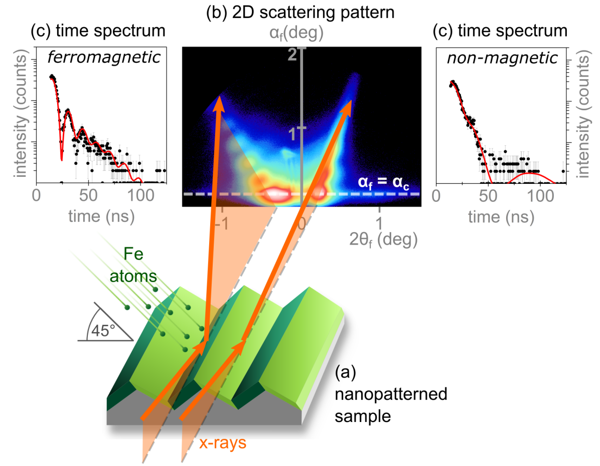
We propose a method for investigating nano-scaled objects, which can simultaneously deliver morphological parameters with sub-nanometer lateral precision and the static and dynamic magnetic characteristics of these structures under in-situ conditions of growth, high temperatures, reactive environments, or strong magnetic fields. To achieve this, we combine two X-ray scattering techniques into a single experiment, namely Grazing Incidence Small Angle X-ray Scattering (GISAXS) and Nuclear Resonant Scattering (NRS). GISAXS provides morphological characterization of nanometer-sized surface features, based on the angular distribution of scattered photons depending on the sample structure. Thus, three-dimensional shapes and lateral arrangements of nanostructures supported on surfaces or buried in thin films are obtained with nanometer resolution. NRS yields information on magnetic ordering, enables precise determination of in-plane and out-of-plane magnetic moment orientation and allows for sensitive detection of magnetization dynamics with an accuracy of a few degrees and sub-microsecond time resolution Röhlsberger (1999); Röhlsberger et al. (2003); Schlage and Röhlsberger (2013); Bocklage et al. (2015) by probing the coherent elastic resonant scattering of photons from Mössbauer-active nuclei Röhlsberger (2004). Intensity maxima in a GISAXS pattern originate from photons, which are scattered off different periodically repeated structural components of the sample. Photons, which have been resonantly scattered from nuclei, are identified by their time delay with respect to the photons, which have been non-resonantly scattered from electrons. The coherent elastic nuclear resonant scattering of photons results in a characteristic time spectrum of the detected intensity. At the specular intensity maximum, this time spectrum reflects the integrated magnetic properties of the entire sample Schlage et al. (2012); Sharma et al. (2015). By placing the detector at selected off-specular intensity maxima within the pattern, however, one obtains information on the magnetic properties of the specific structural component of the sample which the selected intensity maximum is related to. Thus, both structural information and site-specific magnetic characteristics are gathered simultaneously, directly revealing the correlations between these properties.
From merging GISAXS and NRS into a single technique referred to as Grazing Incidence Nuclear Small Angle X-ray Scattering (GINSAXS), comprehensive structural and magnetic information on heterogeneous systems with periodic nanoscopic surface morphology can be obtained. In this paper we provide an experimental proof of principle for the aforementioned concept under two different in-situ conditions. While we apply GINSAXS to disentangle heterogeneous magnetic properties, the method could also be employed to elucidate heterogeneous chemical composition or crystal structure: The hyperfine interactions probed by NRS also characterize local chemical environments and local lattice structures of resonant nuclei Vogl et al. (2009); Potapkin et al. (2013); Freindl et al. (2013). Generally, a combination of GISAXS with NRS is applicable to samples with non-planar surface or interface morphologies, such as periodic arrangements of uniform nanostructures with tilted or curved surfaces. Such a technique could be highly beneficial for studying facet-selective adsorption Chiu et al. (2013); Mdluli et al. (2011), overgrowth Sun et al. (2012); Liu (2011); Mankin et al. (2015), or reactivity Harn et al. (2015); Li et al. (2013). It could also help to clarify the development of magnetic order and the magnetization reversal in faceted nanoparticles with magnetic shells Nasirpouri et al. (2011), or serve to study the magnetic properties of nanoparticle-based mesocrystals Disch et al. (2011); Chen et al. (2013); Josten et al. (2017).
We demonstrate the capability of GINSAXS by investigating a nanostructured sample system with periodically varying morphological and magnetic properties under in-situ conditions requiring ultra-high vacuum and external magnetic fields, respectively. The sample consists of an -Al2O3 substrate with parallel nanometer-scale facets (see Supplemental Material Sup and Heffelfinger and Carter (1997)), supporting a thin continuous Fe film. The substrate has an average facet height of nm and period of nm, the average facet tilt angles are and . From these values, average facet widths of nm and nm are calculated. The Fe film is grown by room temperature sputter deposition from a polar angle of and an azimuthal angle of with respect to the facet edges. Here, the R-plane facets are facing the sputtering source, so that the deposition rate is higher on these facets than on the S-plane facet, which are avert from the source. Consequently, the Fe film consists of thicker regions (18 nm) on the narrower R-plane facets and thinner regions (13 nm) on the wider S-plane facets (see Fig. 1(a)). As determined from a GISAXS pattern recorded with the facet edges aligned perpendicular to the incident X-ray beam, the Fe film is polycrystalline with a crystallite size of approximately 5 nm (see Supplemental Material Sup ). To prevent oxidation of the Fe film, the sample was capped with a Cr layer. The corrugated shape of the Fe film induces a uniaxial magnetic anisotropy with the easy axis of magnetization parallel to the substrate facet edges Liedke et al. (2013).
As summarized in Fig. 1, GINSAXS is conducted in the following sequence: First, a conventional 2D GISAXS pattern is recorded at a suitable angle of incidence using an area detector. With the facet edges aligned parallel to the incident X-ray beam, the GISAXS pattern is characterized by two crystal truncation rods (CTRs) originating from the R-plane and S-plane facets, respectively Rauscher et al. (1999). Second, the positions of intensity maxima which are specific for certain structural units of the sample are selected from the GISAXS pattern. Here, these are the positions of highest intensity along the two CTRs. The angle of incidence is adjusted to maximize the resonantly scattered intensity. Third, using a time-resolving point detector (avalanche photo diode, APD) nuclear resonant time spectra are recorded at the selected positions. The angular distribution of scattered intensity measured in the GISAXS pattern is a signature of the nanometer-scale surface morphology of the sample: the two tilted scattering rods correspond to the Fe film regions supported by the substrate facets with R-plane and S-plane orientation, respectively. Intensity modulations along the CTRs are related to the Fe film thicknesses on the respective facet surfaces (similar to Kiessig fringes). The film thicknesses and geometrical parameters describing the sample morphology are obtained by simulating the GISAXS patterns using the program FitGISAXS Babonneau (2010). The nuclear resonant time spectra serve as fingerprints of the magnetic characteristics of the different repeat units of the sample, conveying information on the degree of magnetic order and the magnetization orientation. The shape of a time spectrum correlates with these properties via strength and orientation, respectively, of the magnetic hyperfine field at the Fe nuclei. Time spectra were fitted using the program CONUSS Sturhahn (2000) (see Supplemental Material Sup ).
Following the procedure described above, GINSAXS was performed at an X-ray energy of 14.4 keV, i.e. the resonance energy of 57Fe, at the high resolution dynamics beamline P01 at PETRA III and the nuclear resonance beamline ID18 at ESRF (see Supplemental Material Sup ). We performed two independent in-situ GINSAXS experiments on a sample system which is structurally and magnetically heterogeneous on the nanoscale. The experiments provide spatially resolved information on the magnetization reversal and allow correlating film growth and development of ferromagnetic ordering.
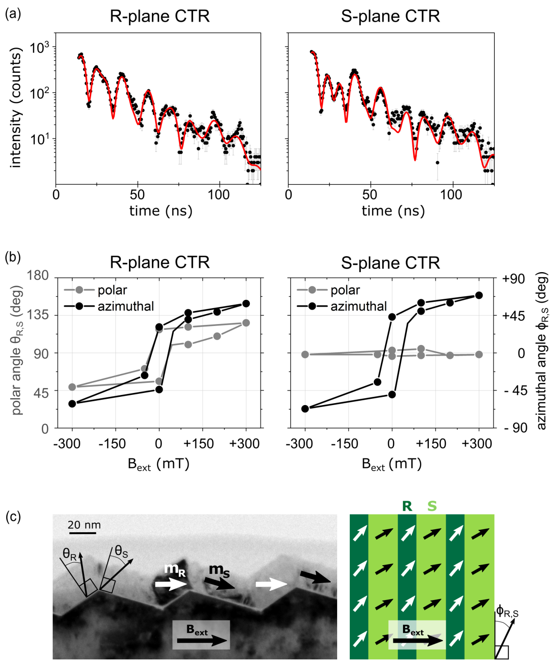
In the first experiment we resolve the heterogeneous magnetization reversal in the nanostructured Fe film upon applying an external magnetic field. The polar and azimuthal angles and of the magnetization as extracted from fitting the NRS time spectra for both R-plane and S-plane film regions are plotted as functions of the external magnetic field in Fig. 2(b). The angles are defined independently for R-plane and S-plane film regions, such that the magnetization is in the plane of the respective film region for a polar angle of and parallel to the facet edges for an azimuthal angle of . The hysteretic behavior of the azimuthal magnetization orientation is similar for the R-plane and S-plane regions of the Fe film: the magnetization is displaced from the easy axis parallel to the facet edges to similar extents but does not align fully with the orientation of the external magnetic field. This can be accounted for by the pronounced uniaxial in-plane magnetic anisotropy of the uniaxially corrugated film. The R-plane and S-plane regions of the Fe film differ markedly, however, in the polar magnetization orientation Liedke et al. (2013): In the S-plane Fe film regions the polar magnetization orientation remains almost constant at , i.e. it remains parallel to the film plane of these regions even at highest field strength. In contrast, the magnetization in the R-plane regions is deflected to an orientation in between the direction of the external magnetic field at and , respectively, and the magnetization in the S-plane film regions at and , respectively. The film regions on the S-plane facets are both thinner and wider than those on the R-plane facets. Thus, the shape anisotropy is more pronounced and in-plane orientation of the magnetization is preferred in the S-plane film regions. Furthermore, the external magnetic field is applied parallel to the average sample surface and does not enclose the same angle with the R-plane and S-plane facets. Consequently, the magnitude of the external magnetic field component normal to the film plane is by approximately 70% larger for the R-plane film regions on the R-plane facets. Interface coupling between the Fe film and the antiferromagnetic Cr capping layer with high magnetic anisotropy may be a cause for the inertness observed in the magnetization returning to its easy axis orientation at remanence. The influence of this effect on the measurement is strong due to the surface sensitivity of NRS at . NRS spectra taken at higher incidence angles indicate a spring-like magnetization structure of the Fe film, where the top layers of the Fe film are coupled to the Cr capping layer, while the bottom layers are free to relax toward the easy axis orientation when no external field is applied (see Supplemental Material Sup ).
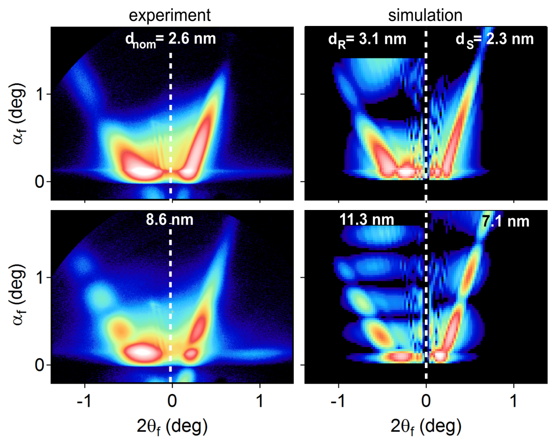
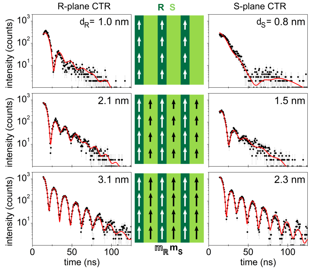
Furthermore, we observed the correlation of growth and magnetic stabilization in the stripe-like regions of the Fe film in an in-situ experiment during Fe deposition. Exemplary GISAXS patterns taken during growth of the Fe film are compared to the corresponding simulations in Fig. 3 (see Supplemental Material Sup ). The periods of intensity modulation along the CTRs decrease in correspondence to the increasing film thicknesses; the unequal modulation periods for each deposition stage evidence the different thicknesses of the Fe film on S-plane and R-plane facets. Simultaneously to the film growth observed in the GISAXS patterns, the evolution of magnetic properties is evidenced by characteristic changes in the time spectra. Fig. 4 depicts sequences of time spectra for the film regions on R-plane and S-plane facets recorded during growth and sketches the corresponding strength and orientation of magnetic moments. A non-magnetic state is characterized by the lack of a beat pattern on the time spectrum, while the time spectrum of a ferromagnetic state exhibits a pronounced beat pattern. The sequences show the successive evolution of beat patterns in the time spectra for the different film regions, thus evidencing a consecutive transition from a non-magnetic to a ferromagnetic state first in the R-plane film regions, then in S-plane regions. Notably, a state is observed in which the S-plane film regions are still non-magnetic, while the R-plane regions already exhibit a magnetization parallel to the facet edges. Full ferromagnetic order is observed at a film thickness of nm for the R-plane Fe film regions. In the S-plane regions, ferromagnetic order is established at a thickness of nm already, which may be due to the direct contact with the fully ferromagnetic R-plane film regions. In polycrystalline Fe thin films with uniaxially corrugated shape, ferromagnetic ordering was observed at film thicknesses 1 nm d 3 nm; the results of this experiment thus agree well with former findings Shiratsuchi et al. (2004). For thicknesses larger than 2.8 nm and 2.3 nm, respectively, all film regions on the R-plane facets have a magnetic hyperfine field magnitude close to the -Fe bulk value. Both the R-plane and S-plane film regions now show time spectra with pronounced beat patterns evidencing ferromagnetically ordered Fe with the magnetization oriented exactly parallel to the direction of the incoming beam, i.e. parallel to the facet edges. Cross-sectional transmission electron microscopy (see Fig.2(c)) confirms that the free surface of the Fe film and its interface with the substrate are parallel as indicated by the sharp intensity modulations in the GISAXS patterns.
In conclusion, GINSAXS, a combination of GISAXS and NRS, allows detecting the magnetic information from different structural units of a nanostructured sample separately. This approach has a great potential for characterizing corrugated magnetic materials, which are of interest for applications in magnetic sensing due to their shape-induced uniaxial magnetic anisotropy. Also standing spin waves in magnetic lattices may be investigated. Furthermore, it is conceivable to employ it for studying supported or buried nanoparticles with faceted or curved surfaces or with core-shell morphologies. The successful demonstration of GINSAXS may also encourage testing the feasibility of combining X-ray Absorption Near Edge Structure (XANES) and GISAXS in an analogous manner: This would be a powerful tool for investigating nanostructures with shape-dependent heterogeneous chemical properties, for instance for in-situ studies of catalytic processes with facet-selective reactivity.
Acknowledgements.
We gratefully acknowledge David Babonneau, Université de Poitiers, for implementing a new form factor in FitGISAXS on our request, which enabled simulating the GISAXS patterns of the Fe film on the faceted substrate. We thank Shengqiang Zhou, Helmholtz-Zentrum Dresden-Rossendorf, for measuring VSM hysteresis curves of the sample.References
- Mehdaoui et al. (2011) B. Mehdaoui, A. Meffre, J. Carrey, S. Lachaize, L.-M. Lacroix, M. Gougeon, B. Chaudret, and M. Respaud, Adv. Funct. Mater. 21, 4573 (2011).
- Reddy et al. (2012) L. H. Reddy, J. L. Arias, J. Nicolas, and P. Couvreur, Chem. Rev. 112, 5818 (2012).
- Topp et al. (2010) J. Topp, D. Heitmann, M. P. Kostylev, and D. Grundler, Phys. Rev. Lett. 104, 207205 (2010).
- Armelles et al. (2013) G. Armelles, A. Cebollada, A. García-Martín, and M. U. González, Adv. Optical Mater. 1, 10 (2013).
- Wettergren et al. (2014) K. Wettergren, F. F. Schweinberger, D. Deiana, C. J. Ridge, A. S. Crampton, M. D. Rötzer, T. W. Hansen, V. P. Zhdanov, U. Heiz, and C. Langhammer, Nano Lett. 14, 5803 (2014).
- Hejral et al. (2016) U. Hejral, P. Müller, O. Balmes, D. Pontoni, and A. Stierle, Nat. Comm. 7, 10964 (2016).
- Li et al. (2012) D. Li, M. H. Nielsen, J. R. I. Lee, C. Frandsen, J. F. Banfield, and J. J. De Yoreo, Science 336, 1014 (2012).
- Rauscher et al. (1999) M. Rauscher, R. Paniago, H. Metzger, Z. Kovats, J. Domke, J. Peisl, H.-D. Pfannes, J. Schulze, and I. Eisele, J. Appl. Phys. 86, 6763 (1999).
- Park et al. (2005) K. Park, D. Lee, A. Rai, D. Mukherjee, and M. R. Zachariah, J. Phys. Chem. B 109, 7290 (2005).
- Pecora (2000) R. Pecora, J. Nanopart. Res. 2, 123 (2000).
- Juvé et al. (2013) V. Juvé, M. F. Cardinal, A. Lombardi, A. Crut, P. Maioli, J. Pérez-Juste, L. M. Liz-Marzán, N. Del Fatti, and F. Vallée, Nano Lett. 13, 2234 (2013).
- Shukla et al. (2003) N. Shukla, C. Liu, P. M. Jones, and D. Weller, J. Magn. Magn. Mater. 266, 178 (2003).
- Amendola and Meneghetti (2009) V. Amendola and M. Meneghetti, J. Phys. Chem. C 113, 4277 (2009).
- Prieto et al. (2012) P. Prieto, V. Nistor, K. Nouneh, M. Oyama, M. Abd-Lefdil, and R. Díaz, Appl. Surf. Sci. 258, 8807 (2012).
- Wiesendanger (2009) R. Wiesendanger, Rev. Mod. Phys. 81, 1495 (2009).
- McCord (2015) J. McCord, J. Phys. D: Appl. Phys. 48, 333001 (2015).
- Koike (2013) K. Koike, Microscopy 62, 177 (2013).
- Cheng and Keavney (2012) X. Cheng and D. Keavney, Rep. Prog. Phys. 75, 026501 (2012).
- Grimsditch and Vavassori (2004) M. Grimsditch and P. Vavassori, J. Phys.: Condens. Mat. 16, R275 (2004).
- Hartmut et al. (2007) H. Hartmut, K. Theis-Bröhl, and B. P. Toperverg, “Polarized neutron reflectivity and scattering from magnetic nanostructures and spintronic materials,” in Handbook of Magnetism and Advanced Magnetic Materials (John Wiley & Sons, Ltd, 2007).
- Stöhr et al. (1993) J. Stöhr, Y. Wu, B. Hermsmeier, M. Samant, G. Harp, S. Koranda, D. Dunham, and B. Tonner, Science 259, 658 (1993).
- Turner et al. (2011) J. Turner, X. Huang, O. Krupin, K. Seu, D. Parks, S. Kevan, E. Lima, K. Kisslinger, I. McNulty, R. Gambino, S. Mangin, S. Roy, and P. Fischer, Phys. Rev. Lett. 107, 033904 (2011).
- Röhlsberger (1999) R. Röhlsberger, Hyperfine Interact. 123/124, 455 (1999).
- Röhlsberger et al. (2003) R. Röhlsberger, J. Bansmann, V. Senz, K. L. Jonas, A. Bettac, K. H. Meiwes-Broer, and O. Leupold, Phys. Rev. B 67, 245412 (2003).
- Schlage and Röhlsberger (2013) K. Schlage and R. Röhlsberger, J. Elect. Spect. Rel. Phenom. 189, 187 (2013).
- Bocklage et al. (2015) L. Bocklage, C. Swoboda, K. Schlage, H.-C. Wille, L. Dzemiantsova, S. Bajt, G. Meier, and R. Röhlsberger, Phys. Rev. Lett. 114, 147601 (2015).
- Röhlsberger (2004) R. Röhlsberger, Springer Tracts in Modern Physics, Vol. 208: Nuclear Condensed Matter Physics with Synchrotron Radiation (Springer, Berlin, 2004).
- Schlage et al. (2012) K. Schlage, S. Couet, S. V. Roth, U. Vainio, R. Rüffer, M. M. A. Kashem, P. Müller-Buschbaum, and R. Röhlsberger, New J. Phys. 14, 043007 (2012).
- Sharma et al. (2015) G. Sharma, A. Gupta, M. Gupta, K. Schlage, and H.-C. Wille, Phys. Rev. B 92, 224403 (2015).
- Vogl et al. (2009) G. Vogl, E. Partyka-Jankowska, M. Zając, and A. I. Chumakov, Phys. Rev. B 80, 115406 (2009).
- Potapkin et al. (2013) V. Potapkin, C. McCammon, K. Glazyrin, A. Kantor, I. Kupenko, C. Prescher, R. Sinmyo, G. V. Smirnov, A. I. Chumakov, R. Rüffer, and L. Dubrovinski, Nature Communications 4, 1427 (2013).
- Freindl et al. (2013) K. Freindl, E. Partyka-Jankowska, W. Karaś, M. Zając, E. Madej, N. Spiridis, M. Ślęzak, T. Ślęzak, D. Wiśnios, and J. Korecki, Surf. Sci. 617, 183 (2013).
- Chiu et al. (2013) C.-Y. Chiu, H. Wu, Z. Yao, F. Zhou, H. Zhang, V. Ozolins, and Y. Huang, JACS 135, 15489 (2013).
- Mdluli et al. (2011) P. S. Mdluli, N. M. Sosibo, P. N. Mashazi, T. Nyokong, R. T. Tshikhudo, A. Skepu, and E. van der Lingen, J. Mol. Struct. 1004, 131 (2011).
- Sun et al. (2012) S. Sun, C. Kong, H. You, X. Song, B. Ding, and Z. Yang, Cryst. Eng. Comm. 14, 40 (2012).
- Liu (2011) X.-W. Liu, Langmuir 27, 9100 (2011).
- Mankin et al. (2015) M. N. Mankin, R. W. Day, R. Gao, Y.-S. No, S.-K. Kim, A. A. McClelland, D. C. Bell, H.-G. Park, and C. M. Lieber, Nano Lett. 15, 4776 (2015).
- Harn et al. (2015) Y.-W. Harn, T.-H. Yang, T.-Y. Tang, M.-C. Chen, and J.-M. Wu, Chem. Cat. Chem. 7, 80 (2015).
- Li et al. (2013) R. Li, F. Zhang, D. Wang, J. Yang, M. Li, J. Zhu, X. Zhou, H. Han, and C. Li, Nat. Comm. 4, 1432 (2013).
- Nasirpouri et al. (2011) F. Nasirpouri, S. J. Bending, L. M. Peter, and H. Fangohr, Thin Solid Films 519, 8320 (2011).
- Disch et al. (2011) S. Disch, E. Wetterskog, R. P. Hermann, G. Salazar-Alvarez, P. Busch, T. Brückel, L. Bergström, and S. Kamali, Nano Lett. 11, 1651 (2011).
- Chen et al. (2013) C.-J. Chen, R.-K. Chiang, and S.-L. Wang, Cryst. Eng. Comm. 15, 9161 (2013).
- Josten et al. (2017) E. Josten, E. Wetterskog, A. Glavic, P. Boesecke, A. Feoktystov, E. Brauweiler-Reuters, U. Rücker, G. Salazar-Alvarez, T. Brückel, and L. Bergström, Sci. Rep. 7, 2808 (2017).
- (44) See Supplemental Material for details on: experimental realization, NRS and GISAXS analysis, magnetic hysteresis and depth dependence of magnetization orientation.
- Heffelfinger and Carter (1997) J. R. Heffelfinger and C. B. Carter, Surf. Sci. 389, 188 (1997).
- Liedke et al. (2013) M. O. Liedke, M. Körner, K. Lenz, M. Fritzsche, M. Ranjan, A. Keller, E. Čižmár, S. A. Zvyagin, S. Facsko, K. Potzger, J. Lindner, and J. Fassbender, Phys. Rev. B 87, 024424 (2013).
- Babonneau (2010) D. Babonneau, J. Appl. Cryst. 43, 929 (2010).
- Sturhahn (2000) W. Sturhahn, Hyperfine Interactions 125, 149 (2000).
- Shiratsuchi et al. (2004) Y. Shiratsuchi, Y. Endo, M. Yamamoto, D. Li, and S. D. Bader, J. Appl. Phys. 95, 6897 (2004).