Imaging ultrafast molecular wavepackets with a single chirped UV pulse
Abstract
We show how to emulate a conventional pump-probe scheme using a single frequency-chirped ultrashort UV pulse to obtain a time-resolved image of molecular ultrafast dynamics. The chirp introduces a spectral phase in time that encodes the delay between the pump and the probe frequencies contained in the pulse. By comparing the results of full dimensional ab initio calculations for the H molecule with those of a simple sequential model, we demonstrate that, by tuning the chirp parameter, two-photon energy-differential ionization probabilities directly map the wave packet dynamics generated in the molecule. As a result, one can also achieve a significant amount of control of the total ionization yields, with a possible enhancement by more than an order of magnitude.
The advent of free-electron-laser facilities and high-harmonic generation has opened the way to the production of intense and ultrashort ultraviolet (UV) pulses with durations in the femtosecond and attosecond range Kling and Vrakking (2008); Krausz and Ivanov (2009); Bostedt et al. (2009, 2016). One of the more awaited capabilities offered by such pulses is to use them to monitor and control electronic and nuclear dynamics, for example within a UV-UV pump-probe scheme, the so-called “holy grail” of attosecond physics. While some progress in this direction has been made Tzallas et al. (2011); Wöstmann et al. (2013); Carpeggiani et al. (2014); Campi et al. (2016); Takanashi et al. (2017), there are many technical challenges still to overcome, such as the limited intensity of the pulses and the fact that few optically active elements exist in the (extreme) ultraviolet. This precludes, for example, the use of pulse-shaping techniques and coherent control approaches that can be applied at optical and infrared frequencies to produce an “optimal” pulse for a desired photo-induced physical process or chemical reaction Assion et al. (1998); Meshulach and Silberberg (1998); Weiner (2000); Levis et al. (2001); Brixner et al. (2004); Chatel and Girard (2005); Djotyan et al. (2004); Weiner (2000); Nuernberger et al. (2009). Consequently, most experiments performed so far with attosecond UV pulses rely instead on an intense infrared pulse for either the pump or the probe step, which can significantly distort the system and alter the dynamics.
In this Letter, we demonstrate that by changing a single parameter, the spectral chirp of an ultrashort UV pulse, we can achieve a significant amount of control over molecular multiphoton ionization, changing the total ionization yield by more than a factor of ten. More importantly, we show how to emulate a conventional pump-probe setup to obtain direct time-resolved imaging of ultrafast molecular dynamics. The spectral chirp is experimentally tuneable both in high harmonic generation and with free electron lasers Chang (2005); Scrinzi et al. (2006); Hofstetter et al. (2011); Bostedt et al. (2016). A similar idea was previously suggested by Yudin et al. Yudin et al. (2006), but only demonstrated for a superposition of two bound states in the hydrogen atom. In the present work, we aim at reconstructing the vibronic wave packet in a small molecule, simultaneously pumped and probed by a single chirped UV pulse.
At optical and infrared frequencies, the effect of frequency-chirped pulses has been actively investigated using theoretical approaches based on second-order time-dependent perturbation theory (TDPT) to treat few-photon excitation processes in atoms Adler et al. (1995); Zhang et al. (2009); Felinto and López (2009); Dudovich et al. (2001); Meshulach and Silberberg (1998, 1999); Chatel et al. (2003); Djotyan et al. (2004); Stowe et al. (2006). These methods have also been applied at ultraviolet frequencies, but, to our knowledge, only to describe the chirp-dependent photoelectron angular distributions in atomic photoionization Pronin et al. (2009); Peng et al. (2009); Pronin et al. (2011). In these approaches, only a limited number of states partake in the dynamics. In contrast, the additional nuclear degrees of freedom in molecular targets, as treated here, induce more complex wavepacket motion characterized by the participation of many vibronic states. We thus directly solve the full-dimensional time-dependent Schrödinger equation (TDSE), , using H as a benchmark target to investigate the coherent manipulation of two-photon molecular photoionization by chirped pulses.
In our approach, the time-dependent wave function is expressed within a single-center expansion, using spherical harmonics to treat the angular components and a finite element discrete variable representation (FEDVR) for the radial coordinates of both the electronic and vibrational degrees of freedom. The full Hamiltonian, , is given by the sum of the Hamiltonian of the isolated molecule, , which depends on both electronic and nuclear coordinates and therefore implicitly incorporates non-adiabatic couplings, and the laser-molecule interaction within the dipole approximation in length gauge, , as the product of the electronic coordinates and the electromagnetic field of the pulse . After solving the TDSE, we implicitly propagate until infinite time and simultaneously Fourier-transform the time-dependent wave function to obtain the scattering function at a given energy Palacios et al. (2007). From the scattering wave function, one can extract the single ionization amplitudes by using the surface integral formalism described in Palacios et al. (2007) and thus obtain the total, energy- and angle-differential ionization probabilities. The electromagnetic field of a (linearly polarized) chirped Gaussian pulse can be written as Yudin et al. (2006); Nakajima (2007); Peng et al. (2009):
| (1) |
where the instantaneous phase is given by
| (2) |
and the temporal envelope is described by a Gaussian function:
| (3) |
The field amplitude, , the pulse duration and the instantaneous frequency explicitly depend on the chirp parameter . Note that the spectral chirp (the quadratic term of the spectral phase) of the field defined here is directly proportional to , but the temporal chirp [prefactor of the term in ] has opposite sign in contrast with the definition in Yudin et al. (2006); Nakajima (2007); Peng et al. (2009). For unchirped pulses (), is the peak amplitude, defines the duration of the pulse (FWHM of the field envelope is ), and is the carrier frequency. The parametrization is chosen such that adding a chirp in frequency (), the spectrum remains unchanged. The duration of the pulse then increases to , while the peak amplitude decreases to . In other words, the same frequencies are “stretched” over a longer duration. The Fourier transform of this Gaussian pulse leads to a Gaussian spectral function, with amplitude independent of , but a spectral phase that is quadratic in with a spectral chirp given by .
In Fig. 1(a) we show the energetics of the two-photon ionization process using linearly polarized light parallel to the molecular axis of the H molecule. The one-photon transition from the 1s ground state creates a vibronic wave packet in the dissociative excited states of symmetry. The two-photon transition reaches the ionization potential leading to the Coulomb explosion of the system (H++H++e-). We use pulses whose frequency spectrum corresponds to that of an unchirped pulse () with a FWHM duration of as centered at a.u. and a laser intensity of W/cm2. With these parameters, ionization is solely due to two-photon absorption paths. The energy bandwidth of these pulses is plotted in Fig. 1(a) as an orange shadowed area. The maximum spectral amplitude, 0.6 a.u., lies in between the 2p and 3p states.
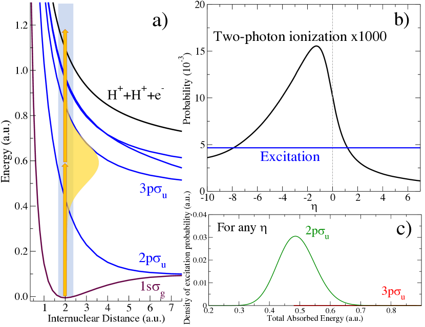
As expected, the excitation probability, shown in Fig. 1(b), is independent of the chirp parameter. As graphically described by Brumer and Shapiro Brumer and Shapiro (1989), the one-photon absorption probability is “an emperor without clothes”, unaffected by the spectral phase, and only depends on the spectral frequency distribution of the pulse. The spectral phase introduced in the excited wave packet can only be captured in a second-order process, for instance the two-photon transition depicted in Fig. 1, where the time evolution of the nuclear wave packet is retrieved through its projection into the electronic continua. The excitation probabilities associated to the 2p and 3p states, which are independent of , are plotted as a function of the vibronic (vibrational+electronic) energy in Fig. 1(c). For the pulses employed here, we can see that the two-photon ionization proceeds almost entirely through the first excited state. First, the photon energies within the pulse are energetically closer to the resonant vertical transition from the ground state to the 2p state. In addition, the dipole coupling to the 3p state is noticeably weaker than that to 2p. As a result, the one-photon excitation probability to the 3p state is three orders of magnitude smaller than that to the 2p state.
The total ionization probability as a function of the parameter is also included in Fig. 1(b). As mentioned above, ionization to the final states of symmetry (even number of absorbed photons) is the dominant process, while the ionization to states of symmetry (odd number of absorbed photons) is negligible. As shown in the figure, by tuning the chirp parameter the total ionization probability can be strongly modified, with a modulation range of more than an order of magnitude. At the H equilibrium distance, the energy difference between the ground and the 2p state is a.u., while the difference between the latter and the Coulomb explosion potential energy curve is a.u.. It is thus expected that the total ionization yield is enhanced for negative values of , i.e. when lower frequencies ( a.u.) arrive earlier and larger frequencies ( a.u.) arrive later. In this way, both transitions, from the ground state to 2p and from 2p to the Coulomb explosion, can take place when the instantaneous frequency is close to resonant, thus maximizing ionization.
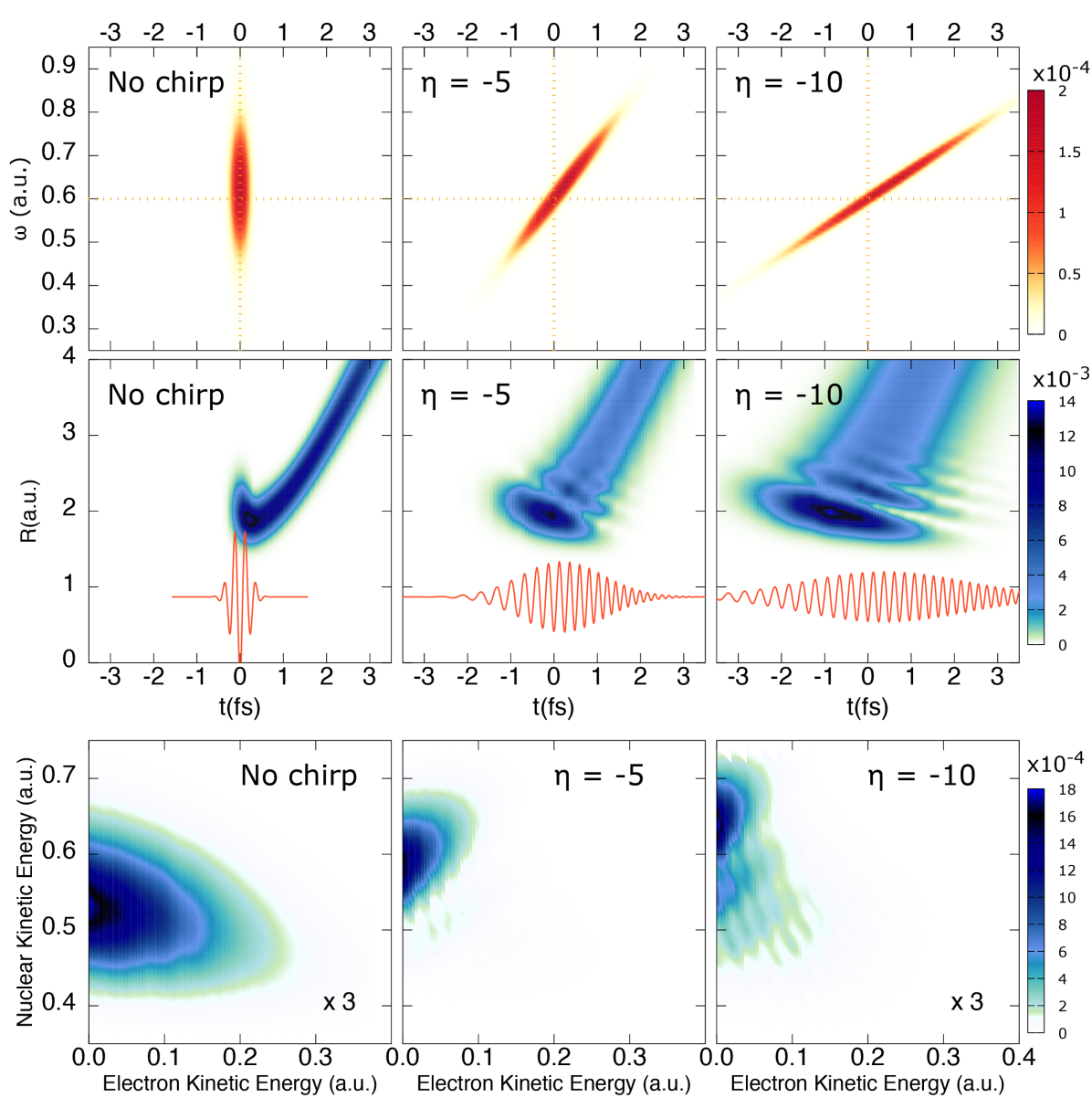
In order to extract dynamical information about the excited wave packet associated to the 2p state, we will study the energy-differential ionization probabilities for different values of the chirp parameter. In the upper row of Fig. 2, we plot the Wigner distributions of the electromagnetic field, which provides a combined time-frequency representation, for three different pulses with , and . For the unchirped pulse, all frequencies reach the target simultaneously. However, for the chirped pulses, the more negative the larger the time delay between the lower and the higher frequencies. In other words, by making more negative, we are creating a nuclear wave packet in the 2p state at earlier times (the direct vertical transition at a.u. occurs earlier), which is probed by promotion into the Coulomb explosion channel at later times (frequencies around a.u. arrive later). Therefore, this is conceptually equivalent to standard pump-probe schemes, where two time-delayed pulses are employed: one pulse launches the dynamics in the target and a second pulse, delayed (and ideally not overlapping) in time, probes the pumped dynamics through promotion to a given final state. In the present case, the time delay is encoded in the chirp parameter.
The middle panels of Fig. 2 show the corresponding nuclear wave packets (NWPs) in the 2p state as a function of time (-axis) and internuclear distance (-axis). In the same subplots, we include the electromagnetic field, , as a red line. We can see that the quadratic spectral phase associated to a given chirp value () introduces structure in the pumped excited wave packet. This is due to interferences resulting from different frequency components with different spectral phases Chatel and Girard (2005). We observe nearly the same wave packet, but stretched in time. As discussed above (cf. Fig. 1c), the energy distribution of the wave packet is identical for all values of . However, as seen in Fig. 2, their spatial structure differs, since for the more negative chirp, the same frequencies are reaching the target with a larger delay between them. This structured wave packet is mapped into the energy differential ionization probabilities upon absorption of a second photon, leading to distinct profiles.
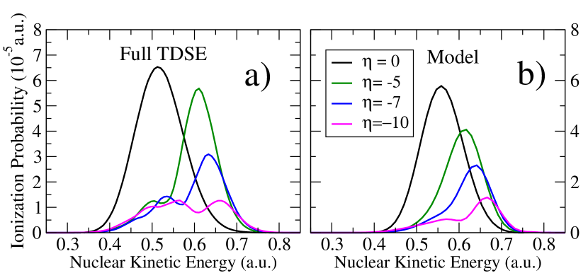
The energy-differential ionization probabilities are shown in the contour plots in the bottom panels of Fig. 2, as a function of the ejected electron energy (-axis) and the nuclear kinetic energy release of the nuclei (-axis). The energy distribution resulting from the interaction with the unchirped pulse is smooth, while the chirped pulses yield distributions shifted towards higher nuclear kinetic energies and with internal structure. For a better visualization, we integrate the ionization probabilities over the electron kinetic energy and obtain the nuclear kinetic energy distributions shown in Fig. 3(a). Here, we have additionally included the results for . These energy distributions actually reflect the dynamics launched in the excited molecule. In order to prove this, in Fig. 3(b), we show the results of a sequential model where the excited NWP created in the 2p state by the lower frequencies is directly projected into the ionization channel. Note that the model qualitatively reproduces the position and the profile of each energy distribution. The model uses as starting point the exact second-order time-dependent perturbation theory expression for the molecular wave packet, , created after two-photon absorption from the ground state, . In the interaction picture, it is given by
| (4) |
| (5) |
where is the driving operator in the interaction picture. The ionization amplitude can be obtained by simply projecting the molecular wave packet, , into the final continuum states, leading to the ionization probabilities in the bottom panel of Fig. 2. The first-order wavepacket corresponds to the nuclear wavepacket after one-photon absorption, as shown in the middle panels of Fig. 2 (note that those NWPs are plotted in the Schrödinger picture and consequently evolve in time even in the absence of the field). In the interaction picture, the wavepackets remain unchanged in time once the frequency components of the driving pulse that are responsible for the transition have been absorbed. The ab initio first-order wavepackets at are shown in Fig. 4(a). For negative chirps, these wavepackets are already fully formed when the second (higher-frequency) photon is absorbed. We can thus use a sequential approximation where the final first-order wavepacket (with ) is used as the source for the second-order wavepacket:
| (6) |
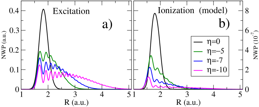
The result of this approximation is plotted in Fig. 4(b), where we can see how the structure of the excited wave packet in Fig. 4(a) is reflected in the ionized wave packet extracted from the model. By using the definition of and Eq. (missing) 1, the corresponding ionization amplitude, i.e. the projection of the approximated second-order wave packet into the final states, , can be written as:
| (7) |
where is the product of the dipole matrix elements involving the ground , intermediate and final states with the chirp-independent spectral amplitude of the pulse at the corresponding transition frequencies , and where results from a Fourier transform of [the c.c. part in Eq. (missing) 1]. The exponential in Eq. (missing) 7 corresponds to the spectral phase of the field. The good agreement between the ionization probabilities resulting from this model [shown in Fig. 3(b)], and the ab initio ones [Fig. 3(a)] validates the use of the sequential approximation to map the wave packet generated by the chirped pulse. More interestingly, Eq. (missing) 7 demonstrates the close relation between the current approach and conventional pump-probe setups Feist et al. (2011); Palacios et al. (2014). In such schemes, the two transitions are driven by two different pulses separated by a time delay , leading to the analog expression for the ionization amplitudes, , but with an important difference: For the single chirped pulse, the relative phase depends quadratically on the intermediate state energy , while it does linearly in a pump-probe scheme. However, if the transition amplitude to intermediate states is peaked around an average value (as in the present case, cf. Fig. 1c), we can bridge this difference and make the analogy even more apparent. Expanding the energy of the intermediate states around this value, , one obtains
| (8) |
where corresponds to an effective time delay, defining and , and the quadratic term can be neglected for sufficiently small . It can be easily shown that for large enough the effective time delay matches the difference between the times when the instantaneous frequency is resonant with the average transition energies and . In summary, these expressions demonstrate that, within the validity of the sequential approximation, the chirped pulse acts like a conventional pump-probe setup, but with an effective time delay given by an average energy difference of the transition of interest.
In conclusion, we have shown that two-photon ionization of molecules can be manipulated by using frequency-chirped femtosecond pulses, leading to modulations of the ionization probability of more than an order of magnitude. We have also shown that chirped pulses can be used to probe the ultrafast molecular dynamics triggered in the excited molecule by just varying the frequency chirp, which is equivalent to varying the time delay in the long awaited UV pump-UV probe schemes. This has been demonstrated by using chirped pulses with the same energy spectrum and a quadratic spectral phase, which as shown in previous works Chang (2005); Scrinzi et al. (2006); Hofstetter et al. (2011); Bostedt et al. (2016), can be easily reproduced in the lab. In this scenario, the energy distribution of the wave packet created by one-photon absorption does not vary with the chirp parameter, while the spatial distribution does. This can be retrieved from its direct mapping into the energy distribution of the charged fragments after Coulomb explosion, and is shown to be formally analogous to a conventional pump-probe scheme. Although applied to H in the present work, the method should also be suitable to probe wave packet dynamics in excited states of more complex molecules. It will not only be easier to implement than UV pump-IR probe methods, where two different pulses must be synchronized, but it will also avoid the significant distortion introduced by the IR probing.
I Acknowledgments
We acknowledge support from the European Research Council under ERC grants no. 290853 XCHEM and 290981 PLASMONANOQUANTA, European COST Action CM1204 XLIC, the Ministerio de Economía y Competitividad projects FIS2013-42002-R, FIS2016-77889-R and the “María de Maeztu” programme for Units of Excellence in R&D (MDM-2014-0377), European grants MC-ITN CORINF and MC-RG ATTOTREND 268284. AP acknowledges a Ramón y Cajal contract from the Ministerio de Economía y Competitividad. The project has been supported with computer time allocated in Mare Nostrum BSC-RES and in the Centro de Computación Científica UAM.
References
- Kling and Vrakking (2008) Matthias F. Kling and Marc J. J. Vrakking, “Attosecond Electron Dynamics,” Annu. Rev. Phys. Chem. 59, 463 (2008).
- Krausz and Ivanov (2009) Ferenc Krausz and Misha Ivanov, “Attosecond physics,” Rev. Mod. Phys. 81, 163 (2009).
- Bostedt et al. (2009) Christoph Bostedt, Henry N. Chapman, John T. Costello, José R. Crespo López-Urrutia, Stefan Düsterer, Sascha W. Epp, Josef Feldhaus, Alexander Föhlisch, Michael Meyer, Thomas Möller, Robert Moshammer, Mathias Richter, Klaus Sokolowski-Tinten, Andrei Sorokin, Kai Tiedtke, Joachim Ullrich, and Wilfried Wurth, “Experiments at FLASH,” Nucl. Inst. Meth. A 601, 108 (2009).
- Bostedt et al. (2016) Christoph Bostedt, Sébastien Boutet, David M. Fritz, Zhirong Huang, Hae Ja Lee, Henrik T. Lemke, Aymeric Robert, William F. Schlotter, Joshua J. Turner, and Garth J. Williams, “Linac Coherent Light Source: The first five years,” Rev. Mod. Phys. 88, 015007 (2016).
- Tzallas et al. (2011) P. Tzallas, E. Skantzakis, L. A. A. Nikolopoulos, G. D. Tsakiris, and D. Charalambidis, “Extreme-ultraviolet pump–probe studies of one-femtosecond-scale electron dynamics,” Nat. Phys. 7, 781 (2011).
- Wöstmann et al. (2013) M. Wöstmann, R. Mitzner, T. Noll, S. Roling, B. Siemer, F. Siewert, S. Eppenhoff, F. Wahlert, and H. Zacharias, “The XUV split-and-delay unit at beamline BL2 at FLASH,” J. Phys. B 46, 164005 (2013).
- Carpeggiani et al. (2014) P. A. Carpeggiani, P. Tzallas, A. Palacios, D. Gray, F. Martín, and D. Charalambidis, “Disclosing intrinsic molecular dynamics on the 1-fs scale through extreme-ultraviolet pump-probe measurements,” Phys. Rev. A 89, 023420 (2014).
- Campi et al. (2016) F. Campi, H. Coudert-Alteirac, M. Miranda, L. Rading, B. Manschwetus, P. Rudawski, A. L’Huillier, and P. Johnsson, “Design and test of a broadband split-and-delay unit for attosecond XUV-XUV pump-probe experiments,” Rev. Sci. Inst. 87, 023106 (2016).
- Takanashi et al. (2017) T. Takanashi, N. V. Golubev, C. Callegari, H. Fukuzawa, K. Motomura, D. Iablonskyi, Y. Kumagai, S. Mondal, T. Tachibana, K. Nagaya, T. Nishiyama, K. Matsunami, P. Johnsson, P. Piseri, G. Sansone, A. Dubrouil, M. Reduzzi, P. Carpeggiani, C. Vozzi, M. Devetta, M. Negro, D. Faccialà, F. Calegari, A. Trabattoni, M. C. Castrovilli, Y. Ovcharenko, M. Mudrich, F. Stienkemeier, M. Coreno, M. Alagia, B. Schütte, N. Berrah, O. Plekan, P. Finetti, C. Spezzani, E. Ferrari, E. Allaria, G. Penco, C. Serpico, G. De Ninno, B. Diviacco, S. Di Mitri, L. Giannessi, G. Jabbari, K. C. Prince, L. S. Cederbaum, Ph. V. Demekhin, A. I. Kuleff, and K. Ueda, “Time-Resolved Measurement of Interatomic Coulombic Decay Induced by Two-Photon Double Excitation of Ne2,” Phys. Rev. Lett. 118, 033202 (2017).
- Assion et al. (1998) A. Assion, T. Baumert, M. Bergt, T. Brixner, B. Kiefer, V. Seyfried, M. Strehle, and G. Gerber, “Control of Chemical Reactions by Feedback-Optimized Phase-Shaped Femtosecond Laser Pulses,” Science 282, 919 (1998).
- Meshulach and Silberberg (1998) Doron Meshulach and Yaron Silberberg, “Coherent quantum control of two-photon transitions by a femtosecond laser pulse,” Nature 396, 239 (1998).
- Weiner (2000) A. M. Weiner, “Femtosecond pulse shaping using spatial light modulators,” Rev. Sci. Inst. 71, 1929 (2000).
- Levis et al. (2001) Robert J. Levis, Getahun M. Menkir, and Herschel Rabitz, “Selective Bond Dissociation and Rearrangement with Optimally Tailored, Strong-Field Laser Pulses,” Science 292, 709 (2001).
- Brixner et al. (2004) T. Brixner, G. Krampert, T. Pfeifer, R. Selle, G. Gerber, M. Wollenhaupt, O. Graefe, C. Horn, D. Liese, and T. Baumert, “Quantum Control by Ultrafast Polarization Shaping,” Phys. Rev. Lett. 92, 208301 (2004).
- Chatel and Girard (2005) Béatrice Chatel and Bertrand Girard, “Coherent Control of Atomic Dynamics with Chirped and Shaped Pulses,” in Femtosecond Laser Spectroscopy (Springer-Verlag, New York, 2005) pp. 267–304.
- Djotyan et al. (2004) G. P. Djotyan, J. S. Bakos, Zs. Sörlei, and J. Szigeti, “Coherent control of atomic quantum states by single frequency-chirped laser pulses,” Phys. Rev. A 70, 063406 (2004).
- Nuernberger et al. (2009) Patrick Nuernberger, Reimer Selle, Florian Langhojer, Frank Dimler, Susanne Fechner, Gustav Gerber, and Tobias Brixner, “Polarization-shaped femtosecond laser pulses in the ultraviolet,” J. Opt. A 11, 085202 (2009).
- Chang (2005) Zenghu Chang, “Chirp of the single attosecond pulse generated by a polarization gating,” Phys. Rev. A 71, 023813 (2005).
- Scrinzi et al. (2006) A. Scrinzi, M. Yu. Ivanov, R. Kienberger, and D. M. Villeneuve, “Attosecond physics,” J. Phys. B 39, R1 (2006).
- Hofstetter et al. (2011) Michael Hofstetter, Martin Schultze, Markus Fieß, Benjamin Dennhardt, Alexander Guggenmos, Justin Gagnon, Vladislav S. Yakovlev, Eleftherios Goulielmakis, Reinhard Kienberger, Eric M. Gullikson, Ferenc Krausz, and Ulf Kleineberg, “Attosecond dispersion control by extreme ultraviolet multilayer mirrors,” Opt. Expr. 19, 1767 (2011).
- Yudin et al. (2006) G. Yudin, A. Bandrauk, and P. Corkum, “Chirped Attosecond Photoelectron Spectroscopy,” Phys. Rev. Lett. 96, 063002 (2006).
- Adler et al. (1995) Andre Adler, Alain Rachman, and Edward J. Robinson, “A time-dependent theory of resonant multiphoton ionization of atoms by short pulses. I. Weak-field results for the two-photon ionization of caesium,” J. Phys. B 28, 5057 (1995).
- Zhang et al. (2009) Shian Zhang, Hui Zhang, Tianqing Jia, Zugeng Wang, and Zhenrong Sun, “Coherent control of two-photon transitions in a two-level system with broadband absorption,” Phys. Rev. A 80, 043402 (2009).
- Felinto and López (2009) Daniel Felinto and Carlos E. E. López, “Theory for direct frequency-comb spectroscopy,” Phys. Rev. A 80, 013419 (2009).
- Dudovich et al. (2001) Nirit Dudovich, Barak Dayan, Sarah Gallagher Faeder, and Yaron Silberberg, “Transform-Limited Pulses Are Not Optimal for Resonant Multiphoton Transitions,” Phys. Rev. Lett. 86, 47 (2001).
- Meshulach and Silberberg (1999) Doron Meshulach and Yaron Silberberg, “Coherent quantum control of multiphoton transitions by shaped ultrashort optical pulses,” Phys. Rev. A 60, 1287 (1999).
- Chatel et al. (2003) Béatrice Chatel, Jérôme Degert, Sabine Stock, and Bertrand Girard, “Competition between sequential and direct paths in a two-photon transition,” Phys. Rev. A 68, 041402 (2003).
- Stowe et al. (2006) Matthew C. Stowe, Flavio C. Cruz, Adela Marian, and Jun Ye, “High Resolution Atomic Coherent Control via Spectral Phase Manipulation of an Optical Frequency Comb,” Phys. Rev. Lett. 96, 153001 (2006).
- Pronin et al. (2009) E. A. Pronin, Anthony F. Starace, M. V. Frolov, and N. L. Manakov, “Perturbation theory analysis of attosecond photoionization,” Phys. Rev. A 80, 063403 (2009).
- Peng et al. (2009) Liang-You Peng, Fang Tan, Qihuang Gong, Evgeny A. Pronin, and Anthony F. Starace, “Few-cycle attosecond pulse chirp effects on asymmetries in ionized electron momentum distributions,” Phys. Rev. A 80, 013407 (2009).
- Pronin et al. (2011) E. A. Pronin, Anthony F. Starace, and Liang-You Peng, “Perturbation-theory analysis of ionization by a chirped few-cycle attosecond pulse,” Phys. Rev. A 84, 013417 (2011).
- Palacios et al. (2007) A. Palacios, C. W. McCurdy, and T. N. Rescigno, “Extracting amplitudes for single and double ionization from a time-dependent wave packet,” Phys. Rev. A 76, 043420 (2007).
- Nakajima (2007) Takashi Nakajima, “Above-threshold ionization by chirped laser pulses,” Phys. Rev. A 75, 053409 (2007).
- Brumer and Shapiro (1989) Paul Brumer and Moshe Shapiro, “One photon mode selective control of reactions by rapid or shaped laser pulses: An emperor without clothes?” Chem. Phys. 139, 221 (1989).
- Feist et al. (2011) J. Feist, S. Nagele, C. Ticknor, B. I. Schneider, L. A. Collins, and J. Burgdörfer, “Attosecond Two-Photon Interferometry for Doubly Excited States of Helium,” Phys. Rev. Lett. 107, 093005 (2011).
- Palacios et al. (2014) Alicia Palacios, Alberto González-Castrillo, and Fernando Martín, “Molecular interferometer to decode attosecond electron-nuclear dynamics.” Proc. Natl. Acad. Sci. 111, 3973 (2014).