Magnetite nano-islands on silicon-carbide with graphene
Abstract
X-ray magnetic circular dichroism (XMCD) measurements of iron nano-islands grown on graphene and covered with a Au film for passivation reveal that the oxidation through defects in the Au film spontaneously leads to the formation of magnetite nano-particles (i.e, \ceFe3O4). The Fe nano-islands (20 and 75 monolayers; MLs) are grown on epitaxial graphene formed by thermally annealing 6H-SiC(0001) and subsequently covered, in the growth chamber, with nominal 20 layers of Au. Our X-ray absorption spectroscopy and XMCD measurements at applied magnetic fields show that the thin film (20 ML) is totally converted to magnetite whereas the thicker film (75 ML) exhibits properties of magnetite but also those of pure metallic iron. Temperature dependence of the XMCD signal (of both samples) shows a clear transition at K consistent with the Verwey transition of bulk magnetite. These results have implications on the synthesis of magnetite nano-crystals and also on their regular arrangements on functional substrates such as graphene.
Introduction
Creating magnetic nano-crystals (NCs), and in particular magnetic oxides, is by now a common practice; however, organizing them in a regular structure on a functional substrate, such as graphene, is still a challenge. Magnetite, the naturally-occurring magnet, and its derivatives have been produced in NC forms by a few methods that include co-precipitation, thermal decomposition and/or reduction, micelle synthesis, hydrothermal synthesis, laser pyrolysis, and othersLu et al. (2007); Wu et al. (2015); Xu et al. (2014). In nature, NCs magnetites (100-200 nm in size) are found to be embedded in lodestone surrounded by \ceFe2TiO4Harrison et al. (2002). Another intriguing natural occurrence of magnetite NCs (50 - 100 nm in size) is in magneto-tactic bacteria that utilize them for orientation with respect to geomagnetic fields presumably for navigation to oxygen rich aqueous regionsBlakemore (1975). Recently, a membrane protein that promotes the growth of magnetite has been used to mimic this biomineralization process in the laboratoryWang et al. (2012). Assembling other transition-metal oxides on graphene has gained impetus in recent yearsWang et al. (2010). Self-assembled graphene/\ceFe3O4 hydrogels with robust interconnected 3D networks have been fabricated on a large scale induced by Fe(II) ions at different pH values,Zhou et al. (2010) and a related study describes the fabrication of graphene nanosheets that are decorated with \ceFe3O4 particles by in-situ reduction of iron hydroxide to form anode materials for Li batteriesCong et al. (2012). Graphene is chosen as a substrate for Fe NCs growth because of its potential applications in microelectronics, catalysis, and spintronicsHershberger et al. (2013); Dankert et al. (2014). Here, we report on the chemical and magnetic properties of pure iron-metal nano-islands, discretely arranged on graphene and capped with a gold film for protection against oxidation when removed from the growth chamber and transferred in air for subsequent experiments. We employ X-ray absorption spectroscopy (XAS) and X-ray magnetic circular dichroism (XMCD) to determine specifically the chemical species and their magnetic properties.
Experimental Details
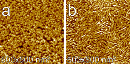
Samples are prepared by depositing Fe on graphene grown on a SiC substrateHershberger et al. (2013) using a molecular beam source held at a temperature of 700 K with a flux rate of 0.1 - 0.2 monolayers (ML)/min. The Fe source is degassed during the bake-out for several hours, so that during deposition the pressure remains below Torr. Figure 1 shows STM images of the (a) thin (20 ML) and (b) thick (75 ML) samples of Fe on grapheneHershberger et al. (2013). As shown in the STM images, islands of approximately 10 nm diameter are formed in the shape of pillars of roughly uniform height. The average number of Fe-MLs in each island is determined by evaluating each island’s volume within a given area after correcting for finite size tip effects. After the STM imaging and while still under ultra-high vacuum, both samples were capped with nominal 20 layers of Au for protection against oxidation during sample transport (in air) for the XMCD experiments. XMCD measurements were performed at the 4-ID-C beamline at the Advanced Photon Source (Argonne National Laboratory) in a chamber equipped with a high magnetic field ( T) produced by a split-coil superconducting magnet. Field dependence of the XMCD spectra were collected in helicity-switching mode in external magnetic fields applied parallel to the incident x-ray wave vector at energies that covered the Fe (719.9 eV) and (706.8 eV) binding energies. The X-ray incident angle was fixed at degrees with respect to the sample surface and measurements of x-ray absorption spectroscopy (XAS) signals were collected by total electron yield (TEY), reflectivity (REF), and fluorescence. REF is detected with a Si diode and TEY is determined from the drain current to the sample. All detection modes are collected simultaneously. Here, we report TEY results only, however results in other modes were examined for consistency. XMCD signal is obtained from the difference between two XAS spectra of the left- and right-handed helicities, and . All XAS signals values for and are normalized to the incident beam monitor intensity. For detailed data normalization and background evaluation see SI.
Results and Discussion
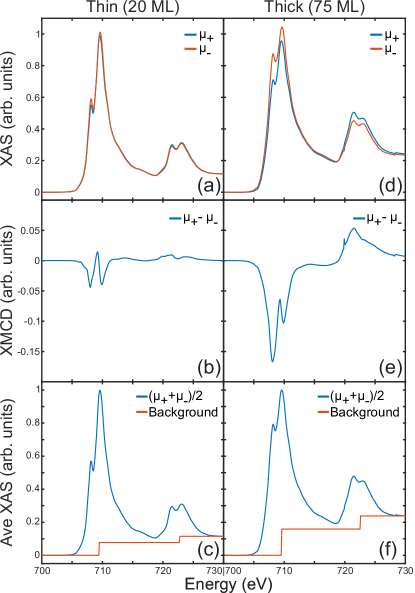
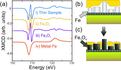
Figure 2 shows normalized XAS and XMCD for both samples (20 and 75 MLs) at T and K over the range of eV. The XAS in the two polarization modes show splittings of the and peaks that are not characteristic of iron metal suggesting the presence of Fe2+ and Fe3+ in the sample. The XMCD spectra for both samples extracted from these two XAS scans (shown in Fig. 2(b and e)) are significantly different from that reported for iron metal.Chen et al. (1995) Thus, despite being covered with gold, our samples show strong features that indicate they are possibly oxidized raising the question of what iron oxide species are formed. It has been shown that both the XAS and the XMCD exhibit distinct features that can distinguish among various iron oxides including FeO, and -\ceFe2O3, Fe3-δO4, and \ceFe3O4.Pellegrain et al. (1999); Brice-Profeta et al. (2005); Huang et al. (2004); Martín-García et al. (2015); Chen et al. (1995) Furthermore, it has been shown that the XMCD spectra of \ceFe3O4 is a superposition of three components corresponding to the three distinct sites of iron in this inverse spinel.Pattrick et al. (2002) In Fig. 3 we compile the XMCD spectra from various iron oxides and iron metal to compare them with that of our thin sample. We note that the two closest spectra to ours, are those of \ceFe3O4 and -\ceFe2O3. Closer inspection of the two spectra shows that the first two minima (at the edge) are reversed in magnitude between the two, indicating that our sample is closer in composition to \ceFe3O4 than to -\ceFe2O3. We therefore conclude that the protection of the nano-islands with 20 MLs of gold does not prevent iron from oxidation, most likely due to defects and incomplete uniform protective layer (see depiction of expected and experimental results in Fig. 3 (b) and (c), respectively). More importantly the Fe nano-islands at this size (20 ML thick) readily convert to a majority magnetite \ceFe3O4 phase with a possible minority phase of maghemite, -\ceFe2O3 (further experimental evidence for the formation of \ceFe3O4 is provided below.) This result, demonstrating that at the nano-scale size (approximately 10 nm in diameter) pure iron if exposed to oxygen transforms spontaneously to magnetite, is extremely important in view of the fact that tremendous efforts have been dedicated to synthesizing magnetite nano-particlesLu et al. (2007); Wu et al. (2015); Xu et al. (2014). This also opens an avenue for assembling magnetite nanoparticles on solid surfaces by first arranging, by deposition, pure iron nano-particles and subsequently exposing them to an oxidative environment.
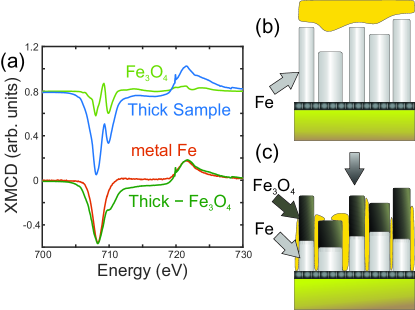
The XMCD for the thick sample (75 MLs; Fig. 4) matches neither that of pure Fe nor that of \ceFe3O4. Due to fast electronic relaxation times, the XMCD features from pure iron (or other good metals) are not split and are slightly broadened over those in insulators and semimetals (i.e., magnetite).Chen et al. (1995) Thus, based on the features and linewidths of peaks observed in the XMCD measurements, we suggest that the islands of the thick sample ( MLs) consist of metallic iron and \ceFe3O4. By applying a scale factor of 1.4 to the XMCD signal from the thin sample (Fig. 2b) and subtracting it from that of the thick sample (Fig. 4) we obtain a curve that resembles that of metallic iron (Fig. 4) albeit slightly wider. We therefore propose that the taller islands are only partially oxidized to form \ceFe3O4 and that metal iron is still present in some parts of the sample (as depicted in Fig. 4 (b) and (c)). The different observations between tall and short islands suggests that the kinetics of oxidation is time dependent (both samples, after being capped by gold, have been in air for about a few weeks outside the ultra-high vacuum chamber). We have, in fact, reexamined the thick sample after seventeen months and found that its XMCD pattern indicates more conversion into magnetite (see Supporting Information). We note that our results on the transformation of pure metal iron to magnetite through defects in a protecting layer are consistent with a recent study that examines the long term efficiency of passivating various transition metal surfaces with a layer of graphene Weatherup et al. (2015). That study shows that for a Ni surface, the graphene protects the surface from oxidation and furthermore, at undesired defects in the graphene layer, the oxidation of Ni is localized and prevents oxidation diffusion into the surface. On the other hand, for iron surfaces it is found that oxidation at defect points is no longer localized and oxidation practically diffuses throughout the surface over long period of time. Whereas that study confirms oxidation of Fe surfaces via graphene defects, it does not provide chemical analysis of the species that are formed at the surface as provided in this study.
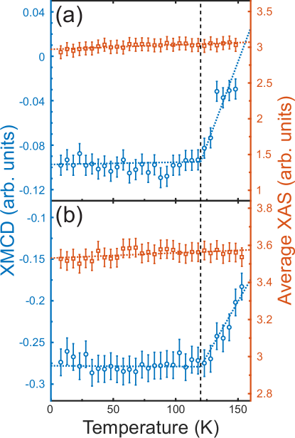
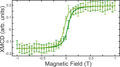
To corroborate our claim on the spontaneous formation of magnetite from pure iron nano-islands by oxidation, we have also systematically looked for signatures typical of magnetite in the temperature dependence of the XAS and XMCD signals from 8 to 153 K. Figure 5 shows the integration of the signal (705-715 eV at T) of the XMCD and XAS versus temperature with a distinct anomaly at 120 K for both samples. This anomaly is consistent with the well documented Verwey transition in magnetite which takes place in \ceFe3O4 at 125 K when undergoing a metal-insulator transition Senn et al. (2012); Yu et al. (2014). The existence of the Verwey transition in nano-size magnetite has been the subject of some debate, however, recently it has been demonstrated that only for particles below nm in size is the transition reduced from its bulk value.Park et al. (2005); Lee et al. (2015) The change in the integrated XMCD signal at results from the change in magnetic ordered moments and orbital ordering, both of which (spin and orbital moments) contribute to the XMCD. We note that whereas both samples show the Verwey transition in the integrated XMCD signals, neither show any change in their XAS signal. The field dependence of the XMCD signal (thick sample) at low temperatures (Fig. 6), which to a good approximation is proportional to the magnetization of the sample, shows a hysteresis loop with coercivity field T. It is well established that for high purity iron (99.95%) TBrown (1958) thus, the observed hysteresis is consistent with that of magnetite. This relatively high value is consistent with similar hysteresis measurements of magnetite that are acicular in shape (needle-like)Morrish and Watt (1958); Okuda and Harada (1985) with similarity to our pillar-like island grown NCs.
Conclusions
Using X-ray magnetic circular dichroism (XMCD) of Au-capped Fe nano-islands, we show that iron-oxidation (by exposure to air) proceeds spontaneously, likely through defects in the gold-film. Furthermore, we find that the iron nano-islands convert to magnetite nano-particles (totally or partially, depending on particle size). The evidence for the transformation of iron nano-islands to magnetite by oxidation is unequivocal as it is also corroborated by the presence of the well known Verwey transition at K as in bulk magnetite. Our results also indicate that oxidation is size and time dependent.
Supplementary Material
See the Supplementary Material for more information on the following: (1) The long-term oxidation time dependence of the thick sample’s XMCD. (2) Selected XMCD spectra above and below the Verwey transition, also used for creating the detailed temperature dependence shown in Figure 5 (3) Details on data normalization and background subtraction.
I Acknowledgments
Ames Laboratory is operated by Iowa State University by support from the U.S. Department of Energy, Office of Basic Energy Sciences, under Contract No. DE-AC02-07CH11358. Use of the Advanced Photon Source, an Office of Science User Facility operated for the U.S. Department of Energy (DOE) Office of Science by Argonne National Laboratory, is supported by the U.S. DOE under Contract No. DE-AC02-06CH11357.
References
- Lu et al. (2007) A.-H. Lu, E. Salabas, and F. Schüth, Angew. Chem. Int. Ed. 46, 1222 (2007).
- Wu et al. (2015) W. Wu, Z. Wu, T. Yu, C. Jiang, and W.-S. Kim, Sci. Technol. Adv. Mater. 16, 023501 (2015).
- Xu et al. (2014) J.-K. Xu, F.-F. Zhang, J.-J. Sun, J. Sheng, F. Wang, and M. Sun, Molecules 19, 21506 (2014).
- Harrison et al. (2002) R. J. Harrison, R. E. Dunin-Borkowski, and A. Putnis, Proc. Natl. Acad. Sci. 99, 16556 (2002).
- Blakemore (1975) R. Blakemore, Science 190, 377 (1975).
- Wang et al. (2012) L. Wang, T. Prozorov, P. E. Palo, X. Liu, D. Vaknin, R. Prozorov, S. Mallapragada, and M. Nilsen-Hamilton, Biomacromolecules 13, 98 (2012).
- Wang et al. (2010) H. Wang, L.-F. Cui, Y. Yang, H. Sanchez Casalongue, J. T. Robinson, Y. Liang, Y. Cui, and H. Dai, J. Am. Chem. Soc. 132, 13978 (2010).
- Zhou et al. (2010) G. Zhou, D.-W. Wang, F. Li, L. Zhang, N. Li, Z.-S. Wu, L. Wen, G. Q. M. Lu, and H.-M. Cheng, Chem. Mater. 22, 5306 (2010).
- Cong et al. (2012) H.-P. Cong, X.-C. Ren, P. Wang, and S.-H. Yu, ACS Nano 6, 2693 (2012).
- Hershberger et al. (2013) M. T. Hershberger, M. Hupalo, P. A. Thiel, and M. C. Tringides, J. Phys. Condens. Matter 25, 225005 (2013).
- Dankert et al. (2014) A. Dankert, M. V. Kamalakar, J. Bergsten, and S. P. Dash, Appl. Phys. Lett. 104, 192403 (2014), arXiv:1405.0836 .
- Chen et al. (1995) C. T. Chen, Y. U. Idzerda, H.-J. Lin, N. V. Smith, G. Meigs, E. Chaban, G. H. Ho, E. Pellegrin, and F. Sette, Phys. Rev. Lett. 75, 152 (1995).
- Pellegrain et al. (1999) E. Pellegrain, M. Hagelstein, S. Doyle, H. O. Moser, J. Fuchs, D. Vollath, S. Schuppler, M. A. James, S. S. Saxena, L. Niesen, O. Rogojanu, G. A. Sawatzky, C. Ferrero, M. Borowski, O. Tjernberg, and N. B. Brookes, phys. stat. sol. (b) 215, 797 (1999).
- Brice-Profeta et al. (2005) S. Brice-Profeta, M. A. Arrio, E. Tronc, N. Menguy, I. Letard, C. Cartier dit Moulin, M. Noguès, C. Chanéac, J. P. Jolivet, and P. Sainctavit, Journal of Magnetism and Magnetic Materials 288, 354 (2005).
- Huang et al. (2004) D. J. Huang, C. F. Chang, H.-T. Jeng, G. Y. Guo, H.-J. Lin, W. B. Wu, H. C. Ku, A. Fujimori, Y. Takahashi, and C. T. Chen, Phys. Rev. Lett. 93 (2004), 10.1103/PhysRevLett.93.077204.
- Martín-García et al. (2015) L. Martín-García, R. Gargallo-Caballero, M. Monti, M. Foerster, J. F. Marco, L. Aballe, and J. de la Figuera, Phys. Rev. B 91 (2015), 10.1103/PhysRevB.91.020408.
- Pattrick et al. (2002) R. A. D. Pattrick, G. Van Der Laan, C. M. B. Henderson, P. Kuiper, E. Dudzik, and D. J. Vaughan, Eur. J. Mineral. 14, 1095 (2002).
- Goering et al. (2007) E. J. Goering, M. Lafkioti, S. Gold, and G. Schuetz, Journal of Magnetism and Magnetic Materials Proceedings of the 17th International Conference on MagnetismThe International Conference on Magnetism, 310, e249 (2007).
- Weatherup et al. (2015) R. S. Weatherup, L. D’Arsié, A. Cabrero-Vilatela, S. Caneva, R. Blume, J. Robertson, R. Schloegl, and S. Hofmann, J. Am. Chem. Soc. 137, 14358 (2015).
- Senn et al. (2012) M. S. Senn, J. P. Wright, and J. P. Attfield, Nature 481, 173 (2012).
- Yu et al. (2014) Q. Yu, A. Mottaghizadeh, H. Wang, C. Ulysse, A. Zimmers, V. Rebuttini, N. Pinna, and H. Aubin, Phys. Rev. B 90, 075122 (2014).
- Park et al. (2005) J. Park, E. Lee, N.-M. Hwang, M. Kang, S. C. Kim, Y. Hwang, J.-G. Park, H.-J. Noh, J.-Y. Kim, J.-H. Park, and T. Hyeon, Angewandte Chemie International Edition 44, 2872 (2005).
- Lee et al. (2015) J. Lee, S. G. Kwon, J.-G. Park, and T. Hyeon, Nano Lett. 15, 4337 (2015).
- Brown (1958) W. F. Brown, Handbook of Chemistry and Physics (McGraw-Hill, 1958).
- Morrish and Watt (1958) A. H. Morrish and L. A. K. Watt, J. Appl. Phys. 29, 1029 (1958).
- Okuda and Harada (1985) Y. Okuda and T. Harada, Process for Producing Acicular Magnetite or Acicular Maghemite U.S. Patent 4,495,164 (10 July 1984).