Phototaxis beyond turning: persistent accumulation and response acclimation of the microalga Chlamydomonas reinhardtii
Abstract
Phototaxis is an important reaction to light displayed by a wide range of motile microorganisms. Flagellated eukaryotic microalgae in particular, like the model organism Chlamydomonas reinhardtii, steer either towards or away from light by a rapid and precisely timed modulation of their flagellar activity. Cell steering, however, is only the beginning of a much longer process which ultimately allows cells to determine their light exposure history. This process is not well understood. Here we present a first quantitative study of the long timescale phototactic motility of Chlamydomonas at both single cell and population levels. Our results reveal that the phototactic strategy adopted by these microorganisms leads to an efficient exposure to light, and that the phototactic response is modulated over typical timescales of tens of seconds. The adaptation dynamics for phototaxis and chlorophyll fluorescence show a striking quantitative agreement, suggesting that photosynthesis controls quantitatively how cells navigate a light field.
The fitness of microorganisms depends critically on their ability to sense dynamic physico-chemical clues from the environment, elaborate the information and respond effectively. Environmental responses range from changes in gene expression Trippens et al. (2012) (typical timescale min); to the activation/deactivation of biochemical processes like chloroplast photoprotection Allorent et al. (2013) (min); to fast movement regulation (s), either active Stocker et al. (2008); Drescher et al. (2010) or passive Arrieta et al. (2015). The best characterised motile response is currently chemotaxis of run-and-tumble bacteria like E. coli Berg (1975), a strategy based on the modulation of tumbling frequency Celani and Vergassola (2010). Chemotaxis features (almost)perfect adaptation to persistent stimuli over intermediate timescales (s) Sourjik and Berg (2002); Meir et al. (2010) and can stimulate/inhibit gene expression through a variety of chemosensory pathways Wadhams and Armitage (2004). This paradigmatic sensory system highlights the important crosstalk happening between responses acting across a wide spectrum of time intervals, and exemplifies the need for a consistent cross-timescale framework to understand motility regulation in microorganisms. In the case of phototaxis, a major response in eukaryotic microalgae Jekely (2009), this framework is lacking.
Among micro-eukaryotes, phototaxis is best characterised in the model system Chlamydomonas reinhardtii (CR) Harris (2009), a green microalga which swims along a helical trajectory by the synchronous breaststroke beating of its flagellar pair Goldstein et al. (2009, 2011). Cell spinning Martinez et al. (2012) induces a periodic modulation of the signal received by the eyespot, a rhodopsin-based light-sensitive organelle Kateriya (2004) featuring a contrast-enhancing dielectric mirror Foster and Smyth (1980). Eyespot stimulation is rapidly relayed via an action-potential-like signal to the flagella (ms)Sineshchekov et al. (1990), and triggers a Ca+2-dependent differential response of their beating Ruffer and Nultsch (1991); Josef et al. (2006) causing cells to steer either towards or away from light Schaller et al. (1997); Yoshimura and Kamiya (2001). Implementation within a minimal model Bennett and Golestanian (2014) confirmed that phototactic steering is robust and can indeed lead to both positive and negative taxis, a property that has been used to achieve photo-hydrodynamic focussing of microalgae Garcia et al. (2013). What happens beyond phototactic steering, however, is not well understood. Phototaxis of microalgae can lead to persistent modification of bioconvective patterns Williams and Bees (2011a, b), and should therefore contribute to the interplay between fluid flow and motility leading to microscale patchiness in the seas Torney and Neufeld (2008); Stocker (2012). At the single cell level, phototaxis will modulate cell irradiance and can therefore be expected to impact both cell metabolism -through chloroplast stimulation- and light-sensitive gene expression Petroutsos et al. (2016). Except for qualitative accounts of red-light control of phototactic sign Takahashi and Watanabe (1993), these links have not been explored.
Here we present the first long-timescale study of phototactic behaviour of CR, as representative of green microalgae. Studying the accumulation dynamics around a localised source, we show that cells use tight circulation around the maximum light intensity as a strategy to maximise their overall light exposure before spontaneously leaving the illuminated region. Periodic exposure experiments reveal that this is accompanied by a decrease in the overall response to light stimuli. The quantitative modulation of phototactic response tracks the dynamics of chlorophyll fluorescence, used here as a proxy for the photosynthetic activity of the cells.
Material and Methods
Chlamydomonas reinhardtii wild type strain CC125 and bald mutant CC2905 were grown axenically at C in Tris-Acetate-Phosphate medium Rochaix et al. (1988) under fluorescent light illumination (OSRAM Fluora, mol/m2s PAR) following a 14h/10h light/dark diurnal cycle. Exponentially growing cells at cells/ml were loaded in the mm diameter circular observation chamber cored out of a mm thick agar pad sandwiched between coverslips. A CCD camera (Pike, AVT) hosted on a continuously focusable objective (InfiniVar CFM-2S, Infinity USA) recorded at fps the phototactic motility of cells within the horizontal sample, visualised through darkfield illumination at nm (FLDR-i70A-R24, Falcon Lighting). Actinic light was provided by a nm LED (Thorlabs M470L2) through a m-diameter multimode optical fibre (FT200EMT, Thorlabs). Approximation of the fibre output by a Gaussian (m, peak intensity mol/m2s) is excellent and will be used throughout the paper. An inverted microscope (TE2000-U, Nikon) fitted with a Plan Apo objective (NA 0.45) and a EMCCD (Evolve, Photometrics) was used to record the chlorophyll fluorescence of CC2905, excited by the epiport-coupled blue LED.
Results and Discussion
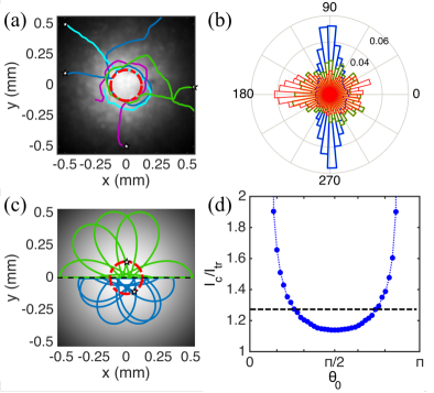
We begin by examining single-cell phototaxis after the light was kept on for min to ensure steady conditions (Fig. 1a). Cells further from the centre than m move inwards along almost radial trajectories as a result of active steering. As they approach the centre, however, individual CRs turn sharply and start circulating around the maximum at an average distance of m. This is confirmed by the azimuthally-averaged probability distribution function of swimming directions in Fig. 1b. Given the average swimming speed m/s, we obtain an angular velocity rad/s which compares well with the average value previously reported for sharp turns (rad/s) where cells achieve their largest angular speeds Polin et al. (2009). Orbiting cells do not show the preference for a particular chirality characteristic of hydrodynamic interactions with the sample surface Frymier et al. (1995); Lauga et al. (2006). Instead, the orbits have a fundamentally phototactic origin. Recorded only episodically in flagellates Rhiel et al. (1988); Figueroa et al. (1998); Matsunaga et al. (2003), orientation perpendicular to light stimulus (diaphototaxis) was reported as an anecdotal curiosity in CR Foster and Smyth (1980). It appears here as a specific modulation of phototaxis allowing CR to dwell in localised light spots.
The position of a cell swimming at constant speed along the direction will evolve according to
| (1) |
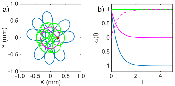
where the angular speed encodes the phototactic response through its (unknown) dependence on the light field. Absent detailed measurements, a common approach Williams and Bees (2011a, b); Furlan et al. (2012) has been to assume proportionality to the local gradient in light intensity, , where the phototactic parameter , possibly dependent on , represents the magnitude of the response.
For CR, the requirement implies . This reasonable model predicts correctly the radial reorientation of cells far from the source, but the incoming trajectories are then expected to overshoot the centre and eventually describe trochoids like to those seen in Fig. 1c. Similar trajectories are indeed seen both in phototactic colloids moving around a diverging laser beam Moyses et al. (2016), and in sea-urchin sperm swimming around a local chemotactic cue Guerrero et al. (2010). Phototactic CR’s however, do not follow trochoids but fall instead onto the tightest closed loops they can achieve around the light source, at an average distance from the centre. This dynamics cannot be reproduced by changing to include a transition between positive and negative phototaxis around (SI Appendix, Fig. S1): it is a fundamentally different type of behaviour that cells follow during positive phototaxis.
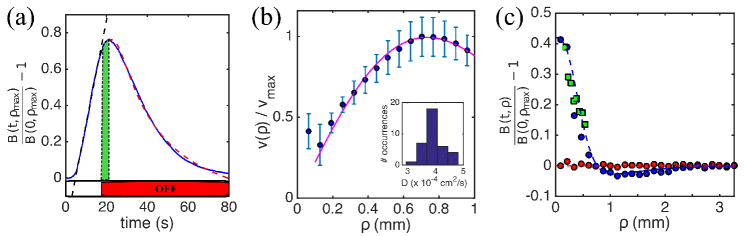
Fig. 1d shows that the real dynamics exposes the microalgae to a larger path-averaged light intensity than the trochoidal case, and therefore appears to be better strategy to optimise light capture by a photosynthetic microswimmer. After s, however, cells stop orbiting and leave the field of view. Consistently observed across the 3290 tracks recorded, this behaviour reflects a clear adaptation of phototactic motility, turning here from positive to negative, and challenge the common assumption of an optimal light intensity which cells simply get attracted to. Flagellar response to light-step-up/step-down stimuli is indeed known to depend -qualitatively- on the choice of pre-stimulus adaptation Ruffer and Nultsch (1991). The adaptive dynamics observed here, however, is a consequence of a history of light-exposure selected autonomously by single cells through their motility.
We now turn to the phototactic behaviour of a population (Fig. 2) to investigate the effect of adaptation over timescales longer than those accessible from the limited field of view of single-cell experiments. Cell concentrations will be kept below cells/ml to prevent effects on either the actinic light field perceived by the algae, or darkfield illumination Schaller et al. (1997). The image brightness is then proportional to the 2D-projected concentration of algae , which integrates the 3D one across approximately the depth of field of the imaging apparatus. Agreement between brightness profiles after prolonged light exposure (s), and the distribution of cell positions from individual tracks (Fig. 2c) confirms the proportionality, and suggests that cell-cell interactions are not important here. Cell accumulation can be characterised through the integrated image brightness where the maximum value m is set by the image size. Initially uniformly distributed, the algae begin to accumulate around the fibre as the light is turned on, causing to increase linearly with time (Fig. 2a, blue solid line). This is a signature of a constant inward flux of cells, proportional to the product of the net phototactic drift at distance , and the geometric factor which takes into account cells moving inwards from deep within the sample. The full curve can then be measured from the initial increase up to a multiplicative constant (Fig. 2a, black dashed line). Figure 2b shows that this is well described by with the exception of the core region m, where we already know that cell behaviour is different.
Switching the light off, the profile relaxes down to the original homogeneous value (Fig. 2a, blue solid line). This dynamics is well characterised by a simple diffusive spreading (Fig. 2b, magenta dashed line) with an effective diffusivity which can be recovered from a one-parameter fit (Fig. 2b inset). The average value cm2/s is in reasonable agreement with the average diffusivity cm2/s reported previously Polin et al. (2009).
The coarse-grained phototactic drift and the effective diffusivity can be used in a Keller-Segel-like continuum model of the phototactic behaviour of a population of CR, in the spirit of previous effective descriptions of phototaxis Furlan et al. (2012); Giometto et al. (2015); Martin et al. (2016). In this model, valid sufficiently far from the source, the local concentration of cells moving in the fibre’s axisymmetric light field obeys the continuity equation
| (2) |
where the extra factor of , non-dimensionalised by the effective thickness has been included to take into account three dimensional effects on our 2D description, as discussed previously. The local phototactic velocity , which incorporates Weber’s law Shoval et al. (2010), is characterised by the phototactic sensitivity of the population, , setting the maximum phototactic drift (). To compare Eq. (2) with experiments, we fix the cell concentration at the sample boundary, , and use the experimentally measured values for the mean swimming velocity and cell diffusivity, light field and phototactic sensitivity. The last parameter is derived from the distribution of single cells’ swimming directions at (Fig. 1b), giving . A one-parameter fit to the long-timescale profile in Fig. 2c (blue circles) sets the value of . The result (dashed blue line) shows that m provides an excellent description of the cell concentration, implying that cells within roughly half of the sample thickness take part in the phototactic accumulation. The model predicts also the presence of a depletion ring at mm responsible for the slight overshoot of experimentally observed right after light-off (Fig. 2a, green bar). Single cell experiments suggest, then, that the measured low phototactic sensitivity results from the balance between inwards/outwards swimming and dwell time, all present in the natural phototactic behaviour of each individual CR cell and modulated by its irradiation history (Fig. 1a).
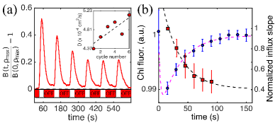
Equipped with an appropriate description of the steady state, we now investigate the adaptation process by characterising the phototactic accumulation of a population of dark-adapted cells to a series of identical light-on/light-off cycles (s on/off; Movie S1 shows one cycle). Figure 3a presents the accumulation dynamics for a representative experiment out of 60, showing a clear dependence on history of light exposure. Accumulation and dispersal phases allow one to measure the time (and light) evolution of both and , and therefore pinpoint the dynamical features responsible for the adaptation. Figure 3a (inset) shows that over the whole experiment increases slightly by , suggesting a increase in (i.e. photokynesis) which, by itself, would lead to an equivalent increase in . Instead, this parameter displays a well defined decrease through the cycles (Fig. 3b, red squares), unequivocally assigning the adaptation to a change in the phototactic sensitivity alone. The evolution of the sensitivity parameter is well described by a single-time adaptation where the adaptation timescale s and are derived from the fit in Fig. 3b (black dashed line). In this analysis, we assumed that evolves only during periods of illumination. Dark re-adaptation was not observed in the experiments; it must happen over significantly longer timescales and therefore was not considered here.
Phototactic adaptation operates on timescales clearly separated from those characterising adaptation of either flagellar photoshock (s) Hegemann and Bruck (1989); Kateriya (2004) or eyespot signalling (ms) Govorunova et al. (1997). Being directly related to cell irradiance, itself relevant for photosynthesis, we therefore wondered whether the dynamics of would contain any signature of light-adaptation by CR’s photosynthetic apparatus. To investigate this, we exposed dark adapted non-swimming CR cells (CC2905) to the sequence of light stimulation used previously (see Fig. 3a), and recorded the evolution of their average chlorophyll fluorescence (nmnm), which can be used as a simple proxy for the activity of the photosynthetic apparatus Maxwell and Johnson (2000).
A homogeneous light field of intensity E/m2s was used (identical results were obtained for and mol/m2s). Figure 3b shows the evolution of the mean during each light-on period (blue circles). Light-off intervals did not induce appreciable dark-adaptation, in line with known differences between light- and dark-adaptation of the photosynthetic apparatus Baker (2008). Chlorophyll fluorescence evolution is well fitted by a simple two-timescale dynamics (Fig. 3b magenta dashed line) with an initial fast response (timescale s) followed by a slow adaptation with timescale s. The exceptional quantitative agreement between and suggests a connection between the two processes, a possibility which would also explain the slow dark-adaptation of phototaxis.
Phototaxis experiments under a simultaneous background illumination have shown that chloroplast stimulation can induce CRs to qualitatively switch their phototactic sign (positive to negative) Takahashi and Watanabe (1993). Our results suggest the intriguing possibility that phototaxis and photosynthesis are in fact connected quantitatively. Although further experiments are needed to firmly establish this layer of control, we propose here the hypothesis that this connection is indeed the major determinant of the phototactic motility of eukaryotic microalgae.
Conclusions
The light-induced steering responses evolved by microorganisms like Chlamydomonas are complex, and have been studied extensively. Ultimately, however, flagellar activity must be integrated into a coherent navigation strategy combining physical stimuli and intracellular requirements: how this is achieved is currently not understood. By shifting the focus to long timescales we start addressing this gap. Our experiments have already revealed a surprisingly rich dynamics, from the ability to increase light exposure by switching to diaphototaxis to the adaptive response of cells which reproduces the slow (re)adaptation of their chlorophyll fluorescence. Future experiments will be needed to systematically explore the role of light intensity and colour; to determine whether phototaxis shares any of the common properties of cellular sensory systems, like exact adaptation Shoval et al. (2010); Lazova et al. (2011); and in particular how these properties are connected with photoprotective dynamics within the chloroplast Allorent et al. (2013) and photosynthetic efficiency Kim et al. (2016).
The authors acknowledge fruitful discussions with Miguel Gonzalez. The work has been partly supported by the Spanish Ministry of Economy and Competitiveness grant No. FIS2013-48444-C2-1-P and the Subprogram Ramón-y-Cajal (IT); the Royal Society Research Grant RG150421 and the University of the Balearic Islands Travel Grant 22/2016 (MP). MP gratefully acknowledges the hospitality of the Mediterranean Institute of Advanced Studies, where part of this work has been performed.
References
- Trippens et al. (2012) J. Trippens, A. Greiner, J. Schellwat, M. Neukam, T. Rottmann, Y. Lu, S. Kateriya, P. Hegemann, and G. Kreimer, The Plant cell 24, 4687 (2012).
- Allorent et al. (2013) G. Allorent, R. Tokutsu, T. Roach, G. Peers, P. Cardol, J. Girard-Bascou, D. Seigneurin-Berny, D. Petroutsos, M. Kuntz, C. Breyton, F. Franck, F.-a. Wollman, K. K. Niyogi, A. Krieger-Liszkay, J. Minagawa, and G. Finazzi, The Plant Cell 25, 545 (2013).
- Stocker et al. (2008) R. Stocker, J. R. Seymour, A. Samadani, D. E. Hunt, and M. F. Polz, Proceedings of the National Academy of Sciences 105, 4209 (2008).
- Drescher et al. (2010) K. Drescher, R. E. Goldstein, and I. Tuval, Proceedings of the National Academy of Sciences of the United States of America 107, 11171 (2010).
- Arrieta et al. (2015) J. Arrieta, A. Barreira, and I. Tuval, Physical Review Letters 114, 128102 (2015).
- Berg (1975) H. C. Berg, Annual review of biophysics and bioengineering 4, 119 (1975).
- Celani and Vergassola (2010) A. Celani and M. Vergassola, Proceedings of the National Academy of Sciences 107, 1391 (2010).
- Sourjik and Berg (2002) V. Sourjik and H. C. Berg, Proceedings of the National Academy of Sciences 99, 123 (2002).
- Meir et al. (2010) Y. Meir, V. Jakovljevic, O. Oleksiuk, V. Sourjik, and N. S. Wingreen, Biophysical Journal 99, 2766 (2010).
- Wadhams and Armitage (2004) G. H. Wadhams and J. P. Armitage, Nature Reviews Molecular Cell Biology 5, 1024 (2004).
- Jekely (2009) G. Jekely, Philosophical Transactions of the Royal Society B: Biological sciences 364, 2795 (2009).
- Harris (2009) E. H. Harris, The Chalmydomonas Sourcebook (2009).
- Goldstein et al. (2009) R. E. Goldstein, M. Polin, and I. Tuval, Physical Review Letters 103, 168103 (2009).
- Goldstein et al. (2011) R. E. Goldstein, M. Polin, and I. Tuval, Physical Review Letters 107, 148103 (2011).
- Martinez et al. (2012) V. a. Martinez, R. Besseling, O. a. Croze, J. Tailleur, M. Reufer, J. Schwarz-Linek, L. G. Wilson, M. A. Bees, and W. C. K. Poon, Biophysical journal 103, 1637 (2012).
- Kateriya (2004) S. Kateriya, News in Physiological Sciences 19, 133 (2004).
- Foster and Smyth (1980) K. W. Foster and R. D. Smyth, Microbiological Review 44, 572 (1980).
- Sineshchekov et al. (1990) O. a. Sineshchekov, F. F. Litvin, and L. Keszthelyi, Biophysical Journal 57, 33 (1990).
- Ruffer and Nultsch (1991) U. Ruffer and W. Nultsch, Cell Motility and the Cytoskeleton 18, 269 (1991).
- Josef et al. (2006) K. Josef, J. Saranak, and K. W. Foster, Cell motility and the cytoskeleton 63, 758 (2006).
- Schaller et al. (1997) K. Schaller, R. David, and R. Uhl, Biophysical Journal 73, 1562 (1997).
- Yoshimura and Kamiya (2001) K. Yoshimura and R. Kamiya, Plant and Cell Physiology 42, 665 (2001).
- Bennett and Golestanian (2014) R. R. Bennett and R. Golestanian, Journal of the Royal Society Interface 12, 20141164 (2014), arXiv:1410.6270 .
- Garcia et al. (2013) X. Garcia, S. Rafaï, and P. Peyla, Physical Review Letters 110, 138106 (2013).
- Williams and Bees (2011a) C. R. Williams and M. A. Bees, The Journal of experimental biology 214, 2398 (2011a).
- Williams and Bees (2011b) C. R. Williams and M. A. Bees, Journal of Fluid Mechanics 678, 41 (2011b).
- Torney and Neufeld (2008) C. Torney and Z. Neufeld, Physical Review Letters 101, 1 (2008).
- Stocker (2012) R. Stocker, Science 338, 628 (2012).
- Petroutsos et al. (2016) D. Petroutsos, R. Tokutsu, S. Maruyama, S. Flori, A. Greiner, L. Magneschi, L. Cusant, T. Kottke, M. Mittag, P. Hegemann, G. Finazzi, and J. Minagawa, Nature 537, 563 (2016).
- Takahashi and Watanabe (1993) T. Takahashi and M. Watanabe, FEBS Letters 336, 516 (1993).
- Rochaix et al. (1988) J. D. Rochaix, S. Mayfield, M. Goldschmidt-Clermont, and J. M. Erickson, Plant Molecular Biology: A Practical Approach, edited by C. H. Schaw (IRL Press, Oxford, England, 1988) pp. 253-275.
- Polin et al. (2009) M. Polin, I. Tuval, K. Drescher, J. P. Gollub, and R. E. Goldstein, Science 325, 487 (2009).
- Frymier et al. (1995) P. D. Frymier, R. M. Ford, H. C. Berg, and P. T. Cummings, Proceedings of the National Academy of Sciences of the United States of America 92, 6195 (1995).
- Lauga et al. (2006) E. Lauga, W. R. DiLuzio, G. M. Whitesides, and H. A. Stone, Biophysical Journal 90, 400 (2006), arXiv:0506675 [cond-mat] .
- Rhiel et al. (1988) E. Rhiel, D.-p. Hader, and W. Wehrmeyer, Plant and Cell Physiology 29, 755 (1988).
- Figueroa et al. (1998) F. L. Figueroa, F. X. Niell, F. G. Figueiras, and M. L. Villarino, Marine Biology 130, 491 (1998).
- Matsunaga et al. (2003) S. Matsunaga, S. Watanabe, S. Sakaushi, S. Miyamura, and T. Hori, Photochemistry and Photobiology 77, 324 (2003).
- Furlan et al. (2012) S. Furlan, D. Comparini, M. Ciszak, L. Beccai, S. Mancuso, and B. Mazzolai, PloS one 7, e38895 (2012).
- Moyses et al. (2016) H. Moyses, J. Palacci, S. Sacanna, and D. G. Grier, Soft Matter 12, 6357 (2016).
- Guerrero et al. (2010) A. Guerrero, T. Nishigaki, J. Carneiro, Yoshiro Tatsu, C. D. Wood, and A. Darszon, Developmental Biology 344, 52 (2010).
- Giometto et al. (2015) A. Giometto, F. Altermatt, A. Maritan, R. Stocker, and A. Rinaldo, Proceedings of the National Academy of Sciences 112, 7045 (2015).
- Martin et al. (2016) M. Martin, A. Barzyk, E. Bertin, P. Peyla, and S. Rafai, Physical Review E 93, 051101 (2016), arXiv:1603.00761 .
- Shoval et al. (2010) O. Shoval, L. Goentoro, Y. Hart, A. Mayo, E. Sontag, and U. Alon, Proceedings of the National Academy of Sciences of the United States of America 107, 15995 (2010).
- Hegemann and Bruck (1989) P. Hegemann and B. Bruck, Cell Motility and the Cytoskeleton 14, 501 (1989).
- Govorunova et al. (1997) E. G. Govorunova, O. A. Sineshchekov, and P. Hegemann, Plant Physiology 115, 633 (1997).
- Maxwell and Johnson (2000) K. Maxwell and G. N. Johnson, Journal of experimental botany 51, 659 (2000).
- Baker (2008) N. R. Baker, Annual review of plant biology 59, 89 (2008).
- Lazova et al. (2011) M. D. Lazova, T. Ahmed, D. Bellomo, R. Stocker, and T. S. Shimizu, Proceedings of the National Academy of Sciences 108, 13870 (2011).
- Kim et al. (2016) J. Y. H. Kim, H. S. Kwak, Y. J. Sung, H. I. Choi, M. E. Hong, H. S. Lim, J.-H. Lee, S. Y. Lee, and S. J. Sim, Scientific Reports 6, 21155 (2016).
I Supplementary Informations
Supplementary movie
The movie Mov1s.avi shows the dynamics of cell accumulation and dispersion in our experiments for a single light on - light off cycle.
Models for single cell phototaxis
The position of a cell swimming at constant speed along the direction will evolve according to
| (3) |
where the angular speed encodes the phototactic response through its (unknown) dependence on the light field. Absent detailed measurements, a common approach has been to assume proportionality to the local gradient in light intensity, , where the phototactic parameter , possibly dependent on , represents the magnitude of the response.
The model used throughout the manuscript uses the simplest description that incorporates constant. Here we address two additional functional forms for chosen to model (a) the transition from positive to negative phototaxis and (b) the transition from positive to dia-phototaxis. For the former, we chose a continuous function of I that changes sign at a prescribed critical intensity; more specifically, we set as seen in Fig. S1a (blue curve). An example of a trajectory corresponding to this choice of is depicted with the same colour in panel (b). For the model to incorporate a transition to diaphototaxis it is necessary to include in the equation for a term proportional to . In our case cells are confined to move on the plane and therefore where is the unit vector in the direction perpendicular to the plane of motion. We have explored as a particular realisation of the phenomenological diaphototactic model the following:
| (4) |
with . The model represents a continuous decay of positive phototaxis to zero for (shown as a magenta line in Fig. S1a) and a concurrent increase of the diaphototactic contribution (dashed magenta line in the same figure). The combined effect of the two contributions leads to the cell circling around the position of as shown for the trajectory plotted with the same colour in panel (b).