Pattern formation in desiccated sessile colloidal droplets with salt admixture: Short review
Abstract
This short review is devoted to the simple process of drying a multi-component droplet consisting of a complex fluid containing a salt. These processes provide a fascinating subject for study. The explanation of the rich variety of patterns formed is not only an academic challenge, but a problem of practical importance, as applications are growing in medical diagnosis and improvement of coating/printing technology. The fundamental scientific problem is the study of the mechanism of micro- and nanoparticle self-organization in open systems. The specific fundamental problem to be solved, related to this system is - the investigation of the mass transfer processes, the formation and evolution of phase fronts and the identification of mechanisms of pattern formation. The drops of liquid containing dissolved substances and suspended particles are assumed to be drying on a horizontal solid substrate. The chemical composition and macroscopic properties of the complex fluid, the concentration and nature of the salt, the surface energy of the substrate and the interaction between the fluid and substrate which determines the wetting, all affect the final morphology of the dried film.
I Introduction
The structures observed after drying biological fluids on a horizontal impenetrable substrate attracted the attention of European researchers as early as the 1950s Koch (1954, 1956, 1957a, 1957b, 1957c, 1957d); Solé (1954, 1955, 1957a, 1957b, 1960). Unfortunately, this series of articles did not attract attention from the physics community. In the 1980s the phenomenon has been reopened in the USSR Rapis (1988). In the 1980s-1990s, doctors of the Soviet Union and the countries of the former Soviet Union began to use the appearance of structures formed by drying droplets of biological fluids for the diagnosis of various diseases. Numerous articles are published in Russian, in medical journals, dissertations are defended, and different methods are patented that are devoted to the diagnosis of diseases on the basis of the structures formed by drying droplets of biological fluids. The relevant references can be, for example, found in the books Savina (1999); Shabalin and Shatohina (2001); Rapis (2003); Vorobiev et al. (2008). Unfortunately, the world scientific community is poorly informed about these works of Soviet and Russian scientists, since only a very few of these articles have been published in English Shabalin and Shatokhina (1996); Yakhno et al. (2003); Shatokhina et al. (2004); Shabalin and Shatokhina (2007); Martusevich and Kamakin (2007); Yakhno et al. (2015).
In the last two decades, interest in the structures arising on drying complex fluids has increased worldwide due to a variety of applications, such as bio-preservation Ragoonanan and Aksan (2008); Less et al. (2013), high-throughput drug screening Takhistov and Chang (2002), fast identification of fluid and substrate chemistry based on automatic pattern recognition of stains Kim et al. (2012), assessment of quality of products Kokornaczyk et al. (2011), and Raman spectroscopy Esmonde-White et al. (2008a, b, 2009a, 2009b); Filik and Stone (2007, 2008, 2009); Zhang et al. (2010); Pearce and Tomlinson (2000); Dingari et al. (2012); Kočišová et al. (2012). During the high-throughput drug screening, pattern formation in the the drying sample is not desirable Takhistov and Chang (2002). Several reviews Routh (2013); Sefiane (2013); Larson (2014); Zhong et al. (2015); Sadek et al. (2015); Chen et al. (2016) and books Lin (2010); Innocenzi et al. (2013); Brutin (2015); Goehring et al. (2015) based on pattern formation during desiccation, published within the short span of just four years confirms the intensely growing interest in this topic.
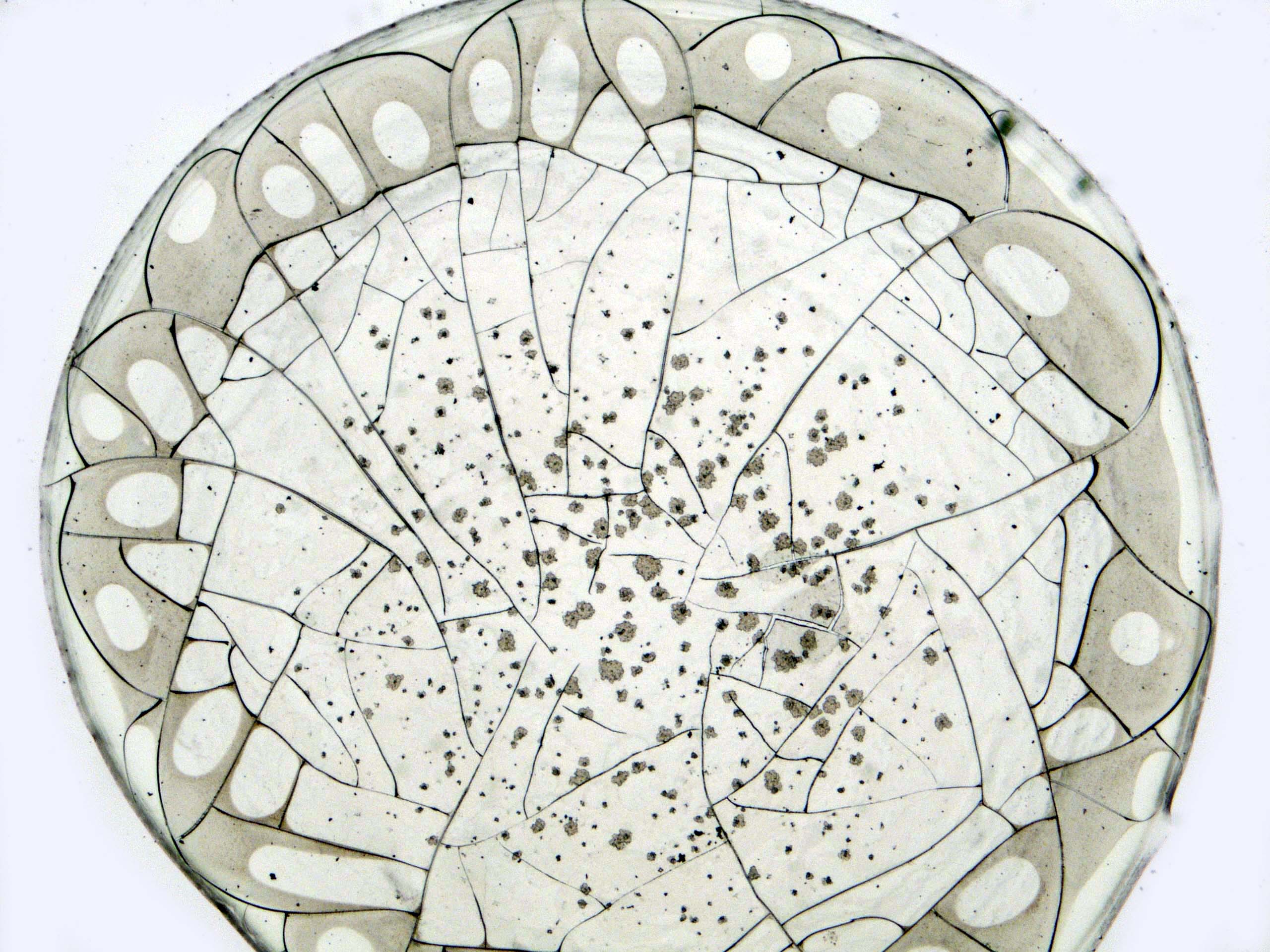
While performing the experiments seems very simple (table top experiments producing interesting patterns can be done even at home), understanding the physics behind the pattern formation phenomena turns out to be extremely complicated and involves a number of interrelated processes of different nature Tarasevich (2004); Yakhno and Yakhno (2009). During desiccation of biological fluids, a sequence of various physical and physico-chemical processes can be observed Yakhno (2008); Yakhno et al. (2011). For example, redistribution of the components occurs. Protein molecules are carried out by flows to the edge of the droplet, and accumulate to form a gel. The salt is distributed over the whole area of the droplet almost uniformly. After complete drying of the droplet, a protein precipitate remains on the substrate in the form of a ring, the width of the ring depends on concentrations of the protein and the salt Shabalin and Shatohina (2001); Prokhorov (2009). Salt crystals can form fractal (dendritic) structures Annarelli et al. (2001); Tarasevich and Ayupova (2003); Gorr et al. (2013); Dutta Choudhury et al. (2013, 2015). In the later stages of drying, a sample may crack Pauchard et al. (1999); Annarelli et al. (2001); Goehring et al. (2015), the characteristic pattern of the cracks also helps in diagnosing diseases which the subject may be suffering from Yakhno et al. (2005a).
Analysis based on a visual comparison of the structures formed by drying a liquid drop Rapis (1988); Savina (1999); Shabalin and Shatohina (2001); Rapis (2003); Yakhno et al. (2003) has significant drawbacks. Conclusions are liable to be subjective, without techniques for defining quantitative parameters to characterize the structures. Computer pattern recognition may be tried to eliminate this shortcoming Buzoverya et al. (2014); Bou Zeid et al. (2013); Kim et al. (2012).
Although pattern formation can be observed during drying of both inorganic and organic colloids, the case of biological fluids is attracting increasingly growing interest from the scientific community in recent years Sobac and Brutin (2011); Brutin et al. (2011a); Sefiane (2010).
Despite the application of the phenomenon, for practical purposes and considerable progress in the understanding of the phenomenon Tarasevich (2004); Yakhno and Yakhno (2009), the theoretical description of the pattern formation in desiccating biological fluids is still incomplete. The physical, biophysical, biochemical, biological, and physico-chemical processes occurring in the dehydration of biological fluids remain largely to be clarified. This explanation is a necessary background for understanding the connections between physico-chemical properties of the biological fluids and observed patterns.
Even though numerous publications in medical literature have been devoted to pattern formation in biological fluids during their drying, physicists, chemists and mathematicians began to pay attention to this problem only recently. During the last decade, several groups of physicists began to actively publish work in this direction Bardakov et al. (2010); Chashechkin and Bardakov (2010); Kistovich et al. (2010); Brutin et al. (2011a). A significant part of the publications from Russia belongs to only two research groups, namely a team from Astrakhan State University Tarasevich and Pravoslavnova (2007a); Tarasevich (2005); Tarasevich et al. (2010, 2009); Tarasevich (2004); Tarasevich and Pravoslavnova (2007b); Vodolazskaya et al. (2010); Tarasevich et al. (2011, 2013) and a team from Insitute of Applied Physics RAS Yakhno et al. (2004); Yakhno and Yakhno (2009); Yakhno et al. (2005b, 2003); Yakhno (2008); Yahno et al. (2007); Yakhno et al. (2005a); Yakhno (2015). At the same time, interest in pattern formation during the drying of biological fluids in the community of Physicists is growing rapidly outside Russia. Recently, a number of works have been published Bou Zeid et al. (2013); Bou Zeid and Brutin (2013); Brutin et al. (2012, 2011a); Gorr et al. (2013); Pradhan et al. (2012). Some researches are dedicated to the similar systems, namely organic Dutta et al. (2013); Dutta Choudhury et al. (2013, 2015); Roy et al. (2015) and inorganic Pauchard et al. (1999) colloids with salt admixtures. A considerable part of publications on desiccation of organic colloids with salt admixtures belongs to the Indian researchers Basu et al. (2012); Dutta Choudhury et al. (2013, 2015); Dutta et al. (2013); Giri et al. (2013); Roy et al. (2015).
Most of the studies consider the following scenario: a droplet of liquid dries on a horizontal hydrophilic substrate with a constant base. Here the fluid-substrate-vapour triple line is pinned and the contact angle decreases Parisse and Allain (1996); Deegan et al. (1997, 2000). Evaporation in the presence of pinning leads to an outward flow within the droplet; this flow carries solute and suspended particles to the edge of the droplet causing a deposit rim to form at the droplet edge.
The processes occurring during the desiccation of the sessile colloidal droplets and morphology of the resulting precipitate depend on many different factors, e.g. the nature and shape of the colloidal particles Yunker et al. (2011) and their initial volume fraction Yakhno (2015), the presence of admixtures (e.g. surfactants) in the solution Yahno et al. (2007); Yakhno et al. (2010); Still et al. (2012); Anyfantakis et al. (2015), ionic strength and pH of the solution Pauchard et al. (1999), the properties of substrate (thermal conductivity, whether hydrophilic/hydrophobic) Ristenpart et al. (2007); Brutin et al. (2012); Dutta Choudhury et al. (2013); Carle and Brutin (2013), evaporation mode Caddock and Hull (2002); Chhasatia et al. (2010); Bou-Zeid and Brutin (2014); Bou Zeid et al. (2013); Bou Zeid and Brutin (2013), etc.
Classification of possible desiccation modes may be done using two characteristic times, namely, the drying time, , and the gelation time, , Pauchard et al. (1999). There are three different modes of colloidal sessile droplet desiccation Pauchard et al. (1999):
-
1.
, where is the gelation time and is the desiccation time. The gelled phase occurs near the droplet edge and moves inward while the central area of the droplet remains liquid.
-
2.
. The gelled skin covers the free droplet surfaces. This thin shell cannot prevent evaporation of the solvent. The buckling instability occurs Pauchard and Allain (2003).
-
3.
. The phase transition from sol to gel in the whole bulk of the droplet is almost instantaneous. The gelled droplet loses solvent via evaporation very slowly.
When , the desiccation process can be divided into several stages (see, e.g., Okuzono et al. (2009); Yakhno (2008); Jung et al. (2009)).
-
1.
Initial single-phase liquid stage. The whole droplet is a sol. The outward flow carries suspended particles to the droplet edge until the volume fraction of the suspended particles, , reaches the critical value, . Note that particle-enriched region is extremely narrow, whereas the particle volume fraction in the central area of the droplet is almost constant along its radius. This stage was simulated in Tarasevich and Pravoslavnova (2007a, b); Tarasevich et al. (2010), as well as in Okuzono et al. (2009).
-
2.
Intermediate two-phase stage. A Gelled ring appears near the droplet edge and grows towards the droplet center. The volume fraction of the colloidal particles is constant inside the foot i.e. the outer gelled band, , and almost constant in the sol, , except for a rather narrow area near the phase front. This stage was simulated in works Vodolazskaya et al. (2010); Tarasevich et al. (2011); Vodolazskaya and Tarasevich (2011), as well as in Ozawa et al. (2005); Okuzono et al. (2009).
-
3.
Final single-phase solid stage. The gelled deposit loses the remaining moisture very slowly. Some real fluids of interest (e.g., biological fluids) can contain both suspended particles and dissolved substances. In this case, the dendritic crystals can occur in the central area of a sample Dutta et al. (2013); Takhistov and Chang (2002); Annarelli et al. (2001). Finally, the desiccation crack patterns appear Pauchard et al. (1999); Annarelli et al. (2001); Leung et al. (2001); Néda et al. (2002); Golbraikh et al. (2003); Bou Zeid et al. (2013); Sobac and Brutin (2014); Goehring et al. (2015).
II 2D models of mass transfer
Modelling of the processes occurring during the drying of colloidal droplet solutions is very complicated, because these processes are extraordinary varied and complex Yakhno (2008). The authors have different views about the driving mechanisms that lead to the formation of the solid phase Deegan (2000); Deegan et al. (1997, 2000); Fischer (2002); Parisse and Allain (1996); Popov (2005). For example, Widjaja and Harris (2008a) considered competition of convection and sedimentation, but Okuzono et al. (2009) considered competition of convection and diffusion. Numerous models were proposed during last two decades. Several models describe some particular processes occurring during the colloidal droplet desiccation, e.g. capillary flow, mass transport processes, etc. Parisse and Allain (1996); Deegan et al. (2000); Fischer (2002); Popov (2005); Ozawa et al. (2005); Widjaja and Harris (2008a); Bhardwaj et al. (2009); Zheng (2009); Witten (2009); Craster et al. (2009); Petsi et al. (2010); Kistovich et al. (2010); Kim et al. (2011). Generally, models are developed for systems with low concentrations of the colloidal particles.
Two very different situations are possible when a colloidal sessile droplet desiccates. In the first case, the particles inside a droplet can interact with each other only mechanically (impacts). In this case, the deposit forms a porous medium. Such a medium prevents neither bulk flow inside it nor evaporation from its surface. Moreover, such a porous medium can enhance evaporation from its surface due to drainage effect Bhardwaj et al. (2009). In the second case, the colloidal particles can form strong inter-particle bonds. In this case, hydrodynamic flows, particle diffusion and solvent evaporation are restricted. The proposed theoretical models mainly deal with the first situation Deegan et al. (2000); Bhardwaj et al. (2009); Craster et al. (2009); Fischer (2002); Kim et al. (2011); Kistovich et al. (2010); Ozawa et al. (2005); Parisse and Allain (1996); Petsi et al. (2010); Popov (2005); Widjaja and Harris (2008a); Witten (2009); Zheng (2009). Only a few models treat the deposit as impenetrable for flows and preventing evaporation Ozawa et al. (2005); Okuzono et al. (2009); Tarasevich et al. (2011); Vodolazskaya and Tarasevich (2011). Nevertheless, the simulation of desiccated colloidal droplets with phase transition is extremely important for high-throughput drug screening Takhistov and Chang (2002), bio-stabilization Ragoonanan and Aksan (2008), identification of fluids Kim et al. (2012), and medical tests Shabalin and Shatokhina (1996); Yakhno et al. (2005b); Killeen et al. (2006); Martusevich et al. (2007). The models Ozawa et al. (2005); Okuzono et al. (2009); Tarasevich et al. (2011); Vodolazskaya and Tarasevich (2011) utilize sets of rather complicated partial differential equations (PDE).
Several models describing desiccated sessile colloidal droplets have been reported recently Ozawa et al. (2005); Okuzono et al. (2009); Tarasevich et al. (2011); Eales et al. (2015a, b). They are based on lubrication approximation Anderson and Davis (1995). This approach has several serious shortcomings Lebovka et al. (2014a) as enumerated below.
-
1.
Only thin films can be considered, all quantities are supposed to be dependent only on one radial coordinate.
-
2.
In fact, a two phase system is considered as one-phase, the gel is assumed to be a liquid with very high viscosity, the hydrodynamic equations are written for the whole droplet desiccation.
-
3.
The mathematical expression for evaporation flux above the free surface is speculative rather than supported by experiments. To our best knowledge, measurements of the vapour flux above a system with sol-gel phase transition are not published yet.
-
4.
Knowledge of the effect of particle concentration on viscosity is needed for calculations. This dependence can be obtained from experiments with rather large volumes of colloid. Viscosity of a small droplet with a large free surface and large contact area with a substrate can deviate from this in a rather complex manner.
-
5.
It is assumed that all the molecules that get to the edge of the droplet pass into the solid phase. Generally, this assumption can be wrong in the presence of convection of any nature in a droplet. An inward flux of particles due to diffusion may also exist.
To overcome the limitations of the listed models, a 3-dimensional (3D) model should be developed and utilized.
III Modelling flow in 3-dimensions
A number of papers devoted to 3D models of processes inside evaporating droplets were published during the few last years. Mostly, the articles considered droplets of pure liquids and simulated flows within them Hu and Larson (2002); Mollaret et al. (2004); Tarasevich (2005); Widjaja and Harris (2008b); Dunn et al. (2008); Barash (2009); Barash et al. (2009). The analytical solutions of the Laplace equation, that describe the velocity field inside evaporating droplets of a non-viscous liquid were obtained for the contact angle of 90∘ by Tarasevish Tarasevich (2005) and for a case of arbitrary contact angle by Masoud Masoud and Felske (2009). Flow inside the boundary line of an evaporating liquid for any contact angle were found using Stokes approach Petsi and Burganos (2008). Numerical calculations of the velocity field within evaporating droplets were performed using Finite Element Method Hu and Larson (2005a, b); Mollaret et al. (2004). Presence of dissolved substances or suspended particles inside the droplets and deposit formation were not taken into account in these models.
IV 3D models of mass transfer
3D models describing the processes inside the particle-laden droplets were developed using both the continuum and discrete approaches. Development of discrete models was initiated by the requirements of modelling of evaporation-driven self-assembly (EDSA) or evaporation-induced self-assembly (EISA) Hsu et al. (2007); Kim et al. (2011); Chen et al. (2013); Crivoi and Duan (2013); Lebedev-Stepanov and Vlasov (2013); Lebovka et al. (2014b); Crivoi and Duan (2014); Fujita et al. (2015); Hwang and Son (2015). Additional references can be found in the review Fujita and Yamaguchi (2010). Recently published models considered the Brownian motion of particles inside the droplets. For instance, in the work of Petsi Petsi et al. (2010) the Brownian motion of the particles is superimposed on the hydrodynamic flow calculated previously Petsi and Burganos (2008). A continuum approach has been applied also in the works Widjaja and Harris (2008a); Bhardwaj et al. (2009); Son (2015).
Unfortunately, the lack of necessary experimental data impedes development of adequate models. Some experimental data show that transfer of the substances to the edge of the drying droplets is possible only when the Marangoni effect is suppressed Hu and Larson (2006). However, other experimental studies demonstrate that the Marangoni effect is the driving force for the formation of a new phase on the edge of a drop. Moreover, the direction of flow can be opposite to a direction that is predicted by calculations for the pure solvent Zaleskiǐ et al. (2004). Independent experiments confirmed that the flows in pure liquids and in liquids with admixtures go in different directions Bardakov et al. (2010); Chashechkin and Bardakov (2010). In the multi-component liquids of biological origin, the thermo-capillary and soluto-capillary effects can eliminate each other Takhistov and Chang (2002). Calculations of various research groups have shown that during evaporation of the droplet of a pure liquid there are circular flows caused by the Marangoni stress. The flow is directed along the droplets base to its edge and along its surface towards to the center of the drop Girard et al. (2006); Hu and Larson (2005b); Mollaret et al. (2004); Barash et al. (2009); Dunn et al. (2008). At the same time, experiments conducted with biological fluids exhibit opposite flow direction Bardakov et al. (2010); Chashechkin and Bardakov (2010); Zaleskiǐ et al. (2004). It has been suggested that during drying of the droplets of biological fluids, generally Marangoni flow cannot occur; the observed circular currents are caused by buoyant convection Kistovich et al. (2010).
V Crystal growth
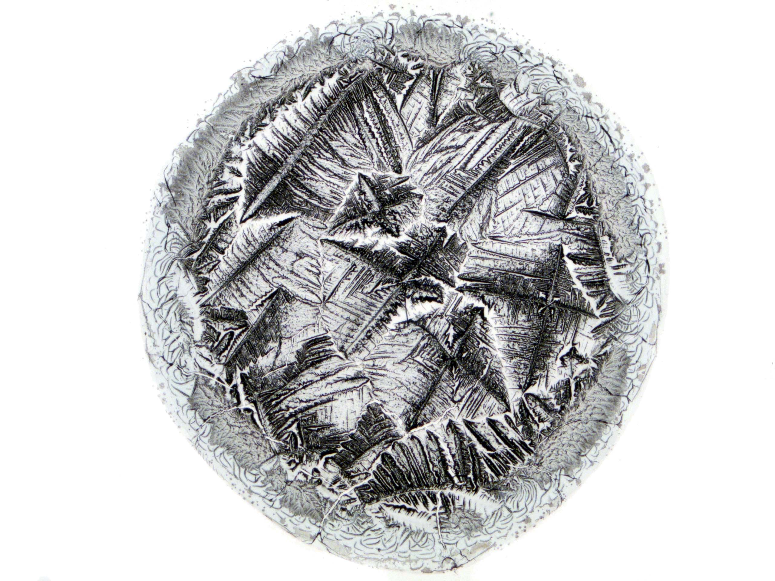
If salts are present in the droplet, they usually crystallize during drying’ Morphology of the salt crystals is very sensitive to the kind of salt, concentration and type of colloidal particles, as well as the rate of evaporation Annarelli et al. (2000, 2001); Basu et al. (2012); Giri et al. (2013); Dutta Choudhury et al. (2013); Dutta et al. (2013); Dutta Choudhury et al. (2015); Roy et al. (2015); Yakhno (2015); Glibitskiy et al. (2015). This sensitiveness allows using the morphology of salt crystals as an indicator, e.g. to diagnose different deceases from biological fluids Martusevich et al. (2007); Martusevich and Kamakin (2007). At the same time, this sensitivity impedes modelling because a lot of different effects have to be taken into account. In fact, all used models should be treated as semi-empirical. The models often utilize the lattice approach Martyushev et al. (1997); Martiouchev et al. (1998); Martyushev and Seleznev (1999); Crivoi and Duan (2012); Dutta Choudhury et al. (2015) and diffusion equation Tarasevich (2001); Dutta et al. (2013). Adequacy of some models Martyushev et al. (1997); Martiouchev et al. (1998); Martyushev and Seleznev (1999) has been questioned Tarasevich (2001). Mainly, dendritic crystal growth can be observed at the final stages of drop desiccation. Both non-equilibrium growth and presence of impurities may produce dendritic shape of crystals Langer (1980); these effects can be reproduced in a simple model Tarasevich et al. (2001). The phase-field method Warren and Boettinger (1995) looks extremely promising for modelling crystal growth in desiccated colloidal droplets with salt admixtures, but it requires a lot of additional information, which is difficult to obtain experimentally.
VI Desiccation crack patterns
Desiccation crack patterns were intensively investigated both experimentally and theoretically Pauchard et al. (1999); Annarelli et al. (2001); Leung et al. (2001); Néda et al. (2002); Caddock and Hull (2002); Jing and Ma (2012); Bou Zeid et al. (2013); Sobac and Brutin (2014); Giorgiutti-Dauphiné and Pauchard (2014); Ghosh et al. (2015); Giorgiutti-Dauphiné and Pauchard (2015); Kim et al. (2015); Bou Zeid et al. (2013); Zhang et al. (2013). State of the art may be found in the recently published book Goehring et al. (2015).

While the study of drying droplets is an interesting subject of research that leads to pattern formation, phase segregation and buckling, in some cases the phenomena also result in cracking of the dried drople Routh (2013); Tsapis et al. (2005); Dutta Choudhury et al. (2013); Annarelli et al. (2001). The simple process of drying a small drop of a multi-component colloidal solution on a flat substrate gives a wealth of data, which is interesting and potentially useful for practical applications. The crack patterns are used to extract valuable information in various fields such as medical diagnosis, forensics related to crime investigation and material science Brutin (2015). The final drying pattern and crack nucleation varies with the kinetics of the evaporation rate. During solvent evaporation, curvature of the solvent-air menisci is responsible for a capillary pressure in the liquid phase. The capillary pressure induces shrinkage of the porous matrix, that is constrained by the adhesion of the deposit to the glass substrate and the evaporation of solvent. As tensile stresses build up, the internal stresses become too great and fractures appear to release mechanical energy. The differences in pattern formation arise due to the competition between the drying process and the adhesion of the matrix on the substrate.
Annarelli et al. Annarelli et al. (2001) worked on the evaporation, gelling and the cracking behaviour of a deposited drop of protein solution, bovine serum albumin. They observed that the cracks appearing at the gelling edge were regularly-spaced, and were a result of the competition between evaporation induced and relaxation induced evolution. When the crack evolution is only evaporation-induced, the mean crack spacing is proportional to the layer thickness. However in the case of a drop of bovine serum albumin, the evolution of cracks has been described in relation to the change with time of the average shrinkage stress. In this case the mean crack spacing was observed to be inversely proportional to the deposit thickness. This is unexpected as normally crack spacing increases with thickness.
Brutin and his group worked on the pattern formation of desiccating droplets of human blood from which the coagulation protein had been removed Brutin (2015); Brutin et al. (2011b). They studied the dynamics of the process of evaporation of a blood droplet using a top-view visualization and the drop mass evolution during the drying process. Sobac and Brutin Brutin et al. (2011b) showed that there are two distinct regimes of evaporation during the drying of whole blood. The first regime is driven by convection, diffusion and gelation, while the second regime is only diffusive in nature. A diffusion model of the drying process allows a prediction of the transition between these two regimes of evaporation. Concentration of the solid mass in the drop was important and fracture occurred at a critical mass concentration of solid in a drying drop of blood. They showed that the final crack patterns formed on drying droplets of blood collected from a healthy person, anaemic person and hyperlipidaemic person are quite different. But drawing conclusions for definite diagnosis is not so straightforward as the crack patterns are strongly affected by external conditions such as the ambient relative humidity and the nature of the substrate.
Brutin et al. conclude that the final drying pattern and crack nucleation varies with the kinetics of the evaporation rate. The transfer of water to air is limited by diffusion and is controlled by the relative humidity in the surrounding air. The drying process of a sessile drop of blood is characterized by an evolution of the solution into a gel saturated with solvent. When the gel is formed, the new porous matrix formed by the aggregation of particles continues to dry by evaporation of the solvent which causes the gel to consolidate. The differences in pattern formation arise due to the competition between the drying process.
Carle and Brutin Carle and Brutin (2013) studied the influence of surface functional groups and substrate surface energy on the formation of crack patterns and on the dry-out shape in drying a water-based droplet of nano-fluid. They have also studied desiccation of blood droplets Brutin et al. (2012) on different substrates such as glass and glass coated with gold or aluminium. They measured the rate of heat transfer from the substrate to the fluid drop. They show that wettability of the substrate by the fluid is the decisive factor, which can account for the differences in the morphology of the desiccated blood drop on different surfaces, rather than the thermal diffusivity which determines rate of heat transfer from the substrate to the drop. On metallic surfaces where the drop is nearly hemispherical and a glassy skin forms on the fluid-air interface, there are hardly any cracks. On a glass surface on the other hand, where the drop is more or less flat, an intricate pattern of cracks form.
While there are several works on desiccating droplets done by different groups, there are very few studies on drying droplets in the presence of a perturbation. The contact angle of a conductive aqueous drop laden with organic or inorganic solutes or ambient oils, changes with the application of Alternating current (AC) voltage during drying. Banpurkar et al. Banpurkar et al. (2008) studied the above effects in experiments to demonstrate the potential of electrowetting-based tensiometry. Contact angle () decreases with increasing amplitude () of AC voltage following the linear relation of with . They applied low frequency AC voltage and obtained interfacial tensions from 5 mJ/m2 to 72 mJ/m2, in close agreement with the macroscopic tensiometry for drop volumes between 20 and 2000 nL. Vancauwenberghe et al. Vancauwenberghe et al. (2013) reviewed the effect of an electric field on a sessile drop. They observed that an external electric field can change the contact angle and shape of a droplet. The electric field also affects the evaporation rate during drying. The contact angle is not always an increasing function of the magnitude of the applied electric field, but may be a decreasing function for some liquid droplets as well.
Khatun et al. Khatun et al. (2013) investigated desiccation cracks on drying droplets of aqueous Laponite solution, in the presence of a static electric field (DC). The electric field had cylindrical geometry, the peripheral electrode being an aluminium wire bent into a circular form of diameter cm. A drop of Laponite gel was deposited inside this wire loop. Another aluminium wire with its tip touching the lower substrate through the centre of the drop, acted as the central electrode. Typical cracks had radial symmetry and were found to emerge always from the positive electrode. With the peripheral electrode positively charged, the final number of cracks appearing on the periphery was measured with the field applied continuously. This was related to the field strength as
| (1) |
where is in the limit of a very strong field and is a constant field. The number of cracks at the centre, when the central electrode is positive, was found to be 3 in most cases and rarely 4.
The time of appearance of the first crack after deposition of the drop, was also found to be a function of the field strength.
| (2) |
Here is the time of appearance of the first crack in the absence of any electric field. is a constant field such that when , falls to .
In another set of experiments, the authors applied the field for a very short time and switched it off before any crack appeared. In this case the final cracks showed the same pattern as when the field was always on. However in this case, the time of appearance of the first crack was delayed to and the final number of cracks reduced to . These quantities were found to obey simple empirical rules on appropriate transformations of the variables. The rules are
| (3) |
and
| (4) |
These relations quantify the strength and duration of the memory of electric field exposure retained by the sample. Following equations (3) and (4), the data for and collapsed on a master graph for different applied voltages. These results are interesting because the collapse of data sets with different applied voltage indicates that the transformed crack appearance times, or rather the extra time needed for crack appearance when the external field is switched off before , increases in proportion to and is a simple function of the non-dimensionalised field exposure time. The same is true of the number of cracks, with a reversal in sign, since the number of cracks for is more than the number when . However, a theoretical interpretation of the scaling relations is not yet understood.
The authors further calculated the energy dissipated in the sample when the field is on and upto the appearance of the first crack, according to
| (5) |
Here is the current through the system at time . was found to be more or less constant for all applied voltages.
Scanning electron microscopy (SEM) was done on different regions of the droplet. The images showed that samples without field exposure or with low field exposure, have more micro-cracks compared to samples exposed to high fields. Field exposure induces large cracks which release stress, so regions between these large cracks are relatively flawless. These studies indicate that direct and alternating electric fields both have a strong effect on the formation of desiccation cracks and may presumably be used for intentionally promoting or suppressing crack growth.
VII Conclusion
There obviously remains much more work to be done in this interesting and useful area of research. Regarding directions of research, some tasks should be especially emphasized
-
•
Obtaining new experimental data, critically needed for the design and development of adequate models.
-
•
The development of 3D models describing the redistribution of the components, the movement of the phase front and the evolution of the profile of the drying colloidal droplets with salt admixtures. In these systems, phase transition from sol to gel is concentration driven. The thermal phase transition from liquid to vapor also takes place in this system. This phase transition leads to a movement of the liquid-vapor phase boundary, i.e. the droplet volume decreases and droplet profile changes.
-
•
Considering additional effects that may be crucial to understanding the processes of pattern formation, but have not yet been included in the model, e.g. variations of the viscosity of a colloid with time and concentration of salts, changes of the vapor flux above the free surface of the droplets when the phase boundary (sol-gel) is moving.
-
•
Analyzing the final pattern through tools such as fractal and multifractal characterization.
Acknowledgements
The reported study was funded by the Ministry of Education and Science of the Russian Federation according to the research Project No. 643 (Yu.T). S. Tarafdar thanks DST, Govt. of India for the grant of a research project (SR/S2/CMP-127/2012).
References
- Koch (1954) C. Koch, “Feinbau und Entstehungsweise von Kristallstrukturen in getrockneten Tropfen hochmolekularsalzhaltiger Flüssigkeiten,” Kolloid-Zeitschrift 138, 81–86 (1954).
- Koch (1956) C. Koch, “Über Austrocknungssprünge,” Kolloid-Zeitschrift 145, 7–14 (1956).
- Koch (1957a) Carlo Koch, “Periodische Erscheinungen an trocknenden Tropfen hochmolekularer Flüssigkeiten,” Kolloid-Zeitschrift 150, 80–83 (1957a).
- Koch (1957b) Carlo Koch, “Über Doppelbildungen (Dubletten) und gleichartige Erscheinungen in trockenen Tropfen hochmolekularer Flüssigkeiten (Dextran, Polyvinylpyrrolidon),” Kolloid-Zeitschrift 151, 62–66 (1957b).
- Koch (1957c) Carlo Koch, “Über Polyvinylpyrrolidonkristallverbände,” Kolloid-Zeitschrift 151, 122–126 (1957c).
- Koch (1957d) C. Koch, “Kristallstrukturen und verwandte Erscheinungen in getrockneten Tropfen hochmolekular-salzhaltiger Flüssigkeiten (Kammerwasser),” Documenta Ophthalmologica 11, 182–189 (1957d).
- Solé (1954) Alphons Solé, “Die rhythmischen Kristallisationen im Influenzstagogramm,” Kolloid-Zeitschrift 137, 15–19 (1954).
- Solé (1955) Alphons Solé, “Die Rhythmik im Influenzstagogramm,” Kolloid-Zeitschrift 143, 73–83 (1955).
- Solé (1957a) Alphons Solé, “Untersuchung über die Bewegung der Teilchen im Stagogramm und Influenzstagogramm,” Kolloid-Zeitschrift 151, 55–62 (1957a).
- Solé (1957b) Alphons Solé, “Stagoskopische Untersuchungen über die Rhythmik einiger Aminosäuren sowie anderer organischer Verbindungen,” Kolloid-Zeitschrift 151, 126–136 (1957b).
- Solé (1960) Alphons Solé, Stagoskopie: Einfuhrung in Methodik, Theorie und Praxis fur Arzte und Naturforscher (Franz Deuticke, Wien, 1960).
- Rapis (1988) E. G. Rapis, Sov. Tech. Phys. Lett. 14, 679 (1988).
- Savina (1999) L. V. Savina, Crystalloscopic Structures of Blood Serum of Healthy People and Patients (Sov. Kuban, Krasnodar, 1999) p. 96, in Russian.
- Shabalin and Shatohina (2001) V. N. Shabalin and S. N. Shatohina, Morphology of Human Biological Fluids (Khrizostom, Moscow, 2001) in Russian.
- Rapis (2003) E. Rapis, Protein and Life (Self-Assembling and Symmetry of Protein Nanostructures) (Philobiblion and Milta-PKPGIT, Jerusalem and Moscow, 2003) in Russian.
- Vorobiev et al. (2008) A. V. Vorobiev, A. K. Martusevich, and S. P. Peretyagin, Crystallogenesis biological fluids and substrates in assessing the state of the organism (FGU ”NNIITO Rosmedtechnology”, Nizhny Novgorod, 2008) in Russian.
- Shabalin and Shatokhina (1996) V. Shabalin and S. Shatokhina, “Autogenous rhythms and self-organization of biological fluids,” Bulletin of Experimental Biology and Medicine 122, 967–973 (1996).
- Yakhno et al. (2003) T. A. Yakhno, O. A. Sedova, A. G. Sanin, and A. S. Pelyushenko, “On the existence of regular structures in liquid human blood serum (plasma) and phase transitions in the course of its drying,” Technical Physics 48, 399–403 (2003).
- Shatokhina et al. (2004) S. N. Shatokhina, V. N. Shabalin, M. E. Buzoverya, and V. T. Punin, “Bio-liquid morphological analysis,” TheScientificWorldJOURNAL 4, 657–661 (2004).
- Shabalin and Shatokhina (2007) V. N. Shabalin and S. N. Shatokhina, “Diagnostic markers in the structures of human biological liquids,” Singapore Medical Journal 48, 440–446 (2007).
- Martusevich and Kamakin (2007) A. K. Martusevich and N. F. Kamakin, “Crystallography of biological fluid as a method for evaluating its physicochemical characteristics,” Bulletin of Experimental Biology and Medicine 143, 385–388 (2007).
- Yakhno et al. (2015) Tatiana A. Yakhno, Anatoly A. Sanin, Robert G. Ilyazov, Gulusa V. Vildanova, Rafat A. Khamzin, Nadezhda P. Astascheva, Mikhail G. Markovsky, Vadim D. Bashirov, and Vladimir G. Yakhno, “Drying drop technology as a possible tool for detection leukemia and tuberculosis in cattle,” Journal of Biomedical Science and Engineering 08, 1–23 (2015).
- Ragoonanan and Aksan (2008) V. Ragoonanan and A. Aksan, “Heterogeneity in desiccated solutions: Implications for biostabilization,” Biophysical Journal 94, 2212–2227 (2008).
- Less et al. (2013) Rebekah Less, Kristin L. M. Boylan, Amy P. N. Skubitz, and Alptekin Aksan, “Isothermal vitrification methodology development for non-cryogenic storage of archival human sera,” Cryobiology 66, 176–185 (2013).
- Takhistov and Chang (2002) P. Takhistov and H.C. Chang, “Complex stain morphologies,” Industrial & Engineering Chemistry Research 41, 6256–6269 (2002).
- Kim et al. (2012) Namwon Kim, Zhenguo Li, Cedric Hurth, Frederic Zenhausern, Shih-Fu Chang, and Daniel Attinger, “Identification of fluid and substrate chemistry based on automatic pattern recognition of stains,” Anal. Methods 4, 50–57 (2012).
- Kokornaczyk et al. (2011) Maria Olga Kokornaczyk, Giovanni Dinelli, Ilaria Marotti, Stefano Benedettelli, Daniele Nani, and Lucietta Betti, “Self-organized crystallization patterns from evaporating droplets of common wheat grain leakages as a potential tool for quality analysis,” TheScientificWorldJOURNAL 11, 1712–1725 (2011).
- Esmonde-White et al. (2008a) Karen A. Esmonde-White, Stephanie V. Le Clair, Blake J. Roessler, and Michael D. Morris, “Effect of conformation and drop properties on surface-enhanced Raman spectroscopy of dried biopolymer drops,” Applied Spectroscopy 62, 503–511 (2008a).
- Esmonde-White et al. (2008b) Karen A. Esmonde-White, Gurjit S. Mandair, Farhang Raaii, Blake J. Roessler, and Michael D. Morris, “Raman spectroscopy of dried synovial fluid droplets as a rapid diagnostic for knee joint damage,” Proc. SPIE 6853, 68530Y (2008b).
- Esmonde-White et al. (2009a) Karen A. Esmonde-White, Gurjit S. Mandair, Farhang Raaii, Jon A. Jacobson, Bruce S. Miller, Andrew G. Urquhart, Blake J. Roessler, and Michael D. Morris, “Raman spectroscopy of synovial fluid as a tool for diagnosing osteoarthritis,” Journal of Biomedical Optics 14, 034013 (2009a).
- Esmonde-White et al. (2009b) Karen A. Esmonde-White, Gurjit S. Mandair, Francis W. L. Esmonde-White, Farhang Raaii, Blake J. Roessler, and Michael D. Morris, “Osteoarthritis screening using raman spectroscopy of dried human synovial fluid drops,” Proc. SPIE 7166, 71660J (2009b).
- Filik and Stone (2007) Jacob Filik and Nicholas Stone, “Drop coating deposition Raman spectroscopy of protein mixtures,” Analyst 132, 544–550 (2007).
- Filik and Stone (2008) Jacob Filik and Nicholas Stone, “Raman point mapping of tear ferning patterns,” Proc. SPIE 6853, 685309 (2008).
- Filik and Stone (2009) Jacob Filik and Nicholas Stone, “Investigation into the protein composition of human tear fluid using centrifugal filters and drop coating deposition Raman spectroscopy,” Journal of Raman Spectroscopy 40, 218–224 (2009).
- Zhang et al. (2010) Dongmao Zhang, Karthikeshwar Vangala, Dongping Jiang, Sige Zou, and Tibor Pechan, “Drop coating deposition raman spectroscopy of fluorescein isothiocyanate labeled protein,” Applied Spectroscopy 64, 1078–1085 (2010).
- Pearce and Tomlinson (2000) E.I. Pearce and A. Tomlinson, “Spatial location studies on the chemical composition of human tear ferns,” Ophthalmic and Physiological Optics 20, 306–313 (2000).
- Dingari et al. (2012) Narahara Chari Dingari, Gary L. Horowitz, Jeon Woong Kang, Ramachandra R. Dasari, and Ishan Barman, “Raman spectroscopy provides a powerful diagnostic tool for accurate determination of albumin glycation,” PLoS ONE 7, e32406 (2012).
- Kočišová et al. (2012) E. Kočišová, A. Vodáková, and M. Procházka, “DCDR spectroscopy as efficient tool for liposome studies: Aspect of preparation procedure parameters,” Spectroscopy: An International Journal 27, 349–353 (2012).
- Routh (2013) Alexander F. Routh, “Drying of thin colloidal films,” Reports on Progress in Physics 76, 046603 (2013).
- Sefiane (2013) Khellil Sefiane, “Patterns from drying drops,” Advances in Colloid and Interface Science (2013), 10.1016/j.cis.2013.05.002.
- Larson (2014) Ronald G. Larson, “Transport and deposition patterns in drying sessile droplets,” AIChE Journal 60, 1538–1571 (2014).
- Zhong et al. (2015) Xin Zhong, Alexandru Crivoi, and Fei Duan, “Sessile nanofluid droplet drying,” Advances in Colloid and Interface Science 217, 13–30 (2015).
- Sadek et al. (2015) Céline Sadek, Pierre Schuck, Yannick Fallourd, Nicolas Pradeau, Cécile Le Floch-Fouéré, and Romain Jeantet, “Drying of a single droplet to investigate process–structure–function relationships: a review,” Dairy Science & Technology 95, 771–794 (2015).
- Chen et al. (2016) Ruoyang Chen, Liyuan Zhang, Duyang Zang, and Wei Shen, “Blood drop patterns: Formation and applications,” Advances in Colloid and Interface Science 231, 1 – 14 (2016).
- Lin (2010) Zhiqun Lin, ed., Evaporative Self-assembly of Ordered Complex Structures (World Scientific Publishing Company, 2010).
- Innocenzi et al. (2013) Plinio Innocenzi, Luca Malfatti, and Paolo Falcaro, Water Droplets to Nanotechnology (The Royal Society of Chemistry, 2013).
- Brutin (2015) David Brutin, ed., Droplet Wetting and Evaporation (Academic Press, Oxford, 2015).
- Goehring et al. (2015) L. Goehring, A. Nakahara, T. Dutta, S. Kitsunezaki, and S. Tarafdar, Desiccation Cracks and their Patterns: Formation and Modelling in Science and Nature, Statistical Physics of Fracture and Breakdown (Wiley, 2015).
- Tarasevich (2004) Yu. Yu. Tarasevich, “Mechanisms and models of the dehydration self-organization in biological fluids,” Physics-Uspekhi 47, 717–728 (2004).
- Yakhno and Yakhno (2009) T. A. Yakhno and V. G. Yakhno, “Structural evolution of drying drops of biological fluids,” Technical Physics 54, 1219–1227 (2009).
- Yakhno (2008) Tatiana Yakhno, “Salt-induced protein phase transitions in drying drops,” Journal of Colloid and Interface Science 318, 225–230 (2008).
- Yakhno et al. (2011) T. A. Yakhno, A. G. Sanin, O. A. Sanina, and V. G. Yakhno, “Dynamics of mechanical properties of drying drops of biological liquids as a reflection of the features of self-organization of their components from nano- to microlevel,” Biophysics 56, 1005–1010 (2011).
- Prokhorov (2009) V. E. Prokhorov, “Some features of the biological liquid droplet revealed from its dehydrated phase structure analysis,” 15 International conference ”Fluxes and structures in fluids: physics of geospheres”. Abstracts 2, 187 (2009).
- Annarelli et al. (2001) C. C. Annarelli, J. Fornazero, J. Bert, and J. Colombani, “Crack patterns in drying protein solution drops,” The European Physical Journal E 5, 599–603 (2001).
- Tarasevich and Ayupova (2003) Yu. Yu. Tarasevich and A. K. Ayupova, “Effect of diffusion on the separation of components in a biological fluid upon wedge-shaped dehydration,” Technical Physics 48, 535–540 (2003).
- Gorr et al. (2013) Heather Meloy Gorr, Joshua M. Zueger, Daniel R. McAdams, and John A. Barnard, “Salt-induced pattern formation in evaporating droplets of lysozyme solutions,” Colloids and Surfaces B: Biointerfaces 103, 59–66 (2013).
- Dutta Choudhury et al. (2013) Moutushi Dutta Choudhury, Tapati Dutta, and Sujata Tarafdar, “Pattern formation in droplets of starch gels containing NaCl dried on different surfaces,” Colloids and Surfaces A: Physicochemical and Engineering Aspects 432, 110–118 (2013).
- Dutta Choudhury et al. (2015) Moutushi Dutta Choudhury, Tapati Dutta, and Sujata Tarafdar, “Growth kinetics of NaCl crystals in a drying drop of gelatin: transition from faceted to dendritic growth,” Soft Matter 11, 6938–6947 (2015).
- Pauchard et al. (1999) L. Pauchard, F. Parisse, and C. Allain, “Influence of salt content on crack patterns formed through colloidal suspension desiccation,” Physical Review E 59, 3737–3740 (1999).
- Yakhno et al. (2005a) T. A. Yakhno, V. G. Yakhno, and A. V. Sokolov, “Shaping processes in drying drops of serum in norm and pathology,” Biophysics 50, 638–645 (2005a).
- Buzoverya et al. (2014) M. E. Buzoverya, Yu. P. Shcherbak, and I. V. Shishpor, “Quantitative estimation of the microstructural inhomogeneity of biological fluid facies,” Technical Physics 59, 1550–1555 (2014).
- Bou Zeid et al. (2013) W. Bou Zeid, J. Vicente, and D. Brutin, “Influence of evaporation rate on cracks’ formation of a drying drop of whole blood,” Colloids and Surfaces A: Physicochemical and Engineering Aspects 432, 139–146 (2013).
- Sobac and Brutin (2011) B. Sobac and D. Brutin, “Structural and evaporative evolutions in desiccating sessile drops of blood,” Phys. Rev. E 84, 011603 (2011).
- Brutin et al. (2011a) D. Brutin, B. Sobac, B. Loquet, and J. Sampol, “Pattern formation in drying drops of blood,” Journal of Fluid Mechanics 667, 85–95 (2011a).
- Sefiane (2010) Khellil Sefiane, “On the formation of regular patterns from drying droplets and their potential use for bio-medical applications,” Journal of Bionic Engineering 7, Supplement, S82–S93 (2010).
- Bardakov et al. (2010) R. N. Bardakov, Yu. D. Chashechkin, and V. V. Shabalin, “Hydrodynamics of a drying multicomponent liquid droplet,” Fluid Dynamics 45, 803–816 (2010).
- Chashechkin and Bardakov (2010) Yu. D. Chashechkin and R. N. Bardakov, “Formation of texture in residue of a drying drop of a multicomponent fluid,” Doklady Physics 55, 68–72 (2010).
- Kistovich et al. (2010) A. V. Kistovich, Yu. D. Chashechkin, and V. V. Shabalin, “Formation mechanism of a circumferential roller in a drying biofluid drop,” Technical Physics 55, 473–478 (2010).
- Tarasevich and Pravoslavnova (2007a) Yu. Yu. Tarasevich and D. M. Pravoslavnova, “Drying of a multicomponent solution drop on a solid substrate: Qualitative analysis,” Technical Physics 52, 159–163 (2007a).
- Tarasevich (2005) Yu. Yu. Tarasevich, “Simple analytical model of capillary flow in an evaporating sessile drop,” Physical Review E 71, 027301 (2005).
- Tarasevich et al. (2010) Yu. Yu. Tarasevich, O. P. Isakova, V. V. Kondukhov, and A. V. Savitskaya, “Effect of evaporation conditions on the spatial redistribution of components in an evaporating liquid drop on a horizontal solid substrate,” Technical Physics 55, 636–644 (2010).
- Tarasevich et al. (2009) Yu. Yu. Tarasevich, I. V. Vodolazskaya, O. P. Isakova, and M. S. Abdel Latif, “Evaporation-induced flow inside circular wells: Analytical results and simulations,” Microgravity Science and Technology 21, 39–44 (2009).
- Tarasevich and Pravoslavnova (2007b) Yu. Yu. Tarasevich and D. M. Pravoslavnova, “Segregation in desiccated sessile drops of biological fluids,” The European Physical Journal E 22, 311–314 (2007b).
- Vodolazskaya et al. (2010) I. V. Vodolazskaya, Yu. Yu. Tarasevich, and O. P. Isakova, “The model of phase boundary motion in drying sessile drop of colloidal solution,” Nonlinear world 8, 142–150 (2010), in Russian.
- Tarasevich et al. (2011) Yuri Tarasevich, Irina Vodolazskaya, and Olga Isakova, “Desiccating colloidal sessile drop: dynamics of shape and concentration,” Colloid & Polymer Science 289, 1015–1023 (2011).
- Tarasevich et al. (2013) Yu. Yu. Tarasevich, I. V. Vodolazskaya, and O. P. Bondarenko, “Modeling of spatial-temporal distribution of the components in the drying sessile droplet of biological fluid,” Colloids and Surfaces A: Physicochemical and Engineering Aspects 432, 99–103 (2013), WETTING AND EVAPORATION: DROPLETS OF PURE AND COMPLEX FLUIDS.
- Yakhno et al. (2004) T. A. Yakhno, V. G. Yakhno, A. G. Sanin, O. A. Sanina, and A. S. Pelyushenko, “Protein and salt: Spatiotemporal dynamics of events in a drying drop,” Technical Physics 49, 1055–1063 (2004).
- Yakhno et al. (2005b) T. Yakhno, V. Yakhno, A. Sanin, O. Sanina, and A. Pelyushenko, “Dynamics of phase transitions in drying drops as an information parameter of liquid structure,” Nonlinear Dynamics 39, 369–374 (2005b).
- Yahno et al. (2007) T. A. Yahno, V. G. Yahno, A. G. Sanin, A. S. Pelyushenko, O. B. Shaposhnikova, and A. S. Chernov, “The phenomenon of drying drops and the possibility of its practical use,” Nonlinear world 5, 54–65 (2007), in Russian.
- Yakhno (2015) T. A. Yakhno, “Sodium chloride crystallization from drying drops of albumin salt solutions with different albumin concentrations,” Technical Physics 60, 1601–1608 (2015).
- Bou Zeid and Brutin (2013) W. Bou Zeid and D. Brutin, “Influence of relative humidity on spreading, pattern formation and adhesion of a drying drop of whole blood,” Colloids and Surfaces A: Physicochemical and Engineering Aspects 430, 1–7 (2013).
- Brutin et al. (2012) David Brutin, Benjamin Sobac, and Céline Nicloux, “Influence of substrate nature on the evaporation of a sessile drop of blood,” Journal of Heat Transfer 134, 061101 (2012).
- Pradhan et al. (2012) T. Pradhan, M. Asfer, and P. K. Panigrahi, “Droplet hydrodynamics during lysozyme protein crystallization,” Phys. Rev. E 86, 051602 (2012).
- Dutta et al. (2013) Tapati Dutta, Abhra Giri, Moutushi Dutta Choudhury, and Sujata Tarafdar, “Experiment and simulation of multifractal growth of crystalline NaCl aggregates in aqueous gelatin medium,” Colloids and Surfaces A: Physicochemical and Engineering Aspects 432, 127–131 (2013).
- Roy et al. (2015) Biswajit Roy, Moutushi Dutta Choudhuri, Tapati Dutta, and Sujata Tarafdar, “Multi-scale patterns formed by sodium sulphate in a drying droplet of gelatin,” Applied Surface Science 357, Part A, 1000–1006 (2015).
- Basu et al. (2012) T. Basu, M. M. Goswami, T.R. Middya, and S. Tarafdar, “Morphology and ion-conductivity of gelatin-LiClO4 films: Factional diffusion analysis,” Journal of Physical Chemistry B 116, 11362–11369 (2012).
- Giri et al. (2013) A. Giri, M. Dutta Choudhury, T. Dutta, and S. Tarafdar, “Multifractal growth of crystalline NaCl aggregates in a gelatin medium,” Crystal Growth and Design 13, 341–345 (2013).
- Parisse and Allain (1996) F. Parisse and C. Allain, “Shape changes of colloidal suspension droplets during drying,” J. Phys. II France 6, 1111–1119 (1996).
- Deegan et al. (1997) R. D. Deegan, O. Bakajin, T. F. Dupont, G. Huber, S. R. Nagel, and T. A. Witten, “Capillary flow as the cause of ring stains from dried liquid drops,” Nature 389, 827–829 (1997).
- Deegan et al. (2000) R. D. Deegan, O. Bakajin, T. F. Dupont, G. Huber, S. R. Nagel, and T. A. Witten, “Contact line deposits in an evaporating drop,” Physical Review E 62, 756–765 (2000).
- Yunker et al. (2011) Peter J. Yunker, Tim Still, Matthew A. Lohr, and A. G. Yodh, “Suppression of the coffee-ring effect by shape-dependent capillary interactions,” Nature 476, 308–311 (2011).
- Yakhno et al. (2010) T. Yakhno, V. Kazakov, O. Sanina, A. Sanin, and V. Yakhno, “Drops of biological fluids drying on a hard substrate: Variation of the morphology, weight, temperature, and mechanical properties,” Technical Physics 55, 929–935 (2010).
- Still et al. (2012) Tim Still, Peter J. Yunker, and Arjun G. Yodh, “Surfactant-induced marangoni eddies alter the coffee-rings of evaporating colloidal drops,” Langmuir 28, 4984–4988 (2012).
- Anyfantakis et al. (2015) Manos Anyfantakis, Zheng Geng, Mathieu Morel, Sergii Rudiuk, and Damien Baigl, “Modulation of the coffee-ring effect in particle/surfactant mixtures: the importance of particle interface interactions,” Langmuir 31, 4113–4120 (2015).
- Ristenpart et al. (2007) W. D. Ristenpart, P. G. Kim, C. Domingues, J. Wan, and H. A. Stone, “Influence of substrate conductivity on circulation reversal in evaporating drops,” Phys. Rev. Lett. 99, 234502 (2007).
- Carle and Brutin (2013) Florian Carle and David Brutin, “How surface functional groups influence fracturation in nanofluid droplet dry-outs,” Langmuir 29, 9962–9966 (2013).
- Caddock and Hull (2002) B. D. Caddock and D. Hull, “Influence of humidity on the cracking patterns formed during the drying of sol-gel drops,” Journal of Materials Science 37, 825–834 (2002).
- Chhasatia et al. (2010) Viral H. Chhasatia, Abhijit S. Joshi, and Ying Sun, “Effect of relative humidity on contact angle and particle deposition morphology of an evaporating colloidal drop,” Appl. Phys. Lett. 97, 231909 (2010).
- Bou-Zeid and Brutin (2014) W. Bou-Zeid and D. Brutin, “Effect of relative humidity on the spreading dynamics of sessile drops of blood,” Colloids and Surfaces A: Physicochemical and Engineering Aspects 456, 273–285 (2014).
- Pauchard and Allain (2003) L. Pauchard and C. Allain, “Mechanical instability induced by complex liquid desiccation,” Comptes Rendus Physique 4, 231–239 (2003).
- Okuzono et al. (2009) T. Okuzono, M. Kobayashi, and M. Doi, “Final shape of a drying thin film,” Physical Review E 80, 021603 (2009).
- Jung et al. (2009) Youngki Jung, Tadashi Kajiya, Tatsuya Yamaue, and Masao Doi, “Film formation kinetics in the drying process of polymer solution enclosed by bank,” Japanese Journal of Applied Physics 48, 031502 (2009).
- Vodolazskaya and Tarasevich (2011) I. V. Vodolazskaya and Yu. Yu. Tarasevich, “The model of drying sessile drop of colloidal solution,” Modern Physics Letters B (MPLB) 25, 1303–1310 (2011).
- Ozawa et al. (2005) Kin’ya Ozawa, Eisuke Nishitani, and Masao Doi, “Modeling of the drying process of liquid droplet to form thin film,” Japanese Journal of Applied Physics 44, 4229–4234 (2005).
- Leung et al. (2001) K.-T. Leung, L. Józsa, M. Ravasz, and Z. Néda, “Pattern formation: Spiral cracks without twisting,” Nature 410, 166 (2001).
- Néda et al. (2002) Z. Néda, K.-t. Leung, L. Józsa, and M. Ravasz, “Spiral cracks in drying precipitates,” Phys. Rev. Lett. 88, 095502 (2002).
- Golbraikh et al. (2003) E. Golbraikh, E.G. Rapis, and S.S. Moiseev, “On the crack pattern formation in a freely drying protein film,” Technical Physics 48, 1333–1337 (2003).
- Sobac and Brutin (2014) B. Sobac and D. Brutin, “Desiccation of a sessile drop of blood: Cracks, folds formation and delamination,” Colloids and Surfaces A: Physicochemical and Engineering Aspects 448, 34–44 (2014).
- Deegan (2000) R. D. Deegan, “Pattern formation in drying drops,” Physical Review E 61, 475–485 (2000).
- Fischer (2002) B. J. Fischer, “Particle convection in an evaporating colloidal droplet,” Langmuir 18, 60–67 (2002).
- Popov (2005) Y. O. Popov, “Evaporative deposition patterns: Spatial dimensions of the deposit,” Physical Review E 71, 036313 (2005).
- Widjaja and Harris (2008a) E. Widjaja and M. Harris, “Particle deposition study during sessile drop evaporation,” AIChE J. 54, 2250–2260 (2008a).
- Bhardwaj et al. (2009) R. Bhardwaj, X. Fang, and D. Attinger, “Pattern formation during the evaporation of a colloidal nanoliter drop: a numerical and experimental study,” New Journal of Physics 11, 075020 (2009).
- Zheng (2009) R. Zheng, “A study of the evaporative deposition process: Pipes and truncated transport dynamics,” The European Physical Journal E: Soft Matter and Biological Physics 29, 205–218 (2009).
- Witten (2009) T. A. Witten, “Robust fadeout profile of an evaporation stain,” EPL (Europhysics Letters) 86, 64002 (2009).
- Craster et al. (2009) R. V. Craster, O. K. Matar, and K. Sefiane, “Pinning, retraction, and terracing of evaporating droplets containing nanoparticles,” Langmuir 25, 3601–3609 (2009).
- Petsi et al. (2010) A. J. Petsi, A. N. Kalarakis, and V. N. Burganos, “Deposition of brownian particles during evaporation of two-dimensional sessile droplets,” Chemical Engineering Science 65, 2978–2989 (2010).
- Kim et al. (2011) Hee-Soo Kim, SungSoo Park, and Frank Hagelberg, “Computational approach to drying a nanoparticle-suspended liquid droplet,” Journal of Nanoparticle Research 13, 59–68 (2011).
- Killeen et al. (2006) Anthony A. Killeen, Natalya Ossina, Ronald C. McGlennen, Sharon Minnerath, John Borgos, Vadim Alexandrov, and Armen Sarvazyan, “Protein self-organization patterns in dried serum reveal changes in b-cell disorders,” Mol. Diag. Ther. 10, 371–380 (2006).
- Martusevich et al. (2007) A. K. Martusevich, Y. Zimin, and A. Bochkareva, “Morphology of dried blood serum specimens of viral hepatitis,” Hep. Mon. 7, 207–210 (2007).
- Eales et al. (2015a) Adam D. Eales, Alexander F. Routh, Nick Dartnell, and Goddard Simon, “Evaporation of pinned droplets containing polymer an examination of the important groups controlling final shape,” AIChE Journal 61, 1759–1767 (2015a).
- Eales et al. (2015b) Adam D. Eales, Nick Dartnell, Simon Goddard, and Alexander F. Routh, “The impact of trough geometry on film shape. A theoretical study of droplets containing polymer, for P-OLED display applications,” Journal of Colloid and Interface Science 458, 53–61 (2015b).
- Anderson and Davis (1995) D. M. Anderson and S. H. Davis, “The spreading of volatile liquid droplets on heated surfaces,” Physics of Fluids 7, 248–265 (1995).
- Lebovka et al. (2014a) N. I. Lebovka, V. A. Gigiberiya, O. S. Lytvyn, Yu. Yu. Tarasevich, I. V. Vodolazskaya, and O. P. Bondarenko, “Drying of sessile droplets of laponite-based aqueous nanofluids,” Colloids and Surfaces A: Physicochemical and Engineering Aspects 462, 52–63 (2014a).
- Hu and Larson (2002) Hua Hu and Ronald G. Larson, “Evaporation of a sessile droplet on a substrate,” The Journal of Physical Chemistry B 106, 1334–1344 (2002).
- Mollaret et al. (2004) R. Mollaret, K. Sefiane, J. R.E. Christy, and D. Veyret, “Experimental and numerical investigation of the evaporation into air of a drop on a heated surface,” in 8th UK National Heat Transfer Conference ”Chemical Engineering Research and Design”, 4, Vol. 82 (2004) pp. 471–480.
- Widjaja and Harris (2008b) Ervina Widjaja and Michael T. Harris, “Numerical study of vapor phase-diffusion driven sessile drop evaporation,” Computers & Chemical Engineering 32, 2169–2178 (2008b).
- Dunn et al. (2008) G. J. Dunn, S. K. Wilson, B. R. Duffy, S. David, and K. Sefiane, “A mathematical model for the evaporation of a thin sessile liquid droplet: Comparison between experiment and theory,” Colloids and Surfaces A: Physicochemical and Engineering Aspects 323, 50–55 (2008), bubble and Drop Interfaces - Selected papers from the International Workshop on Bubble and Drop Interfaces, 25-28 March 2007, Granada, Spain.
- Barash (2009) L. Yu. Barash, “Influence of gravitational forces and fluid flows on the shape of surfaces of a viscous fluid of capillary size,” Physical Review E 79, 025302 (2009).
- Barash et al. (2009) L. Yu. Barash, T. P. Bigioni, V. M. Vinokur, and L. N. Shchur, “Evaporation and fluid dynamics of a sessile drop of capillary size,” Physical Review E 79, 046301 (2009).
- Masoud and Felske (2009) Hassan Masoud and James D. Felske, “Analytical solution for inviscid flow inside an evaporating sessile drop,” Physical Review E 79, 016301 (2009).
- Petsi and Burganos (2008) A. J. Petsi and V. N. Burganos, “Stokes flow inside an evaporating liquid line for any contact angle,” Physical Review E 78, 036324 (2008).
- Hu and Larson (2005a) H. Hu and R. G. Larson, “Analysis of the microfluid flow in an evaporating sessile droplet,” Langmuir 21, 3963–3971 (2005a).
- Hu and Larson (2005b) H. Hu and R. G. Larson, “Analysis of the effects of marangoni stresses on the microflow in an evaporating sessile droplet,” Langmuir 21, 3972–3980 (2005b).
- Hsu et al. (2007) Ching-Ling Hsu, Szu-Ming Chu, Kiwi Wood, and Yi-Rong Yang, “Percolation in two-dimensional nanoparticle films from colloidal self-assembly,” physica status solidi (a) 204, 1856–1862 (2007).
- Chen et al. (2013) Weikang Chen, Joel Koplik, and Ilona Kretzschmar, “Molecular dynamics simulations of the evaporation of particle-laden droplets,” Phys. Rev. E 87, 052404 (2013).
- Crivoi and Duan (2013) A. Crivoi and Fei Duan, “Fingering structures inside the coffee-ring pattern,” Colloids and Surfaces A: Physicochemical and Engineering Aspects 432, 119–126 (2013), WETTING AND EVAPORATION: DROPLETS OF PURE AND COMPLEX FLUIDS.
- Lebedev-Stepanov and Vlasov (2013) Peter Lebedev-Stepanov and Konstantin Vlasov, “Simulation of self-assembly in an evaporating droplet of colloidal solution by dissipative particle dynamics,” Colloids and Surfaces A: Physicochemical and Engineering Aspects 432, 132–138 (2013), WETTING AND EVAPORATION: DROPLETS OF PURE AND COMPLEX FLUIDS.
- Lebovka et al. (2014b) N. I. Lebovka, S. Khrapatiy, R. Melnyk, and M. Vygornitskii, “Effects of hydrodynamic retardation and interparticle interactions on the self-assembly in a drying droplet containing suspended solid particles,” Phys. Rev. E 89, 052307 (2014b).
- Crivoi and Duan (2014) A. Crivoi and Fei Duan, “Three-dimensional Monte Carlo model of the coffee-ring effect in evaporating colloidal droplets,” Scientific Reports 4, 1–6 (2014).
- Fujita et al. (2015) Masahiro Fujita, Osamu Koike, and Yukio Yamaguchi, “Direct simulation of drying colloidal suspension on substrate using immersed free surface model,” Journal of Computational Physics 281, 421–448 (2015).
- Hwang and Son (2015) Hochan Hwang and Gihun Son, “A level-set method for the direct numerical simulation of particle motion in droplet evaporation,” Numerical Heat Transfer, Part B: Fundamentals 68, 479–494 (2015).
- Fujita and Yamaguchi (2010) Masahiro Fujita and Yukio Yamaguchi, “Mesoscale modeling for self-organization of colloidal systems,” Current Opinion in Colloid & Interface Science 15, 8–12 (2010).
- Son (2015) G. Son, “Numerical simulation of particle-laden droplet evaporation with the marangoni effect,” The European Physical Journal Special Topics 224, 401–413 (2015).
- Hu and Larson (2006) Hua Hu and Ronald G. Larson, “Marangoni effect reverses coffee-ring depositions,” The Journal of Physical Chemistry B 110, 7090–7094 (2006).
- Zaleskiǐ et al. (2004) M. G. Zaleskiǐ, V. L. Emanuél’, and M. V. Krasnova, “Physical and chemical characteristics of structurization of a biological fluid drop as examplified by the ”LITOS-system” diagnosticum [fiziko-khimicheskie zakonomernosti strukturizatsii kapli biologicheskoi zhidkosti na primere diagnostikuma ”LITOS-sistema”.],” Klinicheskaia laboratornaia diagnostika , 20–24 (2004), in Russian.
- Girard et al. (2006) F. Girard, M. Antoni, A. Steinchen, and S. Faure, “Numerical study of the evaporating dynamics of a sessile water droplet,” Microgravity Science and Technology 18, 42–46 (2006).
- Annarelli et al. (2000) C. C. Annarelli, L. Reyes, J. Fornazero, J. Bert, R. Cohen, and A. W. Coleman, “Ion and molecular recognition effects on the crystallisation of bovine serum albumin salt mixtures,” Crystal Engineering 3, 173–194 (2000).
- Glibitskiy et al. (2015) Gennadiy Glibitskiy, Dmitriy Glibitskiy, Olga Gorobchenko, Oleg Nikolov, Alexander Roshal, Mikhail Semenov, and Anatoliy Gasan, “Textures on the surface of BSA films with different concentrations of sodium halides and water state in solution,” Nanoscale Research Letters 10, 155 (2015).
- Martyushev et al. (1997) L.M. Martyushev, V.D. Seleznev, and S.A. Skopinov, “Reentrant kinetic phase transitions during dendritic growth of crystals in a two-dimensional medium with phase separation,” Technical Physics Letters 23, 495–497 (1997).
- Martiouchev et al. (1998) L. M. Martiouchev, V. D. Seleznev, and S. A. Skopinov, “Computer simulation of nonequilibrium growth of crystals in a two-dimensional medium with a phase-separating impurity,” Journal of Statistical Physics 90, 1413–1427 (1998).
- Martyushev and Seleznev (1999) L. M. Martyushev and V. D. Seleznev, “Self-similarity in the kinetic growth regime of a crystal in a phase-separating medium,” Technical Physics Letters 25, 833–835 (1999).
- Crivoi and Duan (2012) Alexandru Crivoi and Fei Duan, “Evaporation-induced formation of fractal-like structures from nanofluids,” Phys. Chem. Chem. Phys. 14, 1449–1454 (2012).
- Tarasevich (2001) Yu. Yu. Tarasevich, “Computer simulation of crystal growth from solution,” Technical Physics 46, 627–629 (2001).
- Langer (1980) J. S. Langer, “Instabilities and pattern formation in crystal growth,” Rev. Mod. Phys. 52, 1–28 (1980).
- Tarasevich et al. (2001) Yu. Yu. Tarasevich, V. O. Konstantinov, and A. K. Ayupova, “Simulation of dendritic growth of salt crystals in biological fluids,” Izvestija vuzov. Severo-Kavkazskij region. Estestvennye nauki Special Issue. Math. Mod., 147–149 (2001), in Russian.
- Warren and Boettinger (1995) J. A. Warren and W. J. Boettinger, “Prediction of dendritic growth and microsegregation patterns in a binary alloy using the phase-field method,” Acta Metallurgica et Materialia 43, 689–703 (1995).
- Jing and Ma (2012) Guangyin Jing and Jun Ma, “Formation of circular crack pattern in deposition self-assembled by drying nanoparticle suspension,” The Journal of Physical Chemistry B 116, 6225–6231 (2012).
- Giorgiutti-Dauphiné and Pauchard (2014) F. Giorgiutti-Dauphiné and L. Pauchard, “Elapsed time for crack formation during drying,” The European Physical Journal E 37, 39 (2014), 10.1140/epje/i2014-14039-8.
- Ghosh et al. (2015) Udita Uday Ghosh, Monojit Chakraborty, Aditya Bikram Bhandari, Suman Chakraborty, and Sunando DasGupta, “Effect of surface wettability on crack dynamics and morphology of colloidal films,” Langmuir 31, 6001–6010 (2015).
- Giorgiutti-Dauphiné and Pauchard (2015) F. Giorgiutti-Dauphiné and L. Pauchard, “Striped patterns induced by delamination of drying colloidal films,” Soft Matter 11, 1397–1402 (2015).
- Kim et al. (2015) Jin Young Kim, Kun Cho, Seul-a Ryu, So Youn Kim, and Byung Mook Weon, “Crack formation and prevention in colloidal drops,” Scientific Reports 5, 13166 (2015).
- Zhang et al. (2013) Yongjian Zhang, Zhengtang Liu, Duyang Zang, and Lin Kejun Qian, Yimeng, “Pattern transition and sluggish cracking of colloidal droplet deposition with polymer additives,” Science China Physics, Mechanics & Astronomy 56, 1712–1718 (2013).
- Tsapis et al. (2005) N. Tsapis, E. R. Dufresne, S. S. Sinha, C. S. Riera, J. W. Hutchinson, L. Mahadevan, and D. A. Weitz, “Onset of buckling in drying droplets of colloidal suspensions,” Phys. Rev. Lett. 94, 018302 (2005).
- Brutin et al. (2011b) D. Brutin, B. Sobac, B. Loquet, and J. Sampol, “Pattern formation in drying drops of blood,” Journal of Fluid Mechanics 667, 85–95 (2011b).
- Banpurkar et al. (2008) Arun G. Banpurkar, Kevin P. Nichols, and Frieder Mugele, “Electrowetting-based microdrop tensiometer,” Langmuir 24, 10549–10551 (2008).
- Vancauwenberghe et al. (2013) V. Vancauwenberghe, P. Di Marco, and D. Brutin, “Wetting and evaporation of a sessile drop under an external electrical field: A review,” Colloids and Surfaces A: Physicochemical and Engineering Aspects 432, 50–56 (2013), WETTING AND EVAPORATION: DROPLETS OF PURE AND COMPLEX FLUIDS.
- Khatun et al. (2013) Tajkera Khatun, Tapati Dutta, and Sujata Tarafdar, “Crack formation under an electric field in droplets of laponite gel: Memory effect and scaling relations,” Langmuir 29, 15535–15542 (2013).