Optical probing of mechanical loss of a Si3N4 membrane below 100 mK
Abstract
We report on low mechanical loss in a high-stress silicon nitride (Si3N4) membrane at temperatures below 100 mK. We isolate a membrane via a phononic shield formed within a supporting silicon frame, and measure the mechanical quality factor of a number of high-tension membrane modes as we vary our dilution refrigerator base temperature between 35 mK and 5 K. At the lowest temperatures, we obtain a maximum quality factor () of , corresponding to a -frequency product (QFP) of Hz. These measurements complement the recent observation of improved quality factors of Si3N4 at ultralow temperatures via electrical detection. We also observe a dependence of the quality factor on optical heating of the device. By combining exceptional material properties, high tension, advanced isolation and clamping techniques, high-stress mechanical objects are poised to explore a new regime of exceptional quality factors. Such quality factors combined with an optical probe at cryogenic temperatures will have a direct impact on resonators as quantum objects, as well as force sensors at mK temperatures.
Stoichiometric silicon nitride (Si3N4) films are a unique material in the field of nanomechanics due to their high internal stress that enables high mechanical quality factors along with high resonance frequencies. Thus far, silicon-nitride based resonators have been found to be favorable devices for studying quantum optomechanical phenomena (Thompson et al., 2008), precision force sensing applications (Gavartin et al., 2012; Scozzaro et al., 2016), and bidirectional microwave-optical transducers (Bochmann et al., 2013; Andrews et al., 2014; Bagci et al., 2014). These applications harness extremely high quality factors that are required for observing quantum coherent effects and for precision sensing. The key to the unique nature of membrane mechanics is that stress provides relatively high resonance frequencies, while maintaining low dissipation rate (Unterreithmeier et al., 2010).
Although numerous studies have focused on both internal and external loss mechanisms governing the quality factors of Si3N4-based resonators, the ultimate limit to the QFP has not been identified, especially at cryogenic temperatures (Zwickl et al., 2008; Unterreithmeier et al., 2010; Wilson-Rae et al., 2011; Yu et al., 2012; Chakram et al., 2014; Ghadimi et al., ; Tsaturyan et al., 2016). Specifically, a stringent upper bound for , set by thermoelastic damping of Si3N4, is particularly high, reaching at room-temperature (Norris and Photiadis, 2005; Zwickl et al., 2008). Recently, a square SiN membrane device operated at 20 mK with an electrical readout demonstrated ’s above for a mode frequency of about 250 kHz (Yuan et al., 2015). Their observed increase in with decreasing temperature (below 1 K) was consistent with previous observations of decreased internal mechanical loss in amorphous solids with temperature (Pohl et al., 2002). Recently, it was also shown that by decreasing the external radiation losses as well as the edge clamping losses, room temperature Si3N4 can approach and surpass quality factors of for trampoline and phononic crystal shield designs (Norte et al., 2016; Reinhardt et al., 2016; Tsaturyan et al., 2016). By combining the suppression of internal losses through cryogenic temperatures with advanced designs for isolating the resonator to reduce external losses, further improvements to seem possible.
Here we study the mechanical loss of a MHz frequency Si3N4 membrane at temperatures down to 35 mK using piezoelectric excitation and optical detection. We show that the mechanical loss decreases significantly below 5 K, reaching a maximal measured of for a mode frequency of 1.6 MHz, corresponding to a QFP of Hz. Additionally, the observed quality factors were sensitive to optical probing power, further emphasizing the role of the thermal environment in the mechanical dissipation.
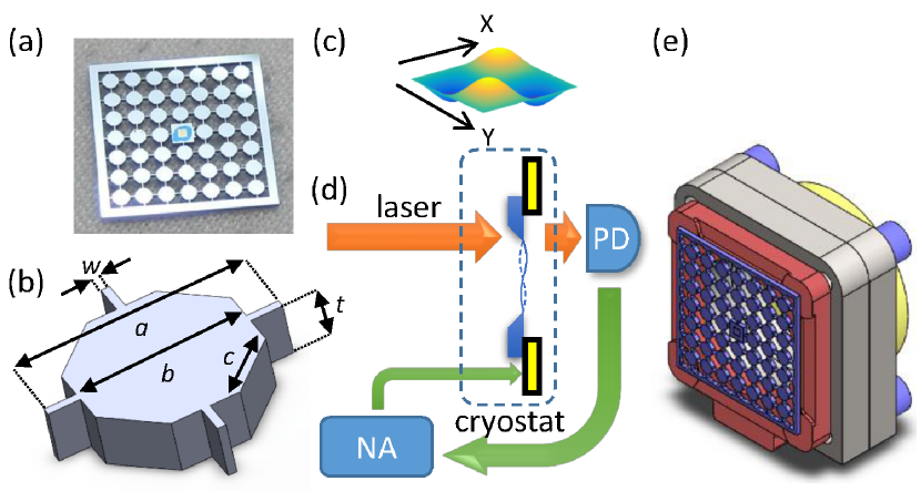
The sample we utilize is a high stress (0.8 GPa) square membrane resonator surrounded by a phononic crystal (PnC) shield. The sample is depicted in Fig. 1(a, b), with the parameter values listed in Table 1. The devices are created starting with a high stress Si3N4 film 100 nm thick grown by low pressure chemical vapor deposition (LPCVD) on a 300 m silicon wafer. The final sample chip is a 10 mm x 10 mm square, with the released Si3N4 membrane in the center. The main aspects of the fabrication process are reported in (Yu et al., 2014). In brief, the membrane is created through a combination of deep reactive ion etching (DRIE), and a final KOH etch to release the membrane. The PnC structure is created by etching all the way through the silicon with DRIE; during the PnC creation the membrane is protected by process adhesive that, for the devices reported here, filled the entire back trench of the released membrane, improving yield. Further, membrane cleanliness was improved by removing the Si3N4 film on the membrane side of the device (outside of the suspended membrane region) prior to creating the PnC. While the the device fabrication is similar to previous work (Yu et al., 2014), there are several important design changes to the PnC which should improve the robustness and reproducibility of the band-gap, see Fig. 1(b). These improvements include narrowing the width of the bridges (47 m compared to 97 m in the previous design) to increase the phononic isolation and designing chamfered corners to better reproduce simulated structures in the fabrication. The design band-gap of the PnC with the dimensions listed in Table 1 spans between 1.1-2.1 MHz, and between 3.1-3.3 MHz. The band-gaps were calculated using Comsol, as described in detail in (Yu et al., 2014).
| Definition | symbol | Device |
|---|---|---|
| chip size | 10 mm | |
| number of unit cells | 4 | |
| unit cell size | 1275 m | |
| block length | 925 m | |
| bridge width | 47 m | |
| wafer thickness | 300 m | |
| membrane length | 516 m | |
| chamfer size | m | |
| membrane frame size | 1125 m | |
| chip frame size | 500 m | |
| membrane thickness | 0.1 m |
The experimental setup to measure the mechanical quality factor is depicted in Fig. 1(d, e). The sample is anchored to the base of a dilution refrigerator, and light is directed to the membrane through a narrow cryogenic beam path designed to filter 300 K blackbody radiation (Kuhn et al., 2014). The laser beam was generated by a diode with a 900 nm wavelength, and detected by a either a Si avalanche or an InGaAs amplified photodiode. An interferometric signal which monitors the position of the membrane is created due to a low finesse cavity between the membrane and a low reflectivity mirror inside the cryostat (Zwickl et al., 2008; Wilson, 2012). Mechanical driving was applied by a piezoelectric ring (Noliac NAC2124). The mechanical ringdown measurements were performed with a network analyzer (HP 4395A), where a continuous wave excitation of the piezoelectric ring at a specific membrane mode frequency was stopped abruptly and the decay of the mechanical oscillation was optically monitored via the interferometric signal. We measure the mechanical decay time constant from the ringdown data , and extract the mechanical loss rate . The quality factor of mode , is the ratio of the mechanical angular frequency , and the loss rate , .
To study the temperature dependence of , we varied the base-plate temperature of the refrigerator from 35 mK to 5 K. The base-plate temperature was measured with a calibrated RuO2 resistive thermometer. The thermal link of the membrane to the base-plate is achieved with a copper foil pressed between the invar adapters, shown in Fig. 1(e). The thermalization efficiency down to 120 mK was confirmed by an independent measurement performed on the same setup and sample, utilizing a 1064 nm resonant high finesse cavity to probe the thermally-induced brownian motion of a membrane. There we obtained the thermal phonon occupancy via optomechanical relations (Peterson et al., 2016) and found good agreement with the base-plate temperature down to 120 mK (Kampel et al., 2016).
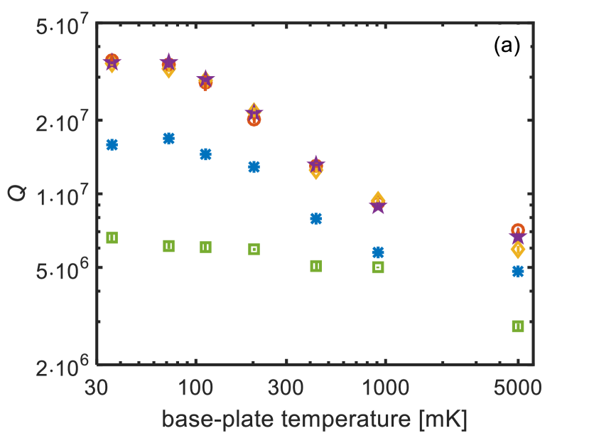
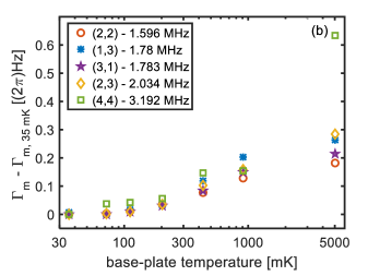
At each temperature, we measured the mechanical of a few membrane modes that displayed values higher than . These mode frequencies were inside the design band-gap of the PnC. Other modes that were outside of the band-gap of the PnC displayed values lower than . As expected, the decrease in the base-plate temperature resulted in an increase in the value of (Fig. 2(a)), and correspondingly a decrease in the loss rate (Fig. 2(b)). In these data, we witness a leveling-off of the -values at the low end of the temperature range. The leveling-off of the between 200 mK to 1 K, does not occur here, in contrast to the results in (Yuan et al., 2015), and continues to drop above 1 K, similar to the results of (Zwickl et al., 2008). To make this feature of the data more apparent, we plot the difference in the loss rate from that at 35 mK in Fig. 2(b). Notice that this quantity increases monotonically above 100 mK
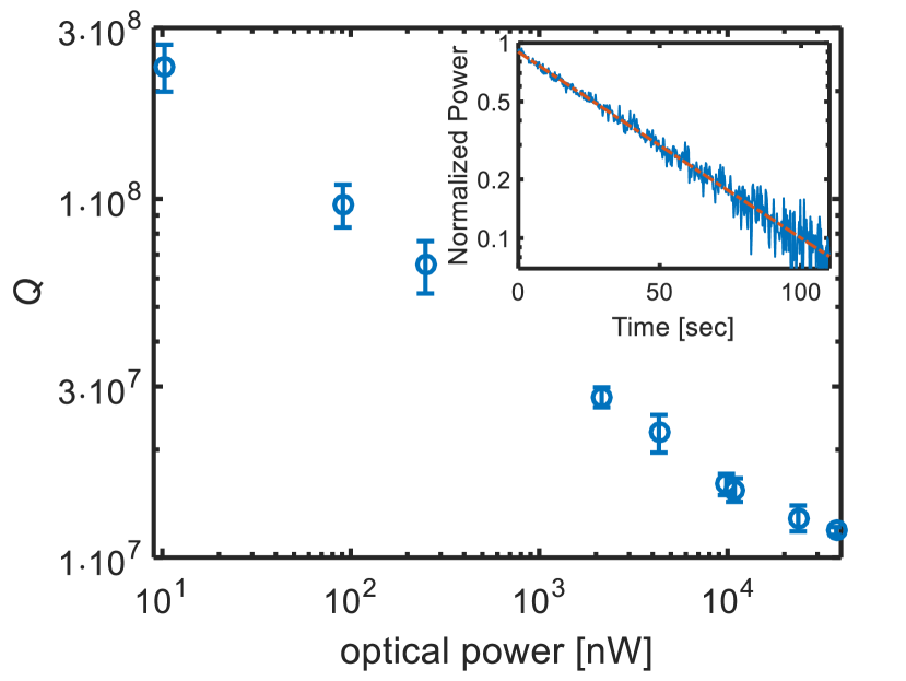
For the above measurements, we used a free space optical probe near 900 nm, operated continuously. By varying the probe’s optical power we examined its effect on the resonator’s dissipation. As we increase the probe’s optical power, we find that the mechanical quality factor decreases significantly (Fig. 3), presumably due to heating resulting from optical absorption. While we do expect that heating effects become more important with the decrease of thermal conductivity of amorphous solids below 1 K (Leivo and Pekola, 1998; Pohl et al., 2002; Wilson et al., 2015), we stress that the measurements here do not represent a quantitative study of the optical properties of Si3N4 itself at cryogenic temperatures; for example, we have not characterized the spatial mode of the optical spot on the membrane. We note the effective power of a 1064 nm beam, probing an optical cavity displaying heating effects, was measured to be considerably higher than for the 900 nm optical probe power for which we see an effect here. Specifically, previous work shows no heating effects at 120 mK (Kampel et al., 2016), and a heat transfer analysis at 35 mK indicates much smaller optical effects than those shown in Fig. 3 would cause significant effects to the Si3N4 alone. Further, we note that optical excitation effect on mechanical quality factor is a complex subject, which cannot be simply described by a heating of a thermal bath (Krause et al., 2015). With the data at hand, a complete picture cannot be discerned. The study of optical properties of Si3N4 membranes will be the subject of a future study.
Nonetheless, the dependence on optical absorption is indicative of variation of an internal dissipation as a function of temperature. Salient features we observe are that at a minimal probe power of 10 nW, and temperature of 35 mK, we find a maximal measured of for the (2,2) mode, corresponding to a QFP of Hz as seen in Fig. 3. The error bar shown is dominated by the low signal to noise level at the light level of the probe. We note the values of Fig. 3 are higher than the values depicted in Fig. 2 and were obtained prior to thermal cycling of the refrigerator. We suspect that the thermal cycling resulted in an added loss that obscured the reduction in dissipation below 100 mK in the data of Fig. 2.
In conclusion, we optically probed the mechanical loss of MHz frequency Si3N4 membranes down to mK temperatures. The highest we measured was at 35 mK, corresponding to a QFP of Hz, the highest value achieved for Si3N4 resonators so far. These results push further the performance of these resonators at cryogenic temperatures, both for quantum studies, as well as for precision measurements. A next step would be to combine cryogenic low-loss membrane resonator with additional geometric design features aiming to further reduce internal and external loss channels to reach even higher levels of isolation. Finally, the low thermal conductivity and the high- of Si3N4 membranes at mK temperatures allows optical heating effects to be measured. This enables a quantitative study of optical properties of Si3N4 membranes.
We thank A. Higginbotham for technical assistance. This work was supported by AFOSR PECASE, ONR DURIP, AFOSR-MURI, Rafael Ltd., the Cottrell Scholar’s program, and the National Science Foundation under grant number 1125844.
Contributions from NIST are not subject to U.S. copyright. Reference to specific products and services does not constitute endorsement by NIST. Other vendors may provide equivalent or better products.
References
- Thompson et al. (2008) J. Thompson, B. Zwickl, A. Jayich, F. Marquardt, S. Girvin, and J. Harris, Nature 452, 72 (2008).
- Gavartin et al. (2012) E. Gavartin, P. Verlot, and T. J. Kippenberg, Nature nanotechnology 7, 509 (2012).
- Scozzaro et al. (2016) N. Scozzaro, W. Ruchotzke, A. Belding, J. Cardellino, E. Blomberg, B. McCullian, V. Bhallamudi, D. Pelekhov, and P. Hammel, Journal of Magnetic Resonance 271, 15 (2016).
- Bochmann et al. (2013) J. Bochmann, A. Vainsencher, D. D. Awschalom, and A. N. Cleland, Nature Physics 9, 712 (2013).
- Andrews et al. (2014) R. W. Andrews, R. W. Peterson, T. P. Purdy, K. Cicak, R. W. Simmonds, C. A. Regal, and K. W. Lehnert, Nature Physics 10, 321 (2014).
- Bagci et al. (2014) T. Bagci, A. Simonsen, S. Schmid, L. G. Villanueva, E. Zeuthen, J. Appel, J. M. Taylor, A. Sorensen, K. Usami, A. Schliesser, and E. S. Polzik, Nature 507, 81 (2014).
- Unterreithmeier et al. (2010) Q. P. Unterreithmeier, T. Faust, and J. P. Kotthaus, Phys. Rev. Lett. 105, 027205 (2010).
- Zwickl et al. (2008) B. M. Zwickl, W. E. Shanks, A. M. Jayich, C. Yang, A. C. B. Jayich, J. D. Thompson, and J. G. E. Harris, Applied Physics Letters 92, 103125 (2008).
- Wilson-Rae et al. (2011) I. Wilson-Rae, R. Barton, S. Verbridge, D. Southworth, B. Ilic, H. Craighead, and J. Parpia, Phys. Rev. Lett. 106, 047205 (2011).
- Yu et al. (2012) P.-L. Yu, T. P. Purdy, and C. A. Regal, Phys. Rev. Lett. 108, 083603 (2012).
- Chakram et al. (2014) S. Chakram, Y. S. Patil, L. Chang, and M. Vengalattore, Phys. Rev. Lett. 112, 127201 (2014).
- (12) A. H. Ghadimi, D. J. Wilson, and T. J. Kippenberg, arxiv:1603.01605v1 .
- Tsaturyan et al. (2016) Y. Tsaturyan, A. Barg, E. S. Polzik, and A. Schliesser, arXiv:1608.00937 (2016).
- Norris and Photiadis (2005) A. N. Norris and D. M. Photiadis, The Quarterly Journal of Mechanics and Applied Mathematics 58, 143 (2005).
- Yuan et al. (2015) M. Yuan, N. A. Cohen, and G. A. Steele, Appl. Phys. Lett. 107 (2015).
- Pohl et al. (2002) R. O. Pohl, X. Liu, and E. Thompson, Rev. Mod. Phys. 74, 991 (2002).
- Norte et al. (2016) R. A. Norte, J. P. Moura, and S. Groblacher, Phys. Rev. Lett. 116 (2016).
- Reinhardt et al. (2016) C. Reinhardt, T. Muller, A. Bourassa, and J. Sankey, Phys. Rev. X 6 (2016).
- Yu et al. (2014) P.-L. Yu, K. Cicak, N. S. Kampel, Y. Tsaturyan, T. P. Purdy, R. W. Simmonds, and C. A. Regal, Appl. Phys. Lett. 104, 023510 (2014).
- Kuhn et al. (2014) A. G. Kuhn, J. Teissier, L. Neuhaus, S. Zerkani, E. van Brackel, S. Deléglise, T. Briant, P.-F. Cohadon, A. Heidmann, C. Michel, L. Pinard, V. Dolique, R. Flaminio, R. Taïbi, C. Chartier, and O. L. Traon, Appl. Phys. Lett. 104, 044102 (2014).
- Wilson (2012) D. J. Wilson, Cavity Optomechanics with High-Stress Silicon Nitride Films, Ph.D. thesis, California Institute of Technology (2012).
- Peterson et al. (2016) R. W. Peterson, T. P. Purdy, N. S. Kampel, R. W. Andrews, P.-L. Yu, K. W. Lehnert, and C. A. Regal, Phys. Rev. Lett. 116, 063601 (2016).
- Kampel et al. (2016) N. S. Kampel, R. W. Peterson, R. Fischer, P.-L. Yu, K. Cicak, R. W. Simmonds, K. W. Lehnert, and C. A. Regal, arXiv:1607.06831 (2016).
- Leivo and Pekola (1998) M. M. Leivo and J. P. Pekola, Appl. Phys. Lett. 72, 1305 (1998).
- Wilson et al. (2015) D. J. Wilson, V. Sudhir, N. Piro, R. Schilling, A. Ghadimi, and T. J. Kippenberg, Nature 524, 325 (2015).
- Krause et al. (2015) A. G. Krause, J. T. Hill, M. Ludwig, A. H. Safavi-Naeini, J. Chan, F. Marquardt, and O. Painter, Phys. Rev. Lett. 115, 233601 (2015).