Further author information: (Send correspondence to G.J.)
G.J.: E-mail: georg.jacob@uni-mainz.de, Telephone: +49 (0)6131 39 23671
K.S.: E-mail: ks@uni-kassel.de, Telephone: +49 (0)561 804-4235
F.S.K.: E-mail: fsk@uni-mainz.de, Telephone: +49 (0)6131 39 26234
Maximizing the Information Gain of a Single Ion Microscope using Bayes Experimental Design
Abstract
We show nanoscopic transmission microscopy, using a deterministic single particle source and compare the resulting images in terms of signal-to-noise ratio, with those of conventional Poissonian sources. Our source is realized by deterministic extraction of laser-cooled calcium ions from a Paul trap. Gating by the extraction event allows for the suppression of detector dark counts by six orders of magnitude. Using the Bayes experimental design method, the deterministic characteristics of this source are harnessed to maximize information gain, when imaging structures with a parametrizable transmission function. We demonstrate such optimized imaging by determining parameter values of one and two dimensional transmissive structures.
keywords:
Ion trapping, Laser cooling, Charged-particle beams, Transmission microscopy, Ion implantation, Information gain optimized imaging.This paper has been published in Proc. SPIE 9900, Quantum Optics, 99001A (April 29, 2016); Copyright 2009 Society of Photo Optical Instrumentation Engineers. One print or electronic copy may be made for personal use only. Systematic electronic or print reproduction and distribution, duplication of any material in this paper for a fee or for commercial purposes, or modification of the content of the paper are prohibited.
1 INTRODUCTION
Trapping and laser-cooling of single ions, has enabled substantial advance in applications as diverse as quantum information processing [1], creation of precise clocks [2, 3] and fundamental research on quantum phenomena[4]. Here we present a new application based on laser-cooled ions which extends the boundaries of nanoscopic imaging to the single particle limit. We implement a transmission microscope using an intrinsically deterministic ion source based on extracting single laser-cooled 40Ca+ ions from a linear segmented Paul trap [5, 6]. This offers several advantages over microscopy with conventional particle sources - in particular, a higher signal-to-noise ratio (SNR), especially at low exposures and an higher information gain per particle. This can be increased even further, when combined with the Bayes experimental design method, which allows for maximizing the information gain for each individual particle probe.
Our apparatus was conceived for ion implantation as well, because besides the favourable statistical properties, it also provides an ultracold monochromatic ion beam where ultimately the phase space occupation in transversal and longitudinal direction is limited by the Heisenberg uncertainty principle. Exactly the same characteristics renders it also favourable for imaging. These different applications are highly complementary, since microscopy - or more precisely alignment by imaging of the sample - is essential for an accurate absolute positioning of dopants, free of parallax errors.
2 Single ion microscope setup
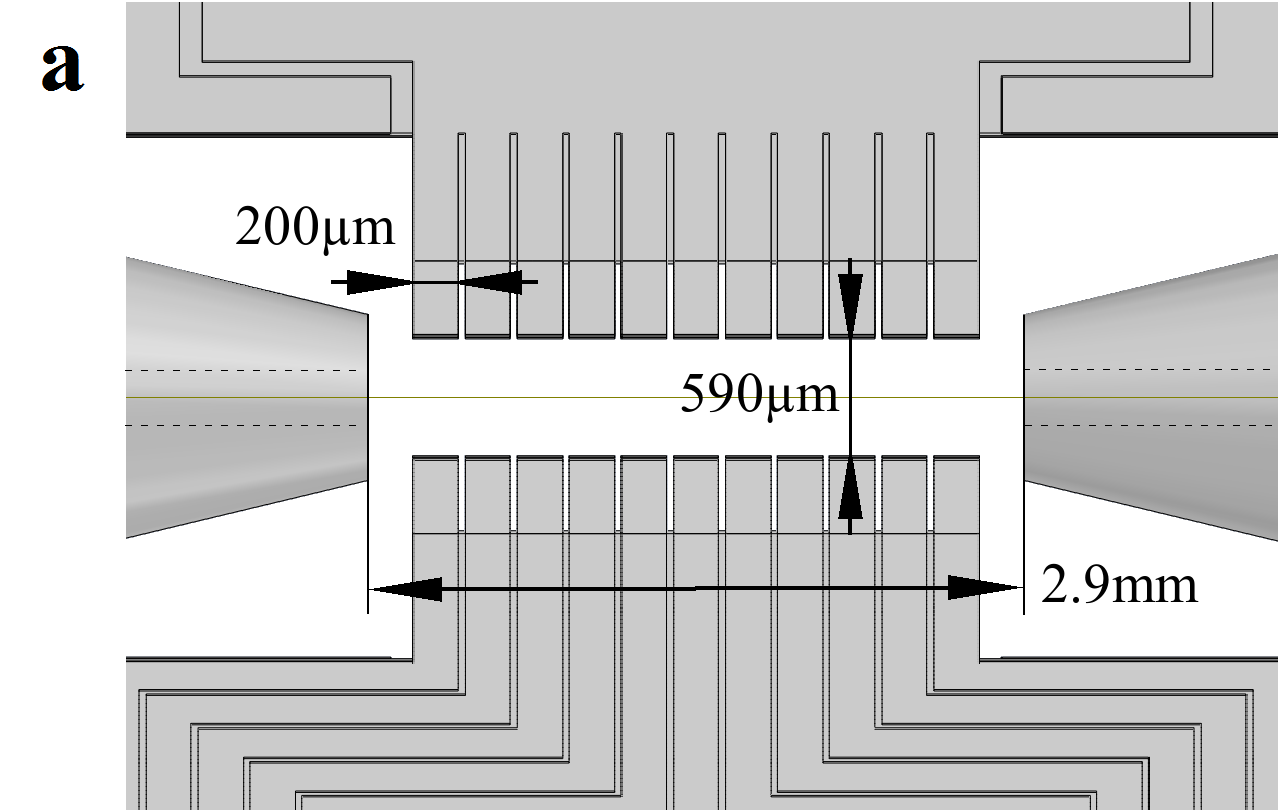
The experimental setup is based on a Paul trap, comprising four micro-fabricated alumina chips, which are arranged in an X-shaped configuration and two pierced end-caps (see Figure 1a). Each chip consists of 11 electrodes to shape the potential in the axial direction. The trap is operated at frequencies MHz and 1.4 MHz for the axial and radial directions, respectively. Calcium ions are generated by photo-ionization and laser cooled on the S1/2 to P1/2 dipole transition. Other ion species are created by a commercial ion gun 111Specs, Ion Source IQE 12/38 which is attached to the vacuum chamber. This allows for direct loading of ions into the trap along the axial direction.


In order to achieve a high repetition rate of the ion extraction, an automated loading of a predefined number of ions is implemented. Initially a random number of ions is trapped and Doppler-cooled using laser light near 397 nm. The ion number is counted by imaging the ion fluorescence on a CCD-camera. Excess ions are removed by lowering the axial trapping potential with a predefined voltage sequence. The cold ions are extracted along the axial direction of the trap by applying an acceleration voltage ranging from 0 to -6 kV, to one end-cap. This voltage is controlled by a fast solid-state switch with a jitter of less than 1 ns. The extraction time is triggered to the phase of the radio-frequency trap-drive (MHz). Extraction rates of single ions of up to 3 Hz, can be achieved, corresponding to an average flux of about 0.5 atto-ampere. The ions leave the trap passing through a hole with a diameter of 200m in the end-cap.
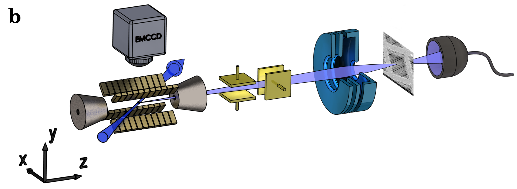
Two pairs of deflection electrodes are used for alignment and scanning of the beam. They are located at a distance of 46 mm and 67 mm from the center of the trap (see Figure 1b). In order to focus the ion beam, a electrostatic einzel-lens is placed 332 mm from the trap. It is constructed from three coaxially arranged ring shaped electrodes with an open aperture of 4 mm. In order to minimize spherical aberration, the geometry parameters are optimized with numerical simulations of the electrostatic field [7]. Chromatic aberration is suppressed because of the narrow velocity distribution the of ions. For the time of flight we measure a half width half maximum spread of ps. This corresponds to a velocity spread of m/s at an typical average speed of about m/s. Microscopy is implemented by placing a partially transmissive object on a three-axis translation stage. This can be either a nano-structured test sample or a profiling edge. The transmitted ions are detected by a secondary electron multiplier. Image information is generated by recording transmission events for a well defined number of extractions while scanning the object position in the focal plane.
3 Imaging with single ions
In electron or ion microscopy poor SNR generally can be overcome by increasing the exposure time or the current. This is a direct consequence of the Poissonian statistics of the sources in use. For some applications however, it is important to minimize the current, as for example where high irradiation causes inconvenient charging [8], contamination or even damage [9] to delicate samples. However, with conventional Poissonian sources the SNR becomes increasingly worse when going to lower currents. Thus effectively lowering the information gain per particle.

In order to solve this problem we employ a deterministic source, which provides an higher SNR compared to Poissonian sources, especially when approaching the single particle regime. This remains valid even if a detector of finite quantum efficiency is used, leading to Binomial statistics. Figure 2 shows how the single ion microscope is implemented in the experiment.
As an example Figure 3a) shows the result of an imaging scan of a optical waveguide-cavity structure made from diamond [10], using exactly one ion at each position, with a resolution of (25x25) nm2.


A quantitative comparison in terms of SNR, of a deterministic sources with a conventional Poissonian source, is presented in Figure 3b). The SNR, calculated as a function of the mean number of extracted ions, is compared for different detection probabilities . Note that the dark count noise is not taken into account in this comparison. For the plot the definition SNR is used, where is the mean value and the standard deviation of the corresponding probability mass function.
4 Bayes experimental Design
We use the Bayes experimental design method[11, 12, 13, 14] to image transmissive structures with optimal efficiency: The algorithm optimizes the probing position for the next probing event by maximizing the expected information gain. This allows to measure parameter values of one or two-dimensional transmissive structures, which are modelled by parametrizing their contour function. The parameter values are determined by incorporating the measurement results, using the Bayes update rule. The model is given as a convolution of this contour function and the beam profile. First we introduce and explain the Bayesian method on an abstract level. Then an example is given by means of a simple profiling scan, demonstrating how the radius and the position of the beam can be obtained by applying this method. In a second example an algorithm is presented which is able to find and determine the exact lateral position of a circular hole structure with optimal efficiency.

In the Bayesian concept of probability, the information or knowledge about the value of a parameter , is represented by a probability distribution function (PDF). In the context of measurement, the Bayes update rule allows for subsequently incorporating new information from the outcome of a measurement, into the prior PDF , which represents the pre-existing information. The resulting PDF is called the posterior:
| (1) |
The right hand side of the equation is the product of the prior PDF and the statistical model of the measurement , which is the probability to observe an outcome given the parameter values and design parameters . contains the free control parameters of the experiment. Normalization is provided by the marginal probability of observing ,
The Bayes experimental design method consists in maximizing the information gain per measurement by means of manipulating the design parameters. The information gain of a measurement with outcome and control parameters is represented by the utility , which is given by the difference between the Shannon entropies of the posterior and the prior PDF:
We obtain a utility, independent of the hitherto unknown observation, by averaging over the measurement outcomes:
| (2) |
The value which yields maximal utility is used to carry out the measurement, ensuring optimal information gain.

For the example of the profiling edge measurement, the design parameter is the profiling edge position, while the parameters to be determined are the beam position , its radius and the detector efficiency , i.e. . The outcome of the measurement is binary, . The measurement is modelled as
which in this case is a convolution of the transmission function of the structure to be imaged and a Gaussian beam profile.
The Bayes experimental design routine (see Figure 4) is implemented as follows. The initial prior, a three dimensional joint PDF of the parameters, position , sigma radius and detector efficiency , is calculated from an initial guess of these parameters. The marginals of the prior can be uniform or an educated guess e.g. a Gaussian distribution. It is implemented numerically, being a three dimensional grid of equidistant, weighted and normalized sampling points. In order to calculate the utility, the posterior PDF is calculated by applying the Bayes update (1) for each sampling point and the integrals are replaced by sums over all sampling points. The maximizing algorithm is now realized by calculating the utility for equally spaced profiling edge positions within the interval of interest, and recursively repeating this calculation for a smaller interval around the position with the highest utility. Five recursions were found to be sufficient to reach the required accuracy without incurring excessive computational expense. Using the measurement outcome of the real experiment performed at the calculated optimal profiling edge position, the Bayesian update (1) is applied to calculate the actual posterior PDF, which assumes the role of the prior PDF for the next iteration. The procedure is repeated until an accuracy goal is reached.


We demonstrate imaging of two-dimensional transmissive structures with a parametrizable transmission function, by determining the parameter values of a circular hole in a diamond sample (see Figure 6) using the Bayes experimental design method. This is also a practical example for sample alignment, since for many applications it is useful to know the exact lateral position of a sample with respect to the beam focus. For this purpose two perpendicular profiling edges as used in the beam radius measurement could equally be employed. However, for practical reasons, it might be more convenient to use a simple hole structure as a marker, which is in close proximity to a structure of interest.
The experiment is parametrized by the lateral position of the center of the circular hole, its radius as well as the 1-radius of the ion beam and the detector efficiency. The radius of the beam and the detector efficiency were kept constant at 25 nm and 95 % respectively. Both values were measured separately in advance. Using 572 ions in total, the position was determined with an accuracy of nm and nm, where the radius was measured to be nm. The systematic errors resulting from the deviations of the shape to the parametrization (ideal circle) are difficult to quantify, since the precise extent of this deviation is unknown. However, the accuracy of the results apply to an ideal circular shape, which could be available in other experiments.
5 outlook: single ion implantation

Besides imaging, the apparatus is also designed for deterministic ion implantation (see Figure 7) on the nanometer scale. This would allow for the fabrication of scalable solid state quantum devices, where individual dopants are coupled by their mutual dipolar magnetic interaction. Our focus is on systems of coupled nitrogen vacancy color centres [15], coupled single phosphorous nuclear spins in silicon [16, 17, 18, 19] and cerium or praseodymium in yttrium orthosilicate [20]. Another promising application of a highly focussed deterministic single ion beam is the doping and structuring of graphene [21].
For applications of ion implantation where absolute positioning is necessary, transmissive structures can be used for referencing. Imaging these markers with the same source which is used for the implantation, allows for the accurate alignment of dopants relative to this markers, free of parallax errors. Depending on the application, it can be necessary to use a different ion species for the imaging to avoid contamination of the sample. For this purpose, it has to be considered that the energy of the different species remain the same, in order to avoid different spatial positions of the beam focus as a result of inconsistent deflection.
Acknowledgements.
The authors acknowledge discussions with S. Prawer and G. Schönhense. The project acknowledges financial support by the Volkswagen-Stiftung, the DFG-Forschergruppe (FOR 1493) and the EU-projects DIAMANT and SIQS (both FP7-ICT). FSK thanks for financial support from the DFG in the DIP program (FO 703/2-1).References
- [1] Blatt, R. and Wineland, D., “Entangled states of trapped atomic ions,” Nature 453(7198), 1008–1015 (2008).
- [2] Rosenband, T., Hume, D., Schmidt, P., Chou, C., Brusch, A., Lorini, L., Oskay, W., Drullinger, R., Fortier, T., Stalnaker, J., et al., “Frequency ratio of al+ and hg+ single-ion optical clocks; metrology at the 17th decimal place,” Science 319(5871), 1808–1812 (2008).
- [3] Ludlow, A. D., Boyd, M. M., Ye, J., Peik, E., and Schmidt, P. O., “Optical atomic clocks,” Reviews of Modern Physics 87(2), 637 (2015).
- [4] Wineland, D. J., “Nobel lecture: Superposition, entanglement, and raising Schrödinger’s cat,” Rev. Mod. Phys. 85, 1103–1114 (Jul 2013).
- [5] Schnitzler, W., Linke, N. M., Fickler, R., Meijer, J., Schmidt-Kaler, F., and Singer, K., “Deterministic ultracold ion source targeting the Heisenberg limit,” Phys. Rev. Lett. 102(7), 070501 (2009).
- [6] Izawa, K., Ito, K., Higaki, H., and Okamoto, H., “Controlled extraction of ultracold ions from a linear Paul trap for nanobeam production,” J. Phys. Soc. Jpn. 79(12), 4502 (2010).
- [7] Singer, K., Poschinger, U. G., Murphy, M., Ivanov, P. A., Ziesel, F., Calarco, T., and Schmidt-Kaler, F., “Colloquium: Trapped ions as quantum bits: Essential numerical tools,” Rev. Mod. Phys. 82, 2609 (2010).
- [8] Kim, Y.-M., Jeong, H. Y., Hong, S.-H., Chung, S.-Y., Lee, J. Y., and Kim, Y.-J., “Practical approaches to mitigation of specimen charging in high-resolution transmission electron microscopy,”
- [9] Prawer, S. and Kalish, R., “Ion-beam-induced transformation of diamond,” Phys. Rev. B 51, 15711–15722 (Jun 1995).
- [10] Riedrich-Möller, J., Kipfstuhl, L., Hepp, C., Neu, E., Pauly, C., Mücklich, F., Baur, A., Wandt, M., Wolff, S., Fischer, M., et al., “One-and two-dimensional photonic crystal microcavities in single crystal diamond,” Nat. Nanotechnol. 7(1), 69–74 (2012).
- [11] Lindley, D. V., “On a measure of the information provided by an experiment,” Ann. Math. Stat. , 986–1005 (1956).
- [12] Guerlin, C., Bernu, J., Deleglise, S., Sayrin, C., Gleyzes, S., Kuhr, S., Brune, M., Raimond, J.-M., and Haroche, S., “Progressive field-state collapse and quantum non-demolition photon counting,” Nature 448(7156), 889–893 (2007).
- [13] Pezzé, L., Smerzi, A., Khoury, G., Hodelin, J. F., and Bouwmeester, D., “Phase detection at the quantum limit with multiphoton Mach-Zehnder interferometry,” Phys. Rev. Lett. 99(22), 223602 (2007).
- [14] Brakhane, S., Alt, W., Kampschulte, T., Martinez-Dorantes, M., Reimann, R., Yoon, S., Widera, A., and Meschede, D., “Bayesian feedback control of a two-atom spin-state in an atom-cavity system,” Phys. Rev. Lett. 109(17), 173601 (2012).
- [15] Dolde, F., Jakobi, I., Naydenov, B., Zhao, N., Pezzagna, S., Trautmann, C., Meijer, J., Neumann, P., Jelezko, F., and Wrachtrup, J., “Room-temperature entanglement between single defect spins in diamond,” Nat. Phys. 9, 139–143 (2013).
- [16] Kane, B. E., “A silicon-based nuclear spin quantum computer,” Nature 393(6681), 133–137 (1998).
- [17] Jamieson, D. N., Yang, C., Hopf, T., Hearne, S., Pakes, C., Prawer, S., Mitic, M., Gauja, E., Andresen, S., Hudson, F., et al., “Controlled shallow single-ion implantation in silicon using an active substrate for sub-20-kev ions,” Applied Physics Letters 86(20), 2101 (2005).
- [18] Pla, J. J., Tan, K. Y., Dehollain, J. P., Lim, W. H., Morton, J. J., Zwanenburg, F. A., Jamieson, D. N., Dzurak, A. S., and Morello, A., “High-fidelity readout and control of a nuclear spin qubit in silicon,” Nature 496(7445), 334–338 (2013).
- [19] Veldhorst, M., Yang, C., Hwang, J., Huang, W., Dehollain, J., Muhonen, J., Simmons, S., Laucht, A., Hudson, F., Itoh, K., Morello, A., and Dzurak, A., “A two-qubit logic gate in silicon,” Nature 526(7573), 410–414 (2015).
- [20] Kolesov, R., Xia, K., Reuter, R., Stöhr, R., Zappe, A., Meijer, J., Hemmer, P., and Wrachtrup, J., “Optical detection of a single rare-earth ion in a crystal,” Nat. Commun. 3, 1029 (2012).
- [21] Kotakoski, J., Brand, C., Lilach, Y., Cheshnovsky, O., Mangler, C., Arndt, M., and Meyer, J. C., “Toward two-dimensional all-carbon heterostructures via ion beam patterning of single-layer graphene,” Nano letters 15(9), 5944–5949 (2015).