A Poroelastic Mixture Model of Mechanobiological
Processes in Tissue Engineering.
Part I: Mathematical Formulation
Abstract.
An adequate control of cell response in tissue engineering applications is of utmost importance to obtain products suitable to clinical practice. This paper is the first part of a series of two connected publications in which we study via mathematical tools the cultivation in bioreactors of articular chondrocytes. The proposed model combines poroelastic theory of mixtures and cellular population models into a framework including stress state and oxygen tension as main determinants of engineered culture evolution. The special mechanosensitivity of articular chondrocytes to the surrounding environment is accounted for in the model through the novel concept of ”force isotropy” acting on the cell which is assumed as the promoting factor of the production of new cells or extracellular matrix.
Keywords: Tissue engineering; mechanobiology; mathematical modeling; mixture growth theory; mass transport; continuum mechanics.
Abbreviations: TE (tissue engineering); ECM (extracellular matrix); ACC (articular chondrocyte cell).
1. Introduction
The poor intrinsic healing capacity of articular cartilage highlights the strong clinical need for reparative therapies. While existing techniques with autograft/allograft transplants have had limited success, artificially engineered cartilage offers a possible alternative. Cartilage TE typically utilizes the seeding of ACCs into polymeric scaffolds. In this arrangement, the ECM concentration is designed to gradually increase and form neo-tissue. Despite the fact that sophisticated scaffold architectures have been devised to optimize ACC growth [25, 50, 17, 29, 38], current culture methodologies present a bottle-neck by being inefficient and sub-optimal. In particular, the cultivation of ACCs is well known to be bioprocess-dependent. This fact is exemplified by the extensive apoptosis of internal regions observed in 3D cellularised constructs maintained in static culture, inside which nutrient levels are well below a critical level [50, 17]. Moreover, limitations exist in maintaining ACCs in cartilaginous ECM-forming state after several passages of monolayer expansion culture [40, 41]. As a matter of fact, ACCs have the potential to proliferate in culture and to express their mature phenotype and, consequently, synthesize cartilaginous ECM [16, 44], but these conditions coexist in the cell culture in a delicate dynamical equilibrium [34]. Such an equilibrium is affected by extrinsic cues, including soluble growth factor signaling, culture substrates, oxygen tension, nutrient/metabolite content and pH (see [32] and the references cited therein). Furthermore, experimental data indicate that early-passage human chondrocytes adapted to fluid-dynamically stimulated culture conditions exhibit differences in transcriptional profiles, growth and matrix deposition compared to statically cultured cells [34, 35]. More in general, mechanical signals are recognized to have a deep influence on ACC behavior both in vivo and in vitro (see [3] and references therein).
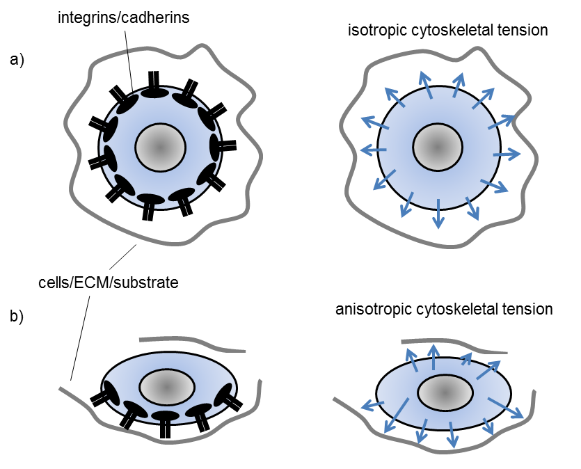
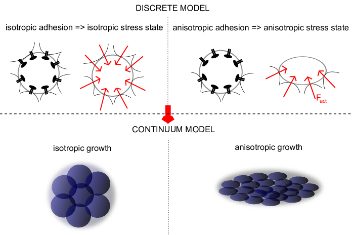
In this work, which is the first part of a series of two connected publications, we focus on the conceptual mechanobiological framework developed in [32, 33], where the idea of “force isotropy” on the cell is introduced. Namely, the complex system of traction forces exerted by the cell on the surrounding environment -substrate, ECM, other cells- induces cytoskeletal tensional states capable of triggering signalling transduction cascades regulating functional cell behavior, for example, the import flow of specific transcription factors in the nucleus. If cell adhesion-mediated traction forces have similar magnitude at varying orientations over the cell surface, the cell nucleus tends to maintain a roundish morphology and this condition is defined as “isotropic cytoskeletal tension” (see Fig. 1(a)). On the contrary, if cell adhesion-mediated traction forces have different magnitudes at varying spatial orientations, then the cell nucleus tends to elongate and this condition is defined as “anisotropic cytoskeletal tension” (see Fig. 1(b)).
To investigate these conditions, due to the continuum-based approach adopted throughout this paper, we do not describe the complex system of adhesion forces exerted on a single cell; rather, we use the isotropic/anisotropic parts of the local stress tensor associated with the biological construct to mathematically represent the isotropic/anisotropic cell adhesion state. To this purpose, we introduce the following qualitative definitions: if the anisotropic part of the local stress tensor is lower than a fixed threshold then the local stress state of the system is isotropic otherwise the local stress state of the system is anisotropic. We refer to Sect. 5 for a quantitative characterization of these concepts.
How a colony of ACCs seeded in a 3D porous scaffold can experience the above mechanical conditions is schematically illustrated in Fig. 2.
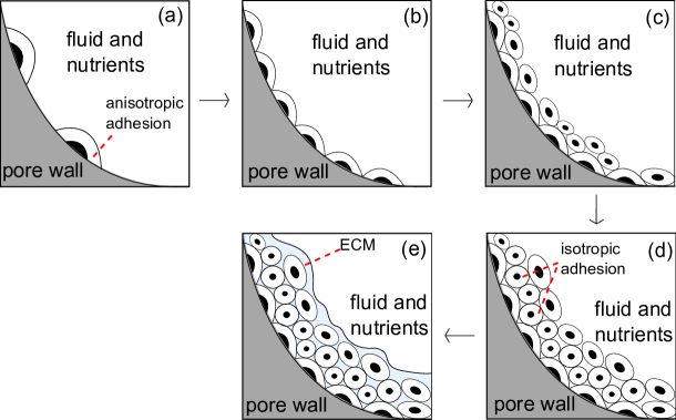
When cells are first seeded in the scaffold, they form a thin layer covering the surface. Since the characteristic dimension of the local scaffold curvature is much larger than cell size, cells find themselves in a local planar condition (see Fig. 2 (a)). Exerting adhesion forces on the scaffold surface, cells tend to assume a spread elongated shape, orienting themselves along a preferred polarization axis. According to the concept of “force isotropy”, this represents an anisotropic stress state. In this condition, the probability that the single cell enters into a proliferative state is enhanced (cf. [32]). The cell entering into the mitotic phase divides along a polarization axis represented by the direction of its long axis. This situation persists until all the pore surface is covered with cells (Fig. 2 (b)). From this moment on, cells start to occupy the empty space of the pore (Fig. 2 (c,d)). Cells in contact with other cells sense an isotropic stress state condition, which drives the cell towards a mature differentiated phenotype, characterized by ECM secretion (Fig.2 (e)). At present, there exists limited knowledge of how different levels of mechanical isotropy are transduced into functional cell fate. Incorporation of this newly-hypothesized mechanobiological mechanism into a computational tool for the prediction of tissue growth could represent a significant step forward towards a better understanding of the underlying mechanisms and consequently to an improvement in bioprocessing methodologies.
An outline of the presentation of theory and results is as follows. In Sect. 2 we state notation and basic assumptions; in Sect. 3 we introduce the basic definitions of the kinematical model; in Sect. 4 we illustrate the conservation laws of the mechanobiological model. In Sect. 5 we describe the mathematical models of production terms; in Sect. 6 we conclude the formulation of our model by providing a mathematical description of the mechanobiological processes involving cell populations and ECM secretion. In the final Sect. 7 we summarize the main modeling contributions proposed in this first part of our study on ACCs and emphasize possible research directions that should be worth investigating in a future work.
2. Multiphase modeling
In this section we develop a mathematical model based on the representation of the ensemble of the growing cartilaginous biomass by the mixture theory. In this framework, equations are postulated for the balance of mass and momentum for each constituent and then for the entire mixture according to the following ideas:
-
a)
the growing biomass is treated as a mixture composed by a multiphase solid mass and an interstitial fluid, this latter representing a fraction of the order of % in mass of the total biomass. The multiphase solid consists of ACCs and of ECM. The cell component includes different ACC populations (proliferative, ECM secreting, quiescent), as in the works of Sengers [45] and Ducrot [15];
- b)
- c)
- d)
-
e)
the mass exchange terms, including the rate of switch of cells from a population to the other, are tuned according to the nutrient level, this latter being itself an unknown of the problem, and to the stress state locally experienced by the mixture, which may drive cells into a certain functional behavior pool.
In the following, we use the term “phase” when we refer to the solid or to the fluid part of the mixture, while the term “component” is used to refer to any of the constituents of the solid phase (cell populations and ECM). When it is not necessary to distinguish between phase and component, we simply use the term “species”. The meaning of the subscripts used throughout the article is as follows: s=solid phase, fl=fluid phase, cells=cell component of the solid phase, ECM= extracellular component of the solid phase.
We let and denote the space and time variables, respectively. We use the convention that the dependence of all variables and model parameters on and is left understood except otherwise stated.
The geometrical configuration of the mixture is identified by the open bounded set ( unless otherwise specified). The domain does not evolve in time, rather, it is the amount of each species at a point that changes with due to cell proliferation and matrix deposition. This is the precise sense of the concept of “growing mixture”. From now on, we denote by the space-time cylinder where the TE problem is studied, being the final time of culture process.
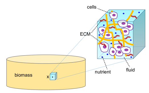
Let us now introduce the basic mathematical elements for developing a theory of the growing mixture. Referring to Fig. 3, for all we associate with a generic point a fixed representative elementary volume (REV) (see [51]) and denote by its -dimensional volume. Then, for each component of the growing mixture, we define the volume fraction
as the time evolving ratio of the volume occupied by the -th component in the REV and the volume of the REV itself. We further have
| (1a) | ||||
According to the biochemical hypothesis a) we consider three ACC populations: proliferating, ECM secreting state or quiescent state, denoted by the letters n, v and q, respectively. Cells in the quiescent state may reversibly lead to the mitotic or ECM-secreting populations or to apoptosis depending on nutrient level. Then, we have
| (1b) |
In the remainder of the article we denote by
the vector-valued function comprising the volume fractions of the fluid phase, the three cellular populations and the ECM.
The following standard assumptions on the mixture are also considered.
Assumption 2.1 (Fully saturated mixture).
The mixture is fully saturated, i.e.
| (1c) |
Relation (1c) is referred to as saturation condition [37, 44, 2] and excludes the possibility of the formation of voids or air bubbles inside the growing mixture.
Assumption 2.2 (Intrinsic incompressibility).
Assumption 2.2 is not, in general, equivalent to assuming that the whole mixture is incompressible (see [37] p. 629).
Assumption 2.3 (Closed mixture).
The mixture is closed, this meaning that the system does not exchange mass with external mass sources or sinks [37].
3. Kinematics of the growing mixture
We consider the so–called intermingled mixture constraint [1], stating that all the solid matrix constituents experience the same overall motion. This hypothesis requires the displacement and velocity vectors of each constituent to be equal to those of the solid matrix. Then, we denote by:
| (2a) | ||||
| (2b) | ||||
| the displacement and velocity at the time level of any point of the solid component of the biomass, and by | ||||
| (2c) | ||||
| the associated infinitesimal deformation of the biomass volume surrounding the point at time . The intermingled mixture constraint yields also the following relation | ||||
| (2d) | ||||
| The growth process of each mixture component (cellular growth and ECM secretion) is taken into account by introducing the following decomposition [24] | ||||
| (2e) | ||||
| where is the infinitesimal growth tensor associated with each solid constituent of the biomass, accounting for the amount and the spatial orientation of the newly deposited mass, and is the elastic accommodation tensor necessary to reinforce at each time level the continuity of the whole solid upon growth. Finally, we denote by | ||||
| (2f) | ||||
the relative (diffusive) velocity [37, 44] of the fluid phase with respect to the solid phase in the biomass, being the velocity of the interstitial fluid. Phenomenological laws for , and are discussed in Sect. 6. For notational brevity, from now on, we simply write and instead of and , respectively.
4. Balance laws for the deformable growing biomass
In this section we illustrate the set of conservation laws that constitute our proposed mathematical picture of the mechanobiological processes regulating biomass tissue growth.
4.1. Mass balance for the growing biomass
The mass balance equation for the growing mixture is expressed by the following coupled system of PDEs in conservation form to be solved in :
| (3a) | ||||
| (3b) | ||||
| (3c) | ||||
| where is the flux matrix and is the net production rate, whose mathematical form is described in detail in Sect. 5. Due to Assumption 2.3, the term satisfies the following constraint | ||||
| (3d) | ||||
| Eq. (3b) represents a phenomenological description of the flux density of each species under the effect of convective transport due to the fluid and solid velocity, respectively. It is worth noting that in cartilage tissue growth, cells do not typically exhibit a significant diffusive motion, rather, they need a solid support for surviving and for developing their functional activities (property of anchorage-dependence [34]). For this reason in the present work we neglect the contribution to the flux density due the the diffusive transport, unlike in other applications where this term plays a significant role [30, 36, 28]. | ||||
4.2. Momentum balance for the growing mixture
Under the assumption of negligible inertial terms and absence of body forces and volumetric fluid mass sources/sinks, the linear momentum balance equation for the solid and fluid phases of the growing mixture is expressed by the following PDEs in conservation form to be solved in :
| (4a) | ||||
| (4b) | ||||
| (4c) | ||||
| (4d) | ||||
| where is the effective stress tensor of the component of the solid phase of the mixture, is the pressure exerted by the fluid phase and is the identity tensor. The isotropic stress accounts for the coupling, typical of poroelasticity, between the flow of the fluid and the deformation of the solid matrix, and in particular describes the contribution to the stress due to the fluid pressure within the structure. | ||||
The quantities , , are the total stress tensors of the solid and fluid phases, while are the interphase forces [27]. As usual, we neglect the effective stress tensor of the fluid, meaning that we assume that the internal fluid viscosity is negligible compared with the friction between the fluid and the solid matrix [4, 18, 37]. For the mathematical characterization of the forces we refer to [27] and [37]. We observe that, for all and at all , it holds
| (4e) |
4.3. Total mass and momentum balance for the growing biomass
The mass balance equation for the whole growing mixture is obtained by summing each component in system (3a) and using Assumptions 2.1 and 2.3, to get:
| (5a) | ||||
| (5b) | ||||
| The vector is the composite velocity of the mixture (cf. Eq. (2.4) of [37]) and Eq. (5a) expresses the conservation of the total mass of the growing tissue. A simple manipulation allows us to write Eq. (5a) as | ||||
| (5c) | ||||
| In a similar manner, summing equations (4) and using (4e), we get | ||||
| (5d) | ||||
| (5e) | . | |||
The quantity is the total stress in the mixture and Eq. (5d) expresses the conservation of the total momentum of the growing tissue.
4.4. Mass balance for nutrient concentration
The mass balance system (3) for the solid and fluid phases of the growing mixture is accompanied by a corresponding continuity equation for the nutrient concentration (oxygen) that is transported throughout the growing on mixture by the interstitial fluid. This continuity equation is expressed by the following PDE in conservation form to be solved in :
| (6a) | ||||
| (6b) | ||||
| the interstitial fluid velocity being computed using (2f) as | ||||
| (6c) | ||||
| The mathematical description of the oxygen diffusion coefficient adopted in this article is the so-called Maxwell model [52], that allows to account, in a volume-averaged sense, for the microscopic composition of the biomass. More precisely, we introduce the effective diffusion coefficient | ||||
| where and represent the nutrient diffusivity in the fluid and solid phase, respectively, while is the coefficient regulating local mass equilibrium between nutrient concentration in the solid and fluid phases (see [52] for a detailed discussion). | ||||
The time rate of oxygen consumed by the cellular populations is modeled by a generalized form of the Michaelis-Menten kinetics
| (6d) |
where , , is the nutrient consumption rate of the cellular population and is the half saturation constant. We refer to [43] and the literature cited therein for a similar treatment of the oxygen consumption term in the framework of a multi-phase growing mixture.
5. Mass exchange pathways
The production terms introduced in Eq. (3c) mathematically describe the mechanisms of addition and/or removal of mass for each species constituting the biomass growing mixture.
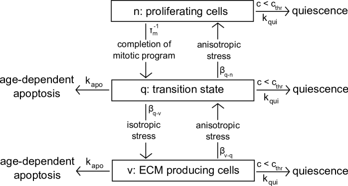
The exchange pathways between the different functional cellular pools are supposed to be mediated by the local stress state, the local nutrient concentration and by natural decay times. Transitions among cellular compartments are modeled by the time rate (probability) of exchange between states and , , while natural decay is modeled by decay rate constants . The scheme of Fig. 4 illustrates the various transitions among constituents, that may occur according to the following cases:
-
-
stress-mediated exchange pathways: cells in state q may switch into proliferative state or ECM secreting with a probability related to the stress state they experience. An anisotropic stress state enhances transition towards the proliferative state, while an isotropic stress state enhances transition towards the ECM secreting state. We quantify this concept as follows. Let be the Heaviside function such that for and for , being a real variable. Then, the stress-state dependent transition rate is represented by
(7a) being a yet unspecified indicator of the isotropy/anisotropy of the local stress state and being a threshold value. An explicit characterization of the local stress indicator will be provided in Part II of the present article. In our modeling description, if the local state of stress can be considered as isotropic, while if the local state of stress can be considered as anisotropic.
-
-
mitosis-mediated exchange pathway: we suppose cell proliferation to be expressed by the following law
(7b) Relation (7b) is a phenomenological law given by the product of two terms: the first term, given by , keeps into account contact inhibition effects, while the term is a nutrient–dependent modulation (Monod law [12]), being the half-saturation constant and the maximum growth rate, respectively;
-
-
decay pathways: all cellular compartments may evolve into quiescent (absence of cell activity due to an insufficient oxygen intake [11, 46]) or apoptotic phases (cellular death). Quiescence occurs if the nutrient concentration falls below a critical level , whereas the apoptotic phase is related to age dependent cell death [42]. The time rates of change between state () and the inactive states (quiescence or apoptosis) are and , respectively. Moreover, the transition rates are regulated by the mitotic characteristic time rate and take the form , where the minus sign is used in the transition, the plus sign in the transition.
For notational brevity we set and . According to the exchange laws illustrated above, the production terms associated with cell populations are defined as:
| (8a) | ||||
| (8b) | ||||
| (8c) | ||||
As for the definition of the production term for ECM mass density, we describe the biosynthesis of the different ECM components, focusing particularly on glycosaminoglycan (GAG) and collagen. We consider here GAG as the main marker for ECM accumulation [34] and we assume a simple proportionality law between the total amount of ECM and the secretion rate of GAG. The constant of proportionality accounts for the heterogeneous composition of cartilagineous ECM (water for 70-80% of its wet weight, collagen fibrils for 10-15% and GAG for 5%) [8]. According to our model, ECM synthesis attains its maximum value when no extracellular matrix is present because more space is available for matrix production. Then, as soon as sythesized matrix accumulates at each point of the biomass construct, the available space diminuishes until reaches a maximum value and matrix secretion ceases. Therefore takes the following form:
| (8d) |
where is the volume of a single cell involved in the ECM secretion process while is the ECM degradation rate [49]. In the description of GAG secretion, we are assuming that at the initial time level, biomass is constituted by a uniform layer of cells and matrix (see [39] for a similar ap- proach). This corresponds to neglecting the very initial phase where the seeded cells proliferate and “pave” the scaffold wall, and is consistent with the mathematical fact that a continuum-based approach does not enable to reproduce the subcellular mechanisms that regulate the early mitotic process. These latter processes should be more properly described by treating seeded cells as individual units that behave according to cellular automata schemes [10, 9, 20, 19].
To conclude the mathematical description of mass exchange terms, we consider the extracellular fluid production . This latter quantity is defined in such a way to satisfy Assumption 2.3 and, consistently, relation (3d). To this purpose, is modeled as
| (8e) |
From a biophysical point of view this is equivalent to assuming that mass exchanges occur only among cells/ECM and fluid, meaning that dead cells and degrading ECM are deteriorated into extracellular fluid, and conversely that the latter is ”consumed” whenever cells duplicate or secrete ECM [48]. From a computational point of view, relation (8e) allows us to eliminate the dependent variable and the corresponding mass balance equation from system (3a) as done in [48], Sect. 2.2, in such a way that the fluid volume fraction can be computed by simple post-processing as
| (8f) |
6. Bio-mechanical models for the deformable growing biomass
In this section we provide a mathematical description of the mechanobiological processes involving cell populations and ECM secretion. To this purpose, we introduce suitable bio-mechanical models for the growth tensors in the decomposition (2e) by extending the theory developed in [23] and [24].
6.1. Growth laws
We propose the following definitions of the growth tensors:
| (9a) | ||||
| (9b) | ||||
| where the symbol represents the tensor dyadic product and , are growth coefficients for which a model equation is provided below. Eqns. (9) state that the mass increment of each mixture solid constituent is isotropically deposited for the v, q and ECM components, while is accumulated along a specific polarization direction, identified by the unit vector , for proliferating cells. Let us now address some biophysical motivations that support our choice of the growth laws (9). Firstly, according to the concept of “force isotropy” on the cell introduced in [32, 33], cells that occupy the bio-synthesizing compartment (v compartment) experience an isotropic adherence condition and consequently tend to assume a spherical shape (see Fig. 5, left) whereas cells that live in the proliferating compartment (n compartment) are subjected to an anisotropic adhesion state and tend to elongate (see Fig. 5, right). Secondly, according to the infinitesimal deformation growth theory developed in [23], the deformation of an infinitesimal sphere of biomass growing into an ellipsoid can be reasonably described by an anisotropic growth tensor, while the deformation of an infinitesimal sphere growing into a larger sphere can be characterized by a isotropic growth tensor. For the sake of simplicity, the infinitesimal growth tensor for the species q and ECM are supposed to be isotropic. | ||||
The definition of in Eq. (9b) and the law for its time evolution is a delicate issue. In [6], is characterized according to the dynamics of the evolution of the cell cytoskeleton, which reorganizes itself according to its mechano–sensing mechanisms. A simplified version of the model proposed in [6], and also adopted in the present work, is represented by the choice
| (9c) |
where is the normalized eigenvector of the infinitesimal strain tensor , associated with the eigenvalue of largest module, which physically corresponds to the maximum principal dilatation of the biomass around such a point [47].
The coefficients , , give a measure of the amount of mass of the cellular population of type deposited at time at point . To determine these quantities, we proceed as in [23] and [24] and require the following growth continuity initial value problem to be satisfied for all :
| (9d) | ||||
| (9e) |
The quantity represents the amount of mass of the cellular population deposited at time at point per unit time and per unit reference mass. According to the general indications illustrated in Sect. 2.2.4 of [24], the growth laws are phenomenological equations that indirectly describe chemical processes responsible for growth and can be typically expressed as ”synthesis” rate minus a ”degradation” rate, that may include a mass conversion rate from one constituent of the mixture to another. Also, the constants that appear in a specific growth law may depend parameterically on biological factors such as, for example, the level of a specific growth factor. Thus, based on the description carried out in Sect. 5, we set
| (9f) |
in such a way that the initial value problems that furnish the characterization of the growth coefficients become:
| (9g) | ||||
| (9h) |
and:
| (9i) | ||||
| (9j) |
having used the identities and .
6.2. Constitutive equations for the mechanical and fluid subsystems
We assume that cells and ECM behave like linear elastic solids, so that the effective stress tensors associated with the solid components of the biomass are defined as
| (10a) | ||||
| Recalling relation (2e), Eq.(10a) can be written as | ||||
| (10b) | ||||
| where and are the Lamé parameters of each component of the solid phase, , and are the growth strain tensors introduced in (9). More sophisticated constitutive models might be adopted [37, 31, 14, 2], but their use is beyond the scope of the present work which is mainly devoted to proposing a computationally feasible mechanobiological model of in vitro cartilage tissue growth. | ||||
We assume the relative velocity in Eq. (5c) to be expressed by the Darcy law (see, e.g., [7] and references cited therein)
| (10c) |
where is the isotropic permeability tensor defined as in [44]
| (10d) |
the quantity being a friction coefficient. To provide a physically consistent characterization of we apply the classic Stokes theory for viscous drag to the biomass mixture and obtain
| (10e) |
and being cell radius and interstitial fluid dynamic viscosity, respectively. Replacing (10e) into (10d) we can express the biomass permeability tensor as
| (10f) |
where
| (10g) |
is a reference value of biomass permeability.
7. Conclusions and research perspectives
In the present article, which is the first part of a series of two distinct but correlated contributions, we propose a novel mathematical formulation based on the continuum assumption to describe the biomechanical sensitivity of articular chondrocytes. The natural application of our model is Tissue Engineering, a continuously growing discipline within the wider area of Regenerative Medicine, in which the control of cell response to multi-factorial stimuli is of utmost importance to obtain products suitable to clinical practice. However, it is worth noting that the proposed scheme may be used as well to describe more general settings in Cellular Biology, for example, the expansion of staminal cells.
The principal novelty of our contribution is the development of a model based on the use of Partial Differential Equations (PDEs) that incorporates the concept of “force isotropy” on the cell within the general and well established framework of poroelastic theory of mixtures and of cell population models. Specifically, the model translates into a simplified mathematical formalism, based on the use of suitably parametrized Heaviside functions, how the induced cytoskeletal tensional states trigger signalling transduction cascades regulating functional cell behavior, for example, the traslocation of specific transcription factors in the nucleus. According to the concept of force isotropy, it turns out that if cell adhesion-mediated traction forces have approximately the same strength over the cell surface, then the cell nucleus tends to maintain a roundish morphology, while if cell adhesion-mediated traction forces have different magnitudes at varying spatial orientations on the cell surface, then the cell nucleus tends to elongate. In the first case, the cell tensile condition is defined as “isotropic cytoskeletal tension” whereas in the second case the cell tensile condition is defined as “anisotropic cytoskeletal tension”.
Having defined the cytoskeletal stress characterization at the single cellular level, the next step of our approach is to build upon the concept of continuum-based approach to extend the above described description to the local stress tensor associated with the biological construct to mathematically represent the isotropic/anisotropic cell adhesion state. To this purpose, we generalize in a natural manner the previous definitions prescribing that if the anisotropic part of the local stress tensor is lower than a fixed threshold then the local stress state of the system is isotropic otherwise the local stress state of the system is anisotropic.
The final step of our model construction is to incorporate the above illustrated mechanobiological scheme within the setting of the theory of poroelasticity of a mixture composed by a solid and a multi-component fluid phases. The mixture represents the cellular construct in which several different cellular populations are well-mixed and oxygen delivery and consumption is taken into account to regulate in a dynamical manner the progressive fate of the evolving (macroscopic) tissue. The overall mathematical formulation consists of a system of conservation laws (mass and linear momentum) for the phases and components of the mixture that includes stress state and oxygen tension as main determinants of cellular culture evolution.
The high level of complexity of our model requires a severe effort for:
-
(1)
its mathematical analysis (well posedness and linear stability);
-
(2)
its numerical simulation (in a truly 3D geometrical configuration).
As far as issue 1. is concerned, we shall illustrate in a subsequent paper a stability analysis looking for homogeneous stable steady states of the dynamical system that describes the conservation of mass of the solid mixture components. Such a study will allow us to characterize the admissible range of values of model constitutive parameters that ensures the biophysical consistency of the proposed mathematical representation, in the same spirit as in [30] and [28].
As far as issue 2. is concerned, we refer the reader to the second part of the present article where a thorough investigation of the PDE system is critically performed in a simplified 1D setting.
Further research effort will be devoted to extend the present formulation by including the following advanced mechanobiological aspects:
- •
- •
Acknowledgements
Chiara Lelli was partially supported by Grant 5 per Mille Junior 2009 ”Computational Models for Heterogeneous Media. Application to Micro Scale Analysis of Tissue-Engineered Constructs” CUPD41J10000490001 Politecnico di Milano. Manuela T. Raimondi has received funding from the European Research Council (ERC) under the European Union’s Horizon 2020 research and innovation program (Grant Agreement No. 646990-NICHOID). These results reflect only the author’s view and the agency is not responsible for any use that may be made of the information contained.
References
- [1] D. Ambrosi and L. Preziosi. On the closure of the mass balance models for tumor growth. Math. Models Methods Appl. Sci., 12:737–754, 2002.
- [2] R. P. Araujo and D. L. Sean McElwain. A mixture theory for the genesis of residual stresses in growing tissues I: a general formulation. SIAM Journal of Applied Mathematics, 65(4):1261–1284, 2005.
- [3] D. L. Bader, D. M. Salter, and T. T. Chowdhury. Biomechanical influence of cartilage homeostasis in health and disease. Arthritis, page Article ID 979032, 2011.
- [4] S. I. Barry and G. N. Mercer. Flow and deformation in poroelasticity - I unusual exact solutions. Mathematical and Computer Modelling, 30:23–29, 1999.
- [5] M. A. Biot. General Theory of Three-Dimensional Consolidation. J. Appl. Phys., 12(2):155–164, 1941.
- [6] C. Borau, R. D. Kamm, and J. M. Garcia-Aznar. Mechano-sensing and cell migration: a 3D model approach. Phys. Biol., 8:1–13, 2011.
- [7] H. Byrne and L. Preziosi. Modelling solid tumour growth using the theory of mixtures. Math Med Biol, 20(4):341–366, December 2003.
- [8] P. Causin, R. Sacco, and M. Verri. A multiscale approach in the computational modeling of the biophysical environment in artificial cartilage tissue regeneration. Biomechanics and Modeling in Mechanobiology, 12(4):763–780, 2013.
- [9] G. Cheng, B. B. Youssef, P. Markenscoff, and K. Zygourakis. Cell population dynamics modulate the rates of tissue growth processes. Biophys J., 90(3):713–724, 2006.
- [10] C. A. Chung, Tze-Hung Lin, Shih-Di Chen, and Hsing-I Huang. Hybrid cellular automaton modeling of nutrient modulated cell growth in tissue engineering constructs. J. Theor. Biol., 262:267–278, 2010.
- [11] H. A. Coller, L. Sang, and J. M. Roberts. A new description of cellular quiescence. PlOs Biol., 4(3):e83, 2006.
- [12] D. E. Contois. Kinetics of bacterial growth: relationship between population density and specific growth rate of continuous cultures. J. Gen. Microbiol., 21:40–50, 1959.
- [13] O. Coussy. Poromechanics. John Wiley & Sons, 2004.
- [14] P.A. DiMilla, K. Barbee, and D.A. Lauffenburger. Mathematical model for the effects of adhesion and mechanics on cell migration speed. Biophysical Journal, 60(1):15–37, 1991.
- [15] A. Ducrot, F. Le Foll, P. Magal, H. Murakawa, J. Pasquier, and G. F. Webb. An in vitro cell population dynamics model incorporating cell size, quiescence, and contact inhibition. Math. Mod. Meth. Appl. S., 21:871–892, 2011.
- [16] L. E. Freed, I. Martin, and G. Vunjak-Novakovic. Frontiers in tissue engineering: In vitro modulation of chondrogenesis. Clin. Orthop., 367S:S46–S58, 1999.
- [17] L.E. Freed and G. Vunjak-Novakovic. Tissue engineering bioreactors. In R. P. Lanza, R. Langer, and J. Vacanti, editors, Principles of tissue engineering. Academic Press, San Diego, 2000.
- [18] A. J. H. Frijns. A four-component mixture theory applied to cartilaginous tissues: Numerical modelling and experiments. ProQuest LLC, Ann Arbor, MI, 2000. Thesis (Dr.ir.)–Technische Universiteit Eindhoven (The Netherlands).
- [19] F. Galbusera, M. Cioffi, and M. T. Raimondi. An in silico bioreactor for simulating laboratory experiments in tissue engineering. Biomedical Microdevices, 10(4):547–554, 2008.
- [20] F. Galbusera, M. Cioffi, M. T. Raimondi, and R. Pietrabissa. Computational modelling of combined cell population dynamics and oxygen transport in engineered tissue subject to interstitial perfusion. Computer Methods in Biomechanics and Biomedical Engineering, 10(4):279–287, 2007.
- [21] S. M. Klisch. Internally constrained mixtures of elastic continua. Mathematics and Mechanics of Solids, 4:481–498, 1999.
- [22] S. M. Klisch, S. S. Chen, R. L. Sah, and A. Hoger. A growth mixture theory for cartilage with application to growth-related experiments on cartilage explants. J. Biomech. Eng., 125:169–179, 2003.
- [23] S. M. Klisch, T. J. Van Dyke, and A. Hoger. A theory of volumetric growth for compressible elastic biological materials. Math. Mech. Solids, 6:551–575, 2001.
- [24] S. M. Klisch, R. L. Sah, and A. Hoger. A cartilage growth mixture model for infinitesimal strains: solutions of boundary-value problems related to in vitro growth experiments. Biomech. Model. Mechanobiol., 3:209–223, 2005.
- [25] R.S. Langer and J.P. Vacanti. Tissue engineering. Science, 260(5110):920–926, 2004.
- [26] G. Lemon and J. R. King. Multiphase modelling of cell behaviour on artficial scaffolds: effects of nutrient depletion and spatially nonuniform porosity. Mathematical Medicine and Biology, 24:57–83, 2007.
- [27] G. Lemon, J. R. King, H. M. Byrne, O. E. Jensen, and K. M. Shakesheff. Mathematical modelling of engineered tissue growth using a multiphase porous flow mixture theory. Journal of Mathematical Biology, 52:571–594, 2006.
- [28] P.K. Maini, J. A. Sherratt, and L. Olsen. Mathematical models for cell-matrix interactions during dermal wound healing. Int. J. Bif. Chaos, 12(9):2021–2029, 2002.
- [29] I. Martin, D. Wendt, and M. Heberer. The role of bioreactors in tissue engineering. Trends Biotechnol., 22(2):80–86, 2004.
- [30] P. Moreo, E. A. Gaffney, J. M. Garcia-Aznar, and M. Doblaré. On the modelling of biological patterns with mechanochemical models: insights from analysis and computation. Bull. Math. Biol., 72:400–431, 2010.
- [31] P. Moreo, J. M. Garcia-Aznar, and M. Doblaré. Modeling mechanosensing and its effect on the migration and proliferation of adherent cells. Acta Biomater., 4:613–621, 2007.
- [32] M.M. Nava, M.T. Raimondi, and R. Pietrabissa. Controlling self-renewal and differentiation of stem cells via mechanical cues. Journal of Biomedicine and Biotechnology, 2012:12, 2012.
- [33] M.M. Nava, M.T. Raimondi, and R. Pietrabissa. Bio-chemo-mechanical models for nuclear deformation in adherent eukaryotic cells. Biomechanics and Modeling in Mechanobiology, 13(5):929–943, 2014.
- [34] N.I. Nikolaev, B. Obradovic, H.K. Versteeg, G. Lemon, and D.J. Williams. A validated model of gag deposition, cell distribution, and growth of tissue engineered cartilage cultured in a rotating bioreactor. Biotechnol Bioeng., 105(4):842–853, 2010.
- [35] J. M. Osborne, R. D. O’Dea, J. P. Whiteley, H. M. Byrneand, and S. L. Waters. The influence of bioreactor geometry and the mechanical environment on engineered tissues. J. Biomech. Eng., 132 (5):051006, 2010.
- [36] G. F. Oster, J. D. Murray, and A. K. Harris. Mechanical aspects of mesenchymal morphogenesis. J. Embryol. exp. Morph., 78:83–125, 1983.
- [37] L. Preziosi and A. Tosin. Multiphase modelling of tumour growth and extracellular matrix interaction: mathematical tools and applications. Journal of Mathematical Biology, 58:625–656, 2009.
- [38] M. T. Raimondi, F. Boschetti, F. Migliavacca, M. Cioffi, and G. Dubini. Micro fluid dynamics in three-dimensional engineered cell systems in bioreactors. In N. Ashammakhi and R. L. Reis, editors, Topics in Tissue Engineering, volume 2, chapter 9. 2005.
- [39] M. T. Raimondi, G. Candiani, M. Cabras, M. Cioffi, M. Laganà, M. Moretti, and R. Pietrabissa. Engineered cartilage constructs subject to very low regimens of interstitial perfusion. Biorheology, 45:471–8, 2008.
- [40] M.T. Raimondi, E. Bonacina, G. Candiani, M. Laganà, E. Rolando, G. Talà, D. Pezzoli, R. D’Anchise, R. Pietrabissa, and M. Moretti. Comparative chondrogenesis of human cells in a 3D integrated experimental–computational mechanobiology model. Biomechanics and Modeling in Mechanobiology, 10(2):259–268, 2011.
- [41] M.T. Raimondi, M. Moretti, M. Cioffi, C. Giordano, F. Boschetti, K. Laganà, and R. Pietrabissa. The effect of hydrodynamic shear on 3D engineered chondrocyte systems subject to direct perfusion. Biorheology, 43(3-4):215–222, 2006.
- [42] Tew S., Redman S., Kwan A., Walker E., Khan I., Dowthwaite G., Thomson B., and Archer C.W. Differences in repair responses between immature and mature cartilage. Clin Orthop Relat Res., 391 Suppl:S142–52, 2001.
- [43] R. Sacco, P. Causin, P. Zunino, and M. T. Raimondi. A multiphysics/multiscale 2D numerical simulation of scaffold-based cartilage regeneration under interstitial perfusion in a bioreactor. Biomech Model Mechanobiol, 10(4):577–589, 2011.
- [44] B. G. Sengers, C. W. J. Oomens, and F. P. T Baaijens. An integrated finite-element approach to mechanics, transport and biosyntheis in tissue engineering. Journal of Biomechanical Engineering, 126(1):82–91, 2004.
- [45] B. G. Sengers, M. Taylor, C. P. Please, and R. O.C. Oreffo. Computational modelling of cell spreading and tissue regeneration in porous scaffolds. Biomaterials, 28:1926–1940, 2007.
- [46] BG Sengers. Modeling the development of tissue engineered cartilage. PhD thesis, Department of Biomedical Engineering, Technische Universiteit Eindhoven, 2005.
- [47] I. S. Sokolnikoff. Mathematical theory of elasticity. Mc Graw-Hill, New York, 1956.
- [48] A. Tosin. Multiphase modeling and qualitative analysis of the growth of tumor cords. Netw. Heterog. Media, 3:43–83, 2008.
- [49] A. J. Trewenack, C. P. Please, and K. A. Landman. A continuum model for the development of tissue-engineered cartilage around a chondrocyte. Mathematical Medicine and Biology, 26:241–262, 2009.
- [50] G. Vunjak-Novakovic, B. Obradovic, I. Martin, P. M. Bursac, R. Langer, and L. E. Freed. Dynamic cell seeding of polymer scaffolds for cartilage tissue engineering. Biotechnology Progress, 14(2):193–202, 1998.
- [51] S. Whitaker. The method of volume averaging. Theory and application of transport in porous media. Kluwer Academic Publishers, 1999.
- [52] B. D. Wood, M. Quintard, and S. Whitaker. Calculation of effective diffusivities for biofilms and tissues. Biotech. and Bioeng., 77(5):495–514, 2002.