Polycrystalline para-terphenyl scintillator adopted in a detecting probe for radio-guided surgery
Abstract
A radio-guided surgery technique exploiting emitters is under development. It aims at a higher target-to-background activity ratio implying both a smaller radiopharmaceutical activity and the possibility of extending the technique to cases with a large uptake of surrounding healthy organs. Such technique requires a dedicated intraoperative probe detecting radiation. A first prototype has been developed relying on the low density and high light yield of the diphenylbutadiene doped para-therphenyl organic scintillator. The scintillation light produced in a cylindrical crystal, 5 mm in diameter and 3 mm in height, is guided to a photo-multiplier tube by optical fibres. The custom readout electronics is designed to optimize its usage in terms of feedback to the surgeon, portability and remote monitoring of the signal. Tests show that with a radiotracer activity comparable to those administered for diagnostic purposes the developed probe can detect a 0.1 ml cancerous residual of meningioma in a few seconds.
1 Introduction
A novel radio-guided surgery (RGS) technique using radiation is being developed [1, 2]. The main advantage with respect to the traditional RGS with radiation [3] is the lower target-to-background activity ratio (TBR). radiation indeed penetrates only a few millimetres of tissue with essentially no contamination being the bremsstrahlung contribution almost negligible. Low background from healthy tissue close to the lesion allows a smaller radiotracer activity to detect tumour remnants, and the possibility of extending the technique to cases with a significant uptake from healthy organs around the lesion.
In the past, the use of decaying tracers was proposed [4], since they are largely diffused in diagnostic applications (Positron Emission Tomography, PET). The emitted positrons have a limited penetration and their detection is local, but at rest annihilate with electrons producing s with energy of 511 keV: the background persists and, actually, increases in energy becoming more penetrating. This approach has been studied in pre-clinical tests but it is not yet in use in clinical practice. The largest limitations range from the activity to be administered to achieve a quick probe response, to the complexity of the probe and to the exposure of the medical personnel.
On the contrary, pure emitting tracers allow to develop a handy and compact probe which, being sensitive only to particles emitted locally and operating in a low background environment, provides a clearer delineation of margins of the lesioned tissue.
Meningioma brain tumour was identified as the first clinical case of interest because of its sensitivity to DOTATOC [5], a synthetic analogue of somatostatin, that can be marked with the pure radionuclide 90Y [6]. This isotope is suitable for RGS because of its half-life (64 h), electron energy spectrum (endpoint: 2.28 MeV) and absence of emission. A study [7] of the potentiality of the proposed RGS technique has been performed on PET diagnostic exams of patients affected by meningioma after administration of 68Ga-DOTATOC (the uptake for the tracer can be assumed independent from the linked radionuclide). The study shows that a TBR greater than 10 is observed in almost all cases, making the effectiveness of the technique very promising. To implement this RGS technique, we are developing an intraoperative probe detecting decays based on para-terphenyl scintillator. In this article we describe the first prototype and its performance.
2 The intraoperative probe
The first prototype has been designed having in mind the constraint coming from the meningioma clinical case. The instrument is optimized to provide a very small device with high sensitivity to low energy electrons, directional detection and a fast response (few seconds) compatible with the surgical activity. The required target spatial resolution is fixed by the typical volume (0.1 ml) of a surgical tumour residual in this clinical case.
The radiation sensitive element of the probe is a small scintillator tip made of commercial poly-crystalline para-terphenyl doped to 0.1% in mass with diphenylbutadiene [9], manufactured by Detec-Europe. This material was adopted after a detailed study [10] due to its high light yield (3 times larger than typical organic scintillators) and non-hygroscopic property, while its low density minimizes the sensitivity to photons. Different detector sizes have been tested (see 2.1), the best configuration is a cylinder of 5 mm in diameter and 3 mm in height. A black PVC ring with external diameter of 11 mm encloses the detector providing a shield against radiation coming from the sides. A 10 m-thick aluminium front-end sheet covers the scintillator to ensure the light tightness. This assembly is mounted on top of an easy-to-handle aluminium cylindrical body (diameter 8 mm and length 14 cm).
In order to avoid risk of patient coming into contact with electrical devices, the scintillation light is guided by four 50 cm long optical fibres outside the probe to a Hamamatsu H10721-210 photo-multiplier tube (PMT). This photo-sensor module has an integrated high voltage power supply circuit requiring an input voltage as low as 5 V, making this device compatible with the surgical environment. The read-out electronics and the logic board are housed in a compact box that wireless connects to a remote monitor to display the counting rate.
2.1 Optimization of the scintillator design
Detection of non-penetrating low energy particles does not require thick scintillators and the high light yield of the para-terphenyl largely compensates its short light attenuation length (about 5 mm [10]).
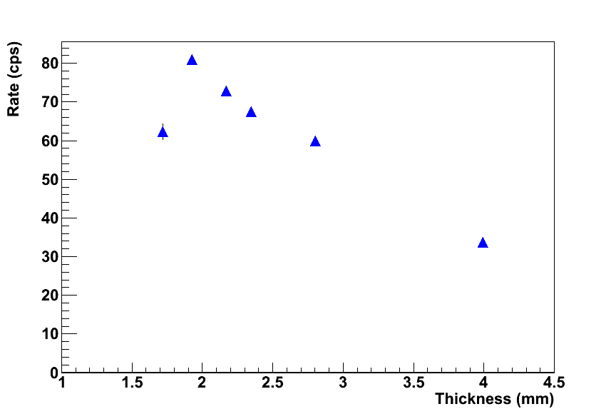
To maximize the sensitivity to particles, the optimal thickness of the scintillator has been determined experimentally. A cylinder of para-terphenyl with diameter of 2.1 mm is connected to the PMT by one optical fibre and the thickness of this scintillator is gradually reduced starting from 4 mm until the counting rate on a point-like 90Sr source with activity of 370 Bq (Sr-source in the following) is maximized. The 90Sr is a long-lived emitter (half-life: 29 years) with decay energy of 546 keV and no gamma radiation. It exists in secular equilibrium with its daughter isotope 90Y resulting in the emission of two particles. The radioactive source is sealed inside a 0.7 mm thick plastic disc. In Fig. 2 the counting rate stored on the Sr-source as a function of the para-terphenyl thickness is shown. The optimal point is found to be around 2 mm.
2.2 The probe working point
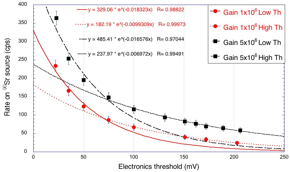
The probe working point is set by looking at the dark count and signal rates measured on the Sr-source by fixing the PMT gain value and varying the electronics threshold that trigger the pulse counter. The results are shown in Fig. 2 for two PMT gain settings: with a gain of 5 and a threshold of 80 mV the contribution of after pulses and dark current is below 0.2 cps with a signal rate of 140 cps.
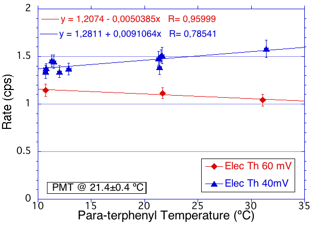
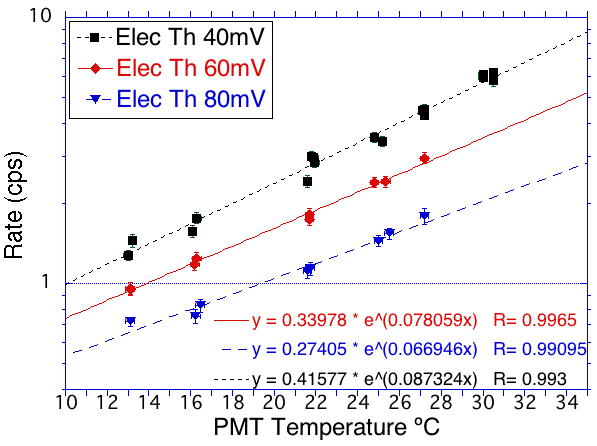
The response stability to temperature variation in the range 10-30 ℃ has been measured separately for the scintillating crystal and the PMT. As shown in Fig. 3 the scintillator is almost insensitive to temperature variations whereas the PMT dark counts increase according to the Hamamatsu specification.
3 detection sensitivity
The probe sensitivity in detecting electrons from decays is measured with the Sr-source (nominal activity 370 Bq). The rate measured by the probe is 3.8105 cps/MBq resulting in a detection efficiency of 40% of the decay products of the chain 90Sr/90Y.
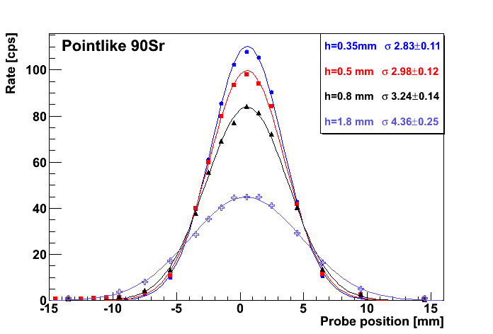
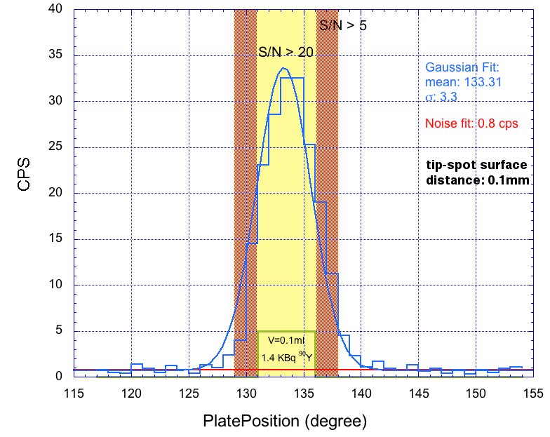
To study the capability of the probe to identify active spots, the detector is mounted on a motorized linear actuator ensuring position accuracy of 1.5 m and horizontal scans are performed over point-like and extended sources. The scan over the point-like Sr-source (Fig. 5) shows how spatial resolution depends on the distance between the probe tip and the surface. The reconstructed profiles measured at distances ranging from 350 m to 1.8 mm are fitted using a Gaussian distribution obtaining between 2.8 and 4.4 mm. The scan over the extended source (Fig. 5), instead, explores the discovery power of the probe in a more realistic simulation of a radio-labelled tumour residual. A cylindrical phantom with volume of 0.1 ml (diameter: 6.0 mm, height: 3.5 mm) is filled with a saline solution with 90Y radionuclide of 1.4 kBq activity, comparable to those administered for diagnostic purposes. The profile reconstructed by the probe is obtained with an horizontal scan of 1.5 mm steps and 10 s per position and a distance between the probe and the phantom surface of 100 m. The probe is able to well identify the active spot with a signal discriminating threshold S/N5, where S is the number of detected events and N is the number of dark counts, and the real phantom width is reconstructed with S/N20.
4 Background rejection
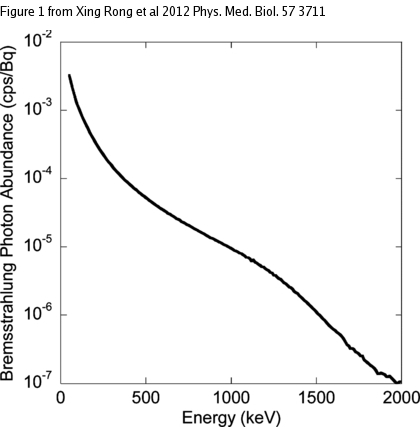
The usage of pure emitting tracers allows to operate with low radiation background, the residual being mainly due to photons coming from the Bremsstrahlung radiation of the electrons penetrating the tissue. The abundance of the expected Bremsstrahlung radiation as a function of the photon energy is shown in Fig. 6 (left) as computed with a simulation of a 90Y source in water [11].
We measure the sensitivity of the probe to these photons using three point-like sources: 133Ba emitting photons with energy ranging from 80 to 350 keV, 137Cs with gamma emission at Eγ=662 keV and 60Co with Eγ1=1170 keV and Eγ2=1330 keV. To avoid signal from electrons in the case of Cs decays, three copper layers with 350 m thickness are inserted in sequence between the source and the probe tip, and the measurements are repeated at each step. The counts as measured by the probe are shown in the Fig. 6 (right) for the three sources. Except for the first measure on the Cs source, introducing the copper absorbers implies a very small decrease in rate compatible with the attenuation in copper and the change in geometrical acceptance.
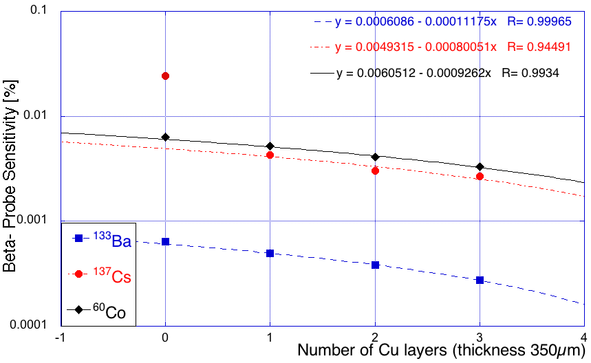
The sensitivity to the photons emitted by the 133Ba source is below 10-5, whereas for the s from the 137Cs and 60Co sources the sensitivity is still below 10-4. These measurements allow to conclude that the intraoperative probe is not sensitive to the Bremsstrahlung photons and therefore the effectiveness of the RGS technique would not be affected by this background.
5 Conclusion
A prototype of the intraoperative probe for RGS exploiting decays based on para-terphenyl scintillator has been developed and tested. The detection efficiency in the energy range of the 90Y emissions is 40%, while the sensitivity to the Bremsstrahlung photons is below 10-4 making this background radiation negligible. A test with a phantom simulating meningioma residual shows that with a radiotracer activity comparable to those administered for diagnostic purposes the probe is able to detect a 0.1 ml active spot in a few seconds.
References
References
- [1] Solfaroli Camillocci E et al 2014 Sci. Rep. 4 4401 DOI:10.1038/srep04401
- [2] Patent PCT/IT2014/000025 deposited by Università degli studi di Roma “La Sapienza”, Istituto Nazionale di Fisica Nucleare and Museo storico della fisica e centro studi e ricerche “E. Fermi”.
- [3] Povoski SP et al 2009 World Journal of Surgical Oncology 7-11
- [4] Bogalhas F et al 2009 Phys Med Biol 54 4439-53
- [5] Bartolomei M et al 2009 Eur J Nucl Med Mol Imaging 36 1407-16
- [6] Heppeler A et al 1999 Chem Eur J 7 1974-81
- [7] Collamati F et al 2015 J Nucl Med 56 1-6 DOI:10.2967/jnumed.114.145995
- [8] Heute D et al 2010 J Nucl Med 51 397-400
- [9] Budakovsky SV, Galunov NZ, Grinyov BV, Kim JK, Kim YK, Tarasenko OA 2009 Functional Materials 16, 1 86-91
- [10] Angelone M et al 2014 IEEE Transactions on Nuclear Science 61, 3 1483-87 DOI:10.1109/TNS.2014.2322106
- [11] Rong X, Du Y, Frey EC 2012 Phys Med Biol 57 3711–25 DOI:10.1088/0031-9155/57/12/3711