Comparing allosteric transitions in the domains of calmodulin through coarse-grained simulations
Abstract
Calmodulin (CaM) is a ubiquitous Ca2+-binding protein consisting of two structurally similar domains with distinct stabilities, binding affinities, and flexibilities. We present coarse grained simulations that suggest the mechanism for the domain’s allosteric transitions between the open and closed conformations depend on subtle differences in the folded state topology of the two domains. Throughout a wide temperature range, the simulated transition mechanism of the N-terminal domain (nCaM) follows a two-state transition mechanism while domain opening in the C-terminal domain (cCaM) involves unfolding and refolding of the tertiary structure. The appearance of the unfolded intermediate occurs at a higher temperature in nCaM than it does in cCaM consistent with nCaM’s higher thermal stability. Under approximate physiological conditions, the simulated unfolded state population of cCaM accounts for 10% of the population with nearly all of the sampled transitions (approximately 95%) unfolding and refolding during the conformational change. Transient unfolding significantly slows the domain opening and closing rates of cCaM. This potentially influences the mechanism of Ca2+-binding to each domain.
Introduction
Allostery is central to the precise molecular control necessary for protein function. Indirect coupling between distant regions of a protein is often provided through a conformational transition between a “closed” (ligand-free) and “open” (ligand-bound) structure upon ligation. NMR experiments that reveal proteins exist in dynamic equilibrium with multiple conformersBarbato et al. (1992); Moy et al. (1994); Evenäs et al. (1999); Evenäs, Malmendal, and Akke (2001); Volkman et al. (2001); Henzler-Wildman et al. (2007) suggest that a protein’s conformational dynamics in the absence of a ligand plays an essential role in allosteric regulation.Ma et al. (1999); Swain and Gierasch (2006); Henzler-Wildman and Kern (2007); Boehr, Nussinov, and Wright (2009) The functional dynamics of a folded protein occurs near the bottom of the funneled energy landscape, a part of the landscape generally more susceptible to perturbations than the self-averaged kinetic bottleneck that determines the mechanism of folding.Bryngelson et al. (1995) This sensitivity, while important for a protein’s ability to dynamically respond to environmental conditions and interaction with ligands, also makes the prospect of general organizing principles for allostery problematic.Zhuravlev and Papoian (2010) In this paper, we explore the sense in which the summarizing statement that native state topology determines the folding mechanism of small single domain proteinsBaker (2000) carries over to large-scale conformational transitions.
Due in part to limitations on computational timescales, much theoretical work modeling largescale conformational transitions in proteins has focused on simplified, coarse-grained models based on the energy basins defined by the open and closed conformations. The Gaussian network and related models describe an allosteric transition as motion along low frequency normal modes of the closed state conformational basin.Bahar, Atilgan, and Erman (1997); Atilgan et al. (2001); Tama and Sanejouand (2001); Bahar and Rader (2005) While the dynamics about a single free energy minimum offers a natural rationale and clear description of the collective motions involved in the conformational change,Yang, Song, and Jernigan (2007); Bahar et al. (2010) a minimal model capable of capturing the transition mechanism must accommodate the change in dynamics as protein moves between the two distinct meta-stable free energy basins. Allosteric transitions have been modeled by several different methods in which two meta-stable basins are coupled through an interpolation based on its energy. For example, minimal energy pathways have been computed for a potential surface based on the strain energies relative to each minimum conformation to predict the transition mechanism.Maragakis and Karplus (2005); Chu and Voth (2007); Das et al. (2014) Structure based simulations that couple two conformational basins have also been developed to understand the mechanism of allosteric transitions.Best, Chen, and Hummer (2005); Okazaki et al. (2006); Okazaki and Takada (2008); Chen and Hummer (2007); Lu and Wang (2008); Yang and Roux (2008) Additionally, transition mechanisms have been described in terms of the evolution of each residue’s local flexibility using a coarse grained variational model. Tripathi and Portman (2008, 2009, 2011, 2013) Itoh and Sasai present an alternative approach to predict allosteric transition mechanisms in which contacts from two meta-stable structures are treated on equal footing rather than through an interpolated energy function.Itoh and Sasai (2010, 2011)
In this paper, we use coupled structure based simulation of the opening transition in the domains of calmodulin (CaM) to explore how subtle differences in the native state topology can lead to qualitative changes in the transition mechanism. This work is motivated in part by an intriguing theoretical predictionTripathi and Portman (2009) that the domain opening mechanism of the C-terminal domain (cCaM) involves local partial unfolding and refolding while the N-terminal domain (nCaM) remains folded throughout the transition. These distinct transition mechanisms are in harmony with the Itoh and Sasai’s model that predicts cCaM has larger fluctuations than nCaM during domain opening.Itoh and Sasai (2011) Local unfolding in cCaM is found to relieve regions of high local strain during the transitionTripathi and Portman (2011) in agreement with the cracking mechanism of allosteric transitions discussed by Miyashita et al.Miyashita, Onuchic, and Wolynes (2003); Miyashita, Wolynes, and Onuchic (2005)
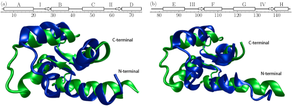
CaM is a ubiquitous Ca2+-binding protein consisting of two structurally similar globular domains connected by a flexible linker. Each domain consists of two helix-loop-helix motifs (the EF-hands) connected by a flexible linker as shown in Fig.1. Although topologically similar, the two CaM domains have distinct flexibilities, melting temperatures and thermodynamic Ca2+-binding properties.Tsalkova and Privalov (1985); Linse, Helmersson, and Forsen (1991); Sorensen and Shea (1998); Masino, Martin, and Bayley (2000) In the absence of Ca2+, the C-terminal domain is particularly dynamicTjandra et al. (1995) and is less stable than the N-terminal domain in the intact protein and when separated into isolated domains.Browne et al. (1997); Sorensen and Shea (1998); Masino, Martin, and Bayley (2000) The C-terminal domain, which has a very low denaturation temperature, is reported to be considerably unfolded under physiological temperature.Rabl et al. (2002) Furthermore, NMR experiments monitoring the open/closed transition of isolated cCaM have revealed local transient unfolding of helix F during domain opening.Lundström, Mulder, and Akke (2005)
The simulations presented in this paper suggest that over a wide range of temperatures, domain opening in cCaM involves global unfolding and refolding, while the unfolded conformations are much less prominent in nCaM’s primarily two-state domain opening mechanism. The appearance of an unfolded intermediate at a sufficiently high temperature is expected and has been reported for similar simulations of the conformational transition of cCaMChen and Hummer (2007) and the homologous protein S100A6,Okazaki et al. (2006) as well as other proteins.Best, Chen, and Hummer (2005); Yang and Roux (2008) Given the structural similarity of the two domains, it is harder to anticipate that the unfolded ensemble becomes locally stable at a significantly higher temperature in nCaM than it does in cCaM. Both the analytic model and simulations suggest that cCaM is more susceptible to unfolding during domain opening, despite employing very different approximations. Nevertheless, the simulated intermediate is globally unfolded in contrast to the local unfolding predicted by the analytic model. In terms of the kinetics, global unfolding and refolding significantly slows the simulated domain opening rate in cCaM which potentially can bias the partitioning of Ca2+-binding kinetics between induced fit and conformational selection for the two domains.
Methods
We use a native-centric model implemented in the Cafemol simulation packageKenzaki et al. (2011) to study the open/closed conformational transitions of the isolated N-terminal and C-terminal domains of CaM. This model couples two energy basins, one biased to the open (holo) conformation and the other to the closed (apo) reference conformation.Okazaki et al. (2006) The open and closed conformations of the domains of CaM are shown in Fig.1.
A conformation in this coarse-grained modelOkazaki et al. (2006) is specified by the position vectors of the C- atoms of the protein backbone, . For an energy basin biased to the reference conformation, , the energy of a configuration can be written as
| (1) |
The first term in Eq. 1 defines the coarse-grained backbone
where , , and denote bond lengths, bond angles, and dihedral angles, respectively. The corresponding values in the native structure are denoted with a superscript: , , and . The non-bonded interaction between neighboring residues in the native structure (native contacts) have short-ranged attraction
| (3) |
while non-native contacts are destabilized through a repulsive potential
| (4) |
Here, is the distance between C- atoms and in a conformation, , and is the corresponding separation distance found in the reference structure, .
The coefficients defining the energy function are set to their default values in Cafemol: , , and , , in units of , and Å. Trajectories are simulated using Langevin dynamics with a friction coefficient of and a timestep of (in coarse-grained units).Okazaki and Takada (2008) With these parameters, the folding transition temperatures of the isolated CaM domains are estimated from equilibrium trajectories to be K and K for the open and closed state of nCaM, and K and K for the open and closed state of cCaM, respectively. Experimentally, the isolated domains have similar folding transition temperatures of approximately KMasino, Martin, and Bayley (2000). Although and as well as and are within 2∘K (with cCaM’s thermal stability slightly below nCaM’s ), coupling the open and closed basins significantly destabilizes cCaM with respect to nCaM (described below). Consequently, the simulations relevant to the domains of intact CaM, for which interactions between the domains, particularly with the linker regionO’Donnell et al. (2009), reduce the folding temperature of the C-terminal domain to roughly 315∘K and increase the folding temperature of N-terminal domain to 328∘K.Tsalkova and Privalov (1985); Sorensen and Shea (1998); Masino, Martin, and Bayley (2000)
To study conformational changes between two meta-stable states, the energies of the corresponding native basins, and , are coupled through an interpolation functionKenzaki et al. (2011)
| (5) |
Here, the interpolation parameters, and , control the barrier height and the relative stability of the two basins. The single basin energies and are computed from Eq. 1 with modifications to some of the reference parameters in the potential in order to minimize conflicts between the two contact maps. (See Ref. Okazaki et al., 2006; Okazaki and Takada, 2008; Kenzaki et al., 2011 for details).
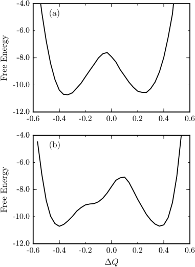
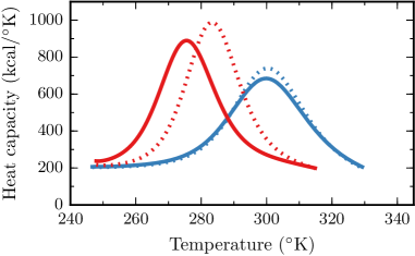
To compare the simulated domain opening mechanisms most clearly, it is convenient to choose coupling parameters and so that the barrier between the two states is low enough to give sufficient sampling of the two states and equal stability of the open and closed conformations (a choice to improve sampling of the equilibrium transition kinetics). With and for nCaM, and and for cCaM, the open and closed states are equally probable with a free energy barrier of as shown in Fig.2. With these parameters, the folding temperature for cCaM is approximately 25 degrees below the folding temperature of nCaM as indicated by the peaks in the heat capacity shown in Fig.3. We report temperatures relative to the simulated folding temperature of cCaM, denoted as K. Although we have explored a wide range of temperatures, most of the results presented in this paper have , a temperature slightly below the folding temperature of cCaM, and significantly below the folding temperature of nCaM.
NMR experiments indicate that the closed state of cCaM is more stable than the open state under physiological conditions, accounting for roughly 90% of the population.Malmendal et al. (1999) Assuming nCaM is similar, we adjust the relative stability of both domains through the coupling parameter to match this stability ( for nCaM, and for cCaM). As shown in Fig.3, the folding temperatures of the domains are sensitive to this destabilization of the open state. The simulated folding temperatures of the two domains differ by approximately K, somewhat larger than the difference in experimental folding temperatures of the domains in intact CaM, approximately K.Rabl et al. (2002) To connect to the domain opening kinetics in intact CaM, we relate the simulated temperatures to the folding temperatures of its N-terminal and C-terminal domains. With this choice, the physiological temperature K corresponds to simulation temperature of 95% of nCaM’s folding temperature, and 98% of cCaM’s folding temperature.
Simulated conformational ensembles are characterized through local and global structural order parameters based on the contacts formed in each sampled conformation. A native contact is considered to be formed if the distance between the residues is closer than 1.2 times the corresponding distance in the native conformation. To characterize structural changes during the conformational transition, it is convenient to separate the set of native contacts in the open (holo) and closed (apo) conformations into three groups: those that occur exclusively in either the open or the closed native reference conformation, and those that are common to both states. For each of these groups, denoted by (open, closed, and ), we define a local order parameter, , as the fraction of native contacts formed involving the residue. Overall native similarity is monitored by corresponding global order parameters, , where the average is taken over the residues of the protein. The free energy parameterized by these global order parameters are used to identify locally stable conformational ensembles such as the open and closed basins.
The transition rates between two coarse-grained ensembles are calculated from equilibrium simulations of length steps typically involving open/closed transitions for nCaM and open/closed transitions for cCaM. The transition rate between two states labeled by and is estimated byBuchete and Hummer (2008)
| (6) |
where is the mean time spent in state between transitions, and are the number of transitions from state to state . When the allosteric transition involves only the open and closed states, Eq. 6 reduces to the two state rates, and , where and are the mean first passage times to leave the open and closed state, respectively.
Conformational transitions of isolated domains
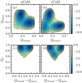
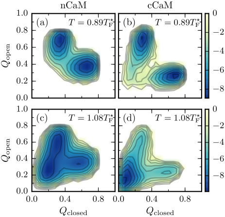
The populations of simulated conformations organized in terms of global order parameters are shown in Fig.4. The free energy as a function of and shows that the nCaM has a two-state domain opening and its conformational transition is sequential. That is, contacts specific to the closed conformation are lost prior to formation of contacts specific to the open conformation which mostly form after transition state region. Fig.4 also shows the free energy projected onto the order parameter monitoring common contacts, , and a progress coordinate for the conformational transition, . The global order parameter monitors the overall structural integrity of the secondary structure as well as tertiary contacts within parts of the protein that do not have large conformational changes during the transition. As shown in Fig.4, the common contacts in nCaM’s transition state ensemble remain largely intact. In contrast, the simulated open/closed free energy for cCaM has a locally stable intermediate state. Simultaneously low values of and (both less than 0.3), and (less than 0.7), indicate that the intermediate has significantly reduced tertiary structure. The probability to form individual contacts in the intermediate (data not shown) verifies that the secondary structure remains intact, though nearly all the tertiary interactions are lost. Since the barrier for the transition () is higher than the barrier for (), the intermediate can be considered to be part of cCaM’s extended open basin.
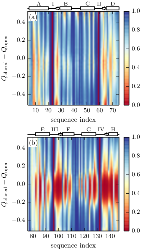
To describe the transition mechanism at the residue level, we consider the local order parameter of each residue as a function of the global progress coordinate . As shown in Fig.5, cCaM’s residues lose the majority of their common contacts upon opening (moving upward in the plot) and regain them later in the transition. Although the folding and refolding of residues in helices E and H are more gradual than other residues, nearly every residue (except the residues in the linker region between helices F and G) looses native tertiary structure. In contrast, the common contacts in nCaM remain intact throughout the transition, though the contacts involving specific residues in helices A and D and the -sheets in the loops are strained. Limited loss of long range common contacts in nCaM reflect an increased flexibility of the folded transition state ensemble.
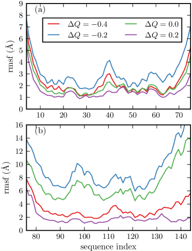
A coarse-grained, analytic model, also predicts distinct transition mechanisms for each domain in which cCaM is susceptible to local unfolding during the open/closed transition, while nCaM remains folded.Tripathi and Portman (2009, 2011) The conformational transition in the analytic model is described as the evolution of local flexibility along the transition route. Fig.6 shows the simulated local flexibility for four discrete values of the progress coordinate, . Although the fluctuations of the residues in both domains increase and then decrease during the transition, the magnitude of the largest fluctuations are much greater in cCaM. In contrast to the global unfolding observed in the simulations, unfolding and refolding of cCaM predicted by the analytic model is localized to particular residues (primarily in the linker between helix F and G).
Exploring a range of temperatures reveals that both domains can exhibit a two-state transition mechanism or a transition mechanism that involves unfolding and refolding depending on the temperature (see Fig.7). The transition mechanism at low temperatures is two state, involving primarily well folded conformational ensembles throughout the transition. Increasing the temperature progressively stabilizes the unfolded ensemble until it becomes locally stable at a spinodal temperature, . Above the spinodal temperature, the transition between the open and closed state involves unfolding and refolding of the domain. At high enough temperatures, the unfolded conformation becomes the most stable state.
Although both domains follow similar transition scenarios as a function of temperature, the domains can have different transition mechanisms from each other because the spinodal temperatures are different. Comparing the two domains, cCaM has a lower spinodal temperature () than nCaM (). For low temperatures, (), both the domains have two state transitions. For intermediate temperatures (), the domain opening transition of nCaM is two state, while the transition of cCaM involves unfolding and refolding. For higher temperatures (), the unfolded ensemble of nCaM is locally stable, but at this temperature the unfolded ensemble of cCaM is stabilized enough to become the global minimum.
Focusing on the scenario when the open state is 10% of the total population and at a simulation temperature corresponding to K (to model intact CaM at physiological conditions), the simulated unfolded population is less than 1% for nCaM, and approximately 9% for cCaM. These equilibrium unfolded populations can be compared to reports of 2% for the N-terminal domain and 24% for the C-terminal domain in intact CaM based on thermodynamic stability measurements.Masino, Martin, and Bayley (2000)
Transition Kinetics
Using Eq. 6 to calculate opening rates for each domain at , we find that unfolding and refolding along the transition route significantly slows cCaM’s domain opening rate compared to nCaM. Quantitatively, the domain opening and closing rates of nCaM, , are 50 times larger than the effective opening and closing rates of cCaM, .
A closer look at cCaM’s kinetic transitions reveals that only 5% of its transition paths proceed through direct transitions from the closed to open state without significant unfolding along the way. The rest of the transitions occur according to the kinetic equation
| (7) |
where , , , and are the corresponding simulated rates between the open, closed, and intermediate states.
Equilibrium between the open and the unfolded intermediate is established quickly on the timescale of the conformational transition so that the unfolded intermediate establishes a steady-state population
| (8) |
where and are the equilibrium populations of the closed and open state respectively. The effective two-state kinetics for open/closed transition can be written as
| (9) |
and
| (10) |
Since , these expressions for the two-state rates can be simplified. The effective domain opening rate is determined by the unfolding of the closed state
| (11) |
and the closing rate can be understood through the equilibration of the intermediate and open state
| (12) |
where is the population of the unfolded intermediate relative to the open state. The simulated effective two state rates for cCaM are consistent with this steady-state description of the kinetics.
The slowing influence of the folding and unfolding transition persists when the open state is destabilized to 10% of the total population, with domain opening approximately 45 times faster in nCaM than in cCaM at simulated temperatures that correspond to K.
Discussion
Although the isolated domains of CaM are topologically similar, the simulated open/closed transition mechanisms are distinct due to the presence of an unfolded intermediate that appears in the free energy landscape at a different temperature for each domain. Two-state transition kinetics persist at higher temperatures in nCaM, whereas the unfolded ensemble is more readily stabilized in cCaM. Above the spinodal temperature, transient unfolding and refolding of the domain occurs through the locally stable unfolded intermediate (exemplified by cCaM at ). Below the spinodal temperature, the transition is two-state like albeit with conformational dynamics that anticipates the unfolded intermediate with high flexibility and stressed tertiary interactions (as in nCaM at ).
The unfolding and refolding along the open and closed transition is reminiscent of the cracking mechanismMiyashita, Onuchic, and Wolynes (2003); Miyashita, Wolynes, and Onuchic (2005); Whitford et al. (2007) in which regions of high local strain are relieved through unfolding and refolding in the transition region. Since the unfolded conformations involved in cracking are typically locally unstable, the domain opening of CaM most closely follows this canonical description at temperatures near the spinodal for the unfolded conformations.
High temperature unfolded intermediates have been reported previously in simulations of the open/closed transition in cCaMChen and Hummer (2007) and the homologous protein S100A6.Okazaki et al. (2006) Chen and Hummer found that the population of the open ensemble is comparable to that of a marginally stable unfolded ensemble within a narrow temperature range. They argue that the sensitive balance between unstable folding and unfolded populations explains why some experiments report an open/closed transition,Evenäs et al. (1999); Malmendal et al. (1999); Vigil et al. (2001); Lundström and Akke (2004); Lundström, Mulder, and Akke (2005) and others report folding/unfolding transition for cCaM under similar conditions.Rabl et al. (2002)
Our simulations suggest that subtle differences in the topology and stability of the two domains can result in distinct transition mechanisms. In particular, we find that the unfolded population is stabilized more readily in cCaM, a result consistent with the prediction that cCaM (and not nCaM) exhibits local folding and unfolding during opening.Tripathi and Portman (2008, 2011) The C-terminal domain’s lower spinodal temperature may reflect its decreased overall relative thermodynamic stability. Indeed, nCaM is measured to be more stable than cCaM in the absence of Ca2+,Sorensen and Shea (1998) with cCaM being significantly unfolded at room temperature (20 – 25∘C).Masino, Martin, and Bayley (2000)
The transient unfolding and refolding observed in the simulations significantly slows the transition kinetics of cCaM. Several key observations of CaM dynamics have been reported, but how the dynamics of the individual domains compare is not clear from the literature. NMR studies of intact CaM in the absence of Ca2+report that cCaM is more dynamic than the nCaM, with an exchange time of for cCaM.Tjandra et al. (1995) This timescale is comparable to the folding and unfolding equilibration time of 200 for cCaM under similar conditions.Rabl et al. (2002) The dynamics of Ca2+-loaded cCaM with a mutation E140Q that stabilizes the open state and prevents binding to loop IV exhibits exchange on the faster timescale of Evenäs, Malmendal, and Akke (2001) and undergoes local transient unfolding.Lundström, Mulder, and Akke (2005) The dynamics of both domains under similar conditions has been reported by Price and co-workers who used fluorescence correlation spectroscopy coupled to Förster Resonance Energy Transfer (FRET) to monitor the intramolecular dynamics of both nCaM and cCaM on the microsecond timescale.Price, Aleksiejew, and Johnson (2011) They report that both domains have fluctuations on the – timescale in the absence of Ca2+. The Ca2+-dependence of the fluctuation amplitude, however, indicates that the observed fluctuations couple to the occupancy of the binding sites (and hence to domain opening) only in nCaM. Taken together, the evidence that the two domains have a different conformational timescale and/or mechanism is intriguing in light of the predictions from the coarse-grained simulations. Nevertheless, understanding how flexibility and transient unfolding influences domain opening dynamics of CaM requires further experimental clarification.
Concluding Remarks
Understanding the open/closed conformational dynamics of CaM is an essential step towards modeling Ca2+-binding. Exploring how transient unfolding in domain opening of CaM influences ligand binding is particularly interesting. Simulations of an extension of this model that includes Ca2+-binding (reported in a separate publication) shows that the two domains differ significantly in their thermodynamic properties such as binding affinity and cooperativity. Nevertheless, these thermodynamic differences seem to depend on the distinct conformational properties of the open and closed ensembles of each domain rather than the presence of an unfolded intermediate. Transient unfolding may still influence binding kinetics due to the slowing of the domain opening rate. This is particularly interesting because the detailed binding mechanism, such as the partitioning of binding kinetics into conformational selected or induced fit binding routesHammes, Chang, and Oas (2009) is thought to be sensitive to the timescale of the open and closed transition.Cai and Zhou (2011) Clarifying how the speed of conformational dynamics influences the kinetic binding mechanism through a molecular model is a rich problem that we wish to explore in the future.
Acknowledgements.
We would like to thank Swarnendu Tripathi for interesting discussions, and Daniel Gavazzi for help in preparing some of the figures. Financial support from the National Science Foundation Grant No. MCB-0951039 is gratefully acknowledged.References
- Barbato et al. (1992) G. Barbato, M. Ikura, L. E. Kay, R. W. Pastor, and A. Bax, Biochemistry 31, 5269 (1992).
- Moy et al. (1994) F. J. Moy, D. F. Lowry, P. Matsumura, F. W. Dahlquist, J. E. Krywko, and P. J. Domaille, Biochemistry 33, 10731 (1994).
- Evenäs et al. (1999) J. Evenäs, S. Forsén, A. Malmendal, and M. Akke, J. Mol. Biol. 289, 603 (1999).
- Evenäs, Malmendal, and Akke (2001) J. Evenäs, A. Malmendal, and M. Akke, Structure 9, 185 (2001).
- Volkman et al. (2001) B. F. Volkman, D. Lipson, D. E. Wemmer, and D. Kern, Science 291, 2429 (2001).
- Henzler-Wildman et al. (2007) K. A. Henzler-Wildman, V. Thai, M. Lei, M. Ott, M. Wolf-Watz, T. Fenn, E. Pozharski, M. A. Wilson, G. A. Petsko, M. Karplus, et al., Nature 450, 838 (2007).
- Ma et al. (1999) B. Ma, S. Kumar, C. J. Tsai, and R. Nussinov, Protein Eng. 12, 713 (1999).
- Swain and Gierasch (2006) J. F. Swain and L. M. Gierasch, Current Opinion in Structural Biology 16, 102 (2006).
- Henzler-Wildman and Kern (2007) K. Henzler-Wildman and D. Kern, Nature 450, 964 (2007).
- Boehr, Nussinov, and Wright (2009) D. D. Boehr, R. Nussinov, and P. E. Wright, Nat. Chem. Biol. 5, 789 (2009).
- Bryngelson et al. (1995) J. D. Bryngelson, J. N. Onuchic, N. D. Socci, and P. G. Wolynes, Proteins 21, 167 (1995).
- Zhuravlev and Papoian (2010) P. I. Zhuravlev and G. A. Papoian, Quarterly Reviews of Biophysics 43, 295 (2010).
- Baker (2000) D. Baker, Nature 405, 39 (2000).
- Bahar, Atilgan, and Erman (1997) I. Bahar, A. R. Atilgan, and B. Erman, Folding and Design 2, 173 (1997).
- Atilgan et al. (2001) A. Atilgan, S. Durell, R. Jernigan, M. Demirel, O. Keskin, and I. Bahar, Biophysical Journal 80, 505 (2001).
- Tama and Sanejouand (2001) F. Tama and Y. Sanejouand, Protein Engineering Design and Selection 14, 1 (2001).
- Bahar and Rader (2005) I. Bahar and A. Rader, Current Opinion in Structural Biology 15, 586 (2005).
- Yang, Song, and Jernigan (2007) L. Yang, G. Song, and R. L. Jernigan, Biophys. J. 93, 920 (2007).
- Bahar et al. (2010) I. Bahar, T. R. Lezon, L. W. Yang, and E. Eyal, Annual Review of Biophysics 39, 23 (2010).
- Maragakis and Karplus (2005) P. Maragakis and M. Karplus, J. Mol. Biol. 352, 807 (2005).
- Chu and Voth (2007) J. Chu and G. Voth, Biophys. J. 93, 3860 (2007).
- Das et al. (2014) A. Das, M. Gur, M. H. Cheng, S. Jo, I. Bahar, and B. Roux, PLoS Comput. Biol. 10, e1003521 (2014).
- Best, Chen, and Hummer (2005) R. B. Best, Y. G. Chen, and G. Hummer, Structure 13, 1755 (2005).
- Okazaki et al. (2006) K. Okazaki, N. Koga, S. Takada, J. N. Onuchic, and P. G. Wolynes, Proc. Natl. Acad. Sci. U. S. A. 103, 11844 (2006).
- Okazaki and Takada (2008) K. Okazaki and S. Takada, Proc. Natl. Acad. Sci. U. S. A. 105, 11182 (2008).
- Chen and Hummer (2007) Y. G. Chen and G. Hummer, J. Am. Chem. Soc. 129, 2414 (2007).
- Lu and Wang (2008) Q. Lu and J. Wang, J. Am. Chem. Soc. 130, 4772 (2008).
- Yang and Roux (2008) S. Yang and B. Roux, PLoS Comput. Biol. 4, e1000047 (2008).
- Tripathi and Portman (2008) S. Tripathi and J. J. Portman, J. Chem. Phys. 128, 205104 (2008).
- Tripathi and Portman (2009) S. Tripathi and J. J. Portman, Proc. Natl. Acad. Sci. U. S. A. 106, 2104 (2009).
- Tripathi and Portman (2011) S. Tripathi and J. J. Portman, J. Chem. Phys. 135, 075104 (2011).
- Tripathi and Portman (2013) S. Tripathi and J. J. Portman, The Journal of Physical Chemistry B 117, 13182 (2013).
- Itoh and Sasai (2010) K. Itoh and M. Sasai, Proc. Natl. Acad. Sci. U. S. A. 107, 7775 (2010).
- Itoh and Sasai (2011) K. Itoh and M. Sasai, J. Chem. Phys. 134, 125102 (2011).
- Miyashita, Onuchic, and Wolynes (2003) O. Miyashita, J. N. Onuchic, and P. G. Wolynes, Proc. Natl. Acad. Sci. U. S. A. 100, 12570 (2003).
- Miyashita, Wolynes, and Onuchic (2005) O. Miyashita, P. G. Wolynes, and J. N. Onuchic, J. Phys. Chem. B 109, 1959 (2005).
- Tsalkova and Privalov (1985) T. N. Tsalkova and P. L. Privalov, J. Mol. Biol. 181, 533 (1985).
- Linse, Helmersson, and Forsen (1991) S. Linse, A. Helmersson, and S. Forsen, Journal of Biological Chemistry 266, 8050 (1991).
- Sorensen and Shea (1998) B. R. Sorensen and M. A. Shea, Biochemistry 37, 4244 (1998).
- Masino, Martin, and Bayley (2000) L. Masino, S. R. Martin, and P. M. Bayley, Protein Sci. 9, 1519 (2000).
- Tjandra et al. (1995) N. Tjandra, H. Kuboniwa, H. Ren, and A. Bax, European Journal of Biochemistry 230, 1014 (1995).
- Browne et al. (1997) J. P. Browne, M. Strom, S. R. Martin, and P. M. Bayley, Biochemistry 36, 9550 (1997).
- Rabl et al. (2002) C. R. Rabl, S. R. Martin, E. Neumann, and P. M. Bayley, Biophys. Chem. 101-102, 553 (2002).
- Lundström, Mulder, and Akke (2005) P. Lundström, F. A. A. Mulder, and M. Akke, Proc. Natl. Acad. Sci. U. S. A. 102, 16984 (2005).
- Kenzaki et al. (2011) H. Kenzaki, N. Koga, N. Hori, R. Kanada, W. Li, K. Okazaki, X. Q. Yao, and S. Takada, Journal of Chemical Theory and Computation 7, 1979 (2011).
- Kuboniwa et al. (1995) H. Kuboniwa, N. Tjandra, S. Grzesiek, H. Ren, C. B. Klee, and A. Bax, Nat. Struct. Biol. 2, 768 (1995).
- Chattopadhyaya et al. (1992) R. Chattopadhyaya, W. E. Meador, A. R. Means, and F. A. Quiocho, J. Mol. Biol. 228, 1177 (1992).
- Humphrey, Dalke, and Schulten (1996) W. Humphrey, A. Dalke, and K. Schulten, Journal of Molecular Graphics 14, 33 (1996).
- O’Donnell et al. (2009) S. E. O’Donnell, R. A. Newman, T. J. Witt, R. Hultman, J. R. Froehlig, A. P. Christensen, and M. A. Shea, Methods in Enzymology 466, 503 (2009).
- Malmendal et al. (1999) A. Malmendal, J. Evenäs, S. Forsén, and M. Akke, J. Mol. Biol. 293, 883 (1999).
- Buchete and Hummer (2008) N. Buchete and G. Hummer, The Journal of Physical Chemistry B 112, 6057 (2008).
- Whitford et al. (2007) P. Whitford, O. Miyashita, Y. Levy, and J. N. Onuchic, J. Mol. Biol. 366, 1661 (2007).
- Vigil et al. (2001) D. Vigil, S. C. Gallagher, J. Trewhella, and A. E. García, Biophys. J. 80, 2082 (2001).
- Lundström and Akke (2004) P. Lundström and M. Akke, J. Am. Chem. Soc. 126, 928 (2004).
- Price, Aleksiejew, and Johnson (2011) E. Price, M. Aleksiejew, and C. Johnson, The Journal of Physical Chemistry B (2011).
- Hammes, Chang, and Oas (2009) G. G. Hammes, Y. C. Chang, and T. G. Oas, Proc. Natl. Acad. Sci. U. S. A. 106, 13737 (2009).
- Cai and Zhou (2011) L. Cai and H. X. Zhou, J. Chem. Phys. 134, 105101 (2011).