Superdiffusive motion of membrane-targeting C2 domains
Abstract
Membrane targeting domains play crucial roles in the association of signalling molecules to the plasma membrane. For most peripheral proteins, the protein-to-membrane interaction is transient. After proteins dissociate from the membrane they have been observed to rebind following brief excursions in the bulk solution. Such membrane hops can have broad implications for the efficiency of reactions on membranes. We study the diffusion of membrane-targeting C2 domains using single-molecule tracking in supported lipid bilayers. The ensemble-averaged mean square displacement (MSD) exhibits superdiffusive behaviour. However, traditional time-averaged MSD analysis of individual trajectories remains linear and it does not reveal superdiffusion. Our observations are explained in terms of bulk excursions that introduce jumps with a heavy-tail distribution. These hopping events allow proteins to explore large areas in a short time. The experimental results are shown to be consistent with analytical models of bulk-mediated diffusion and numerical simulations.
A myriad of signalling proteins are recruited to specific cell membranes via phospholipid-binding domains Hurley2006805 ; lemmon2008membrane . These molecules dock to the surface of specific lipid membranes and undergo two-dimensional diffusion in search of a target. Once the target is located, many proteins either activate or suppress a downstream signalling pathway for various physiological and pathological processes. Examples of membrane-targeting domains include pleckstrin homology (PH) lemmon2000signal and C2 Cho2006838 , which have been identified in hundreds of human signalling molecules as well as in eukaryotic species as diverse as fungi and flies letunic2012smart . PH domains bind specifically to phosphoinositides while C2 domains bind a variety of membranes, and a subset of C2 domains only bind membranes in the presence of calcium and play key roles in signalling pathways. The association to lipid membranes often takes place in response to different extracellular and intracellular stimuli, but typically the residence on the membrane surface is only temporary. The transient nature of peripheral protein-membrane interactions enables a tight temporal regulation of signal transduction. Further, membrane dissociation has also broad implications on the search for the target substrate, but this process is less understood.
Recently, Knight and Falke observed the dissociation of PH domains from supported bilayers followed by rapid rebinding to the surface after a short excursion in the bulk solution knight2009BJ . They proposed that the hopping process may be important in the search for target molecules in eukaryotic cells. Subsequently, Yasui et al. found that PTEN (phosphatase and tensin homologue) molecules hop along the plasma membrane of living cells due to dissociation followed by rebinding Yasui2014 . PTEN is an important protein that suppresses development of cancer. It prevents cells from growing and dividing too rapidly by dephosphorylating phosphoinositide substrates on the plasma membrane. PTEN-membrane affinity is regulated by a C2 domain and it is enhanced by electrostatic interactions. The observed hopping of the C2 domain on the plasma membrane is thus expected to alter the dynamics of the search for a phospholipid substrate.
A straightforward consequence of membrane hopping is that a molecule remains in its immediate vicinity for a short time and then jumps to a location that is further away than expected from two-dimensional diffusion. Therefore, the search process is allowed to explore larger areas and the molecule can bypass diffusion barriers that may be present in the membrane. However, hopping comes at the cost of the search being less exhaustive. We may ask the questions how the dynamics of membrane-targeting domains is affected by such long jumps and how this motion deviates from a simpler two-dimensional diffusion. Such potential complex behaviour can yield anomalous diffusion of membrane-targeting domains, which would alter the outcome of search processes and the sequential molecular reactions.
Anomalous diffusion is widespread in the motion of molecules in biological systems barkai2012PhysToday ; hofling2013review ; metzler2014review ; krapf2015reviewCTM . In general, a particle exhibits anomalous diffusion when the mean square displacement (MSD) scales as a power law with an exponent
| (1) |
where is the generalized diffusion coefficient with units . When the process is subdiffusive and when it is superdiffusive. Subdiffusion in the cytoplasm goldingCox2006 ; tolic2004anomalous ; jeon2011vivo , the nucleus bronstein2009PRL , and the plasma membrane weigel2013pnas ; heinemann2013lateral ; torreno2014enhanced of live cells is caused by crowding banksFradin2005 ; szymanskiWeiss2009 and complex interactions with the cytoskeleton and macromolecular complexes, among others. Similarly, subdiffusion can take place in model membranes due to crowding and packing effects horton2010development ; jeon2012anomalous . The appearance of superdiffusion processes in biomolecular systems is far less common. The archetypal mode of superdiffusive motion is due to active cytoplasmic flows and transport mediated by molecular motors, requiring ATP energy consumption bursac2007cytoskeleton ; Kahana2008 ; bruno2009transition .
From a theoretical point, there are three major mechanisms that can introduce superdiffusion akimoto2012distributional . It can be caused by correlations in the random walk, such as those in fractional Brownian motion with a Hurst index , by persistent directional motions (Lévy walks), and by long jumps (Lévy flights). Active biological transport can be modelled as Lévy walks bruno2009transition . Bulk-mediated diffusion processes, which can be described as Lévy flights, have been observed for transient adsorption on a solid surface where molecules display intermittent behaviour, alternating between periods of immobilization at the solid-liquid interface and periods of diffusion in the bulk fluid skaug2013intermittent ; yu2013single .
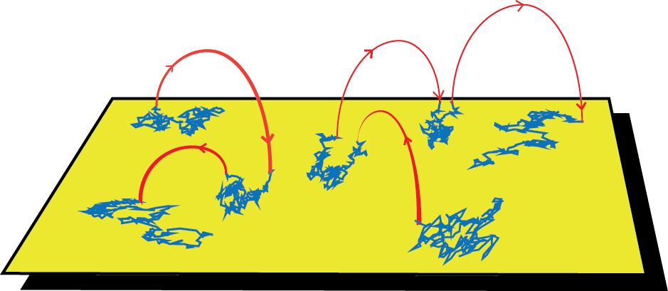
In this article we report the experimental observation of superdiffusive transport of membrane-targeting C2 domains on supported lipid bilayers. Measurements of the diffusion of membrane-targeting domains are performed by single-particle tracking and are compared to both analytical theory and numerical simulations. In stark contrast to active cytoplasmic transport, superdiffusion in model membranes does not require energy. Our data strongly suggests that superdiffusion is caused by bulk-mediated diffusion, namely molecules dissociate from the membrane and perform three-dimensional random walks until they reach the membrane again and readsorb at a new location, as sketched in Figure 1. Interestingly, the motion of membrane-targeting domains shows weak ergodicity breaking, a phenomenon that has recently attracted considerable attention in cellular environments and other complex systems bouchaud1992weak ; bel2005weak ; barkai2012PhysToday ; krapf2013nonergodicity ; metzler2014review . The ergodic hypothesis, which is fundamental to statistical mechanics, states that ensemble averages and long-time averages of individual trajectories are equivalent. The violation of ergodicity has pronounced implications for the dynamics of individual molecules, which can be very different from the ensemble statistics barkai2012PhysToday . In the traditional way of obtaining the MSD, the square displacements are averaged over a large ensemble of molecules at a time since the beginning of the measurement, i.e. an ensemble average. Alternatively, it is possible to perform the average over all the displacements in a lag time of a single trajectory, i.e. a temporal average. For ergodic systems, both averages converge to the same value. However, weak ergodicity breaking can take place as a consequence of kinetics with power-law statistics in the plasma membrane weigel2011PNAS ; manzo2015PRX and in the cytoplasm of live cells jeon2011vivo ; tabei2013intracellular as well as in inorganic complex systems such as quantum dots brokmann2003statistical ; stefani2009beyond and models of glassy dynamics bouchaud1992weak .
Results
Diffusion of membrane targeting proteins on supported lipid bilayers
We tracked the motion of the membrane-targeting C2A domain from synaptotagmin 7 sugita2002synaptotagmins , labelled with Atto-565, in a supported lipid bilayer composed of phosphatidylcholine (PC) and phosphatidylserine (PS) at a 3:1 ratio. The lipid bilayer was self-assembled on a clean coverslip knight2009BJ . Imaging was done in a home-built total internal reflection (TIRF) microscope under continuous illumination at 20 frames/s. Surface densities were kept low enough to enable accurate tracing of trajectories and to allow assignment of connections even after micrometer-long jumps.
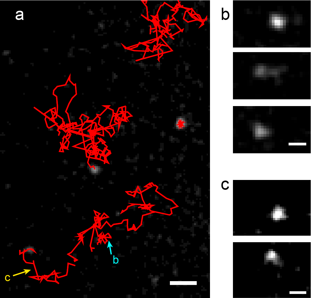
Figure 2a shows an example of trajectories obtained in a 10-s window, overlaid on the last frame. Often, long jumps are observed in the particle trajectories as seen in the examples in Figs. 2b and c. These jumps suggest the C2A molecules detach from the surface and readsorb after brief excursions into the liquid bulk. The motion in the bulk is much faster than diffusion on the viscous membrane and jumps are thus expected to occur instantaneously for all practical purposes. For the C2A domain, the diffusion coefficient in the lipid bilayer is of the order of 2 , but in liquid the diffusion coefficient is estimated to be 100 times higher ziemba2013lateral . As a consequence, when a molecule performs a jump through the bulk it can sometimes be observed at reduced intensity in both the old and new locations within the same imaging frame, as seen in Figure 2b.
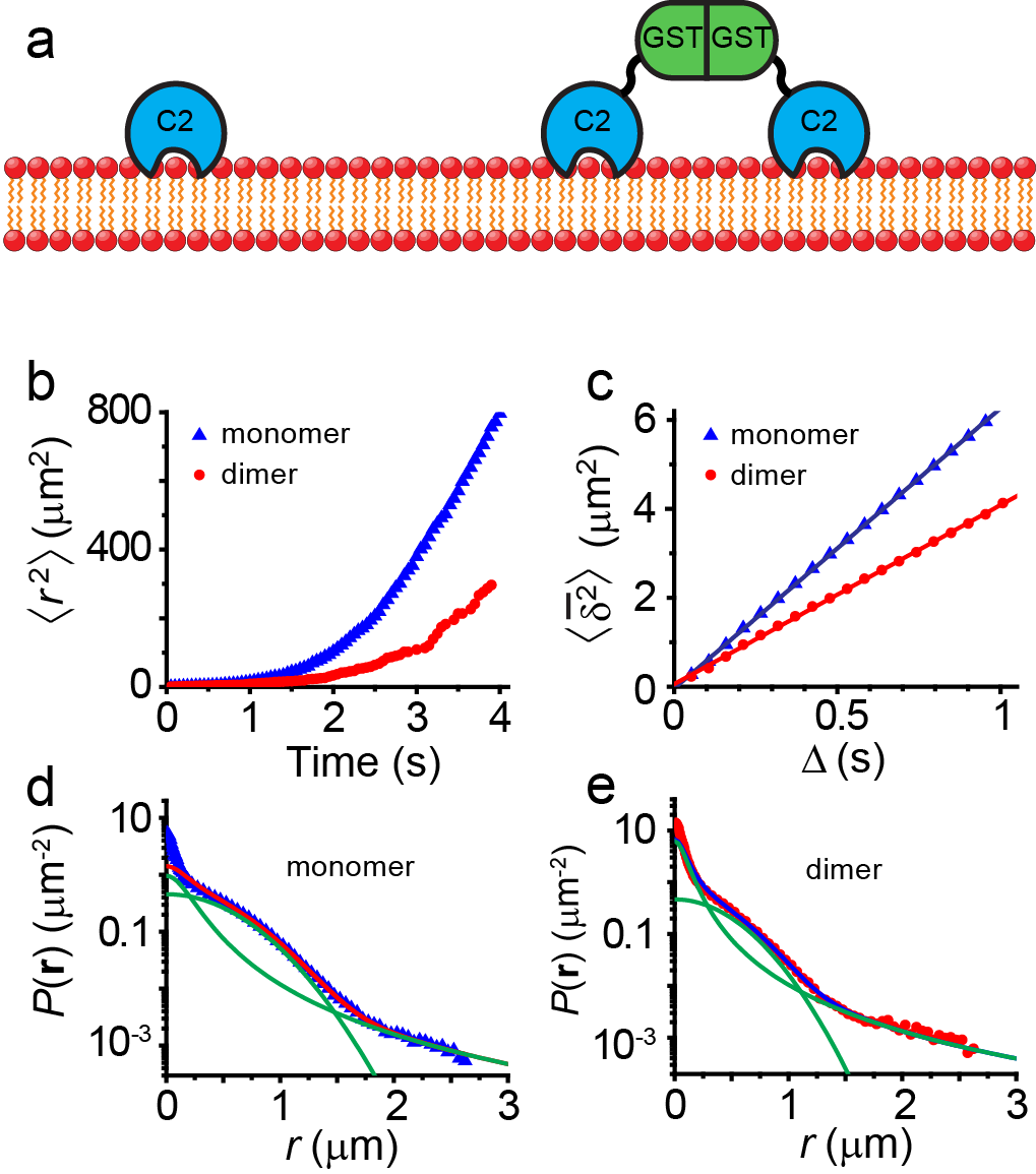
In order to study the effect of the dissociation constant, we also employed a C2A construct fused to a non-membrane interacting glutathione S-transferase (GST), which has a strong tendency to dimerize (Figure 3a). The GST-C2A dimer forms two independent interactions with the membrane and will consequently have a slower dissociation rate than C2A monomer, providing a good comparison for validating our superdiffusion predictions. Additionally, GST-C2A dimer has a higher viscous drag coefficient and, in turn, its diffusion coefficient on the membrane surface is reduced to nearly half knight2010BJ .
We collected 14,000 C2A and 3,600 GST-C2A mobile trajectories. Immobile fluorophores that did not exhibit any apparent diffusive motion were excluded from the analysis. The ensemble-averaged MSD of C2A monomers and dimer-forming GST-C2A are shown in Figure 3b. A deviation from a linear MSD is evident in the figure, showing superdiffusive behaviour. Further, the onset of superdiffusion for GST-C2A occurs at a later stage.
The time-averaged MSD is often used in the analysis of individual trajectories. Throughout this manuscript we will denote the ensemble average of an observable with brackets and the time average with an overbar . For a trajectory with time points,
| (2) |
where is the time interval between consecutive measurements and . This approach is especially useful when a limited number of trajectories is available, as usually occurs in single-molecule studies. Figure 3c shows the time-averaged MSD after it is additionally averaged over all the trajectories. GST-C2A exhibits the expected slower diffusion rate than C2A, based on the MSD slope. As mentioned above, for ergodic processes, the temporal and ensemble averages coincide in the long time limit, . However, the ergodic hypothesis breaks down for C2A molecules. In contrast to the ensemble-averaged MSD, the time-averaged MSD is linear in lag time
| (3) |
Thus, an observer analysing time-averages would reach the misleading conclusion that the diffusion behaviour is not anomalous.
The distribution of displacements at is shown in Figs. 3d and e for C2A and GST-C2A, respectively. The distribution exhibits two different characteristic regimes: a central part up to a distance and a long tail. This behaviour can be understood from the scaling properties of bulk-mediated diffusion as discussed by Bychuk and O’Shaughnessy Bychuk1995 . Once a molecule dissociates from the surface, it performs a three-dimensional random walk until it returns. In the asymptotic limit, the first return time distribution scales as . For any given return time, the surface distance between the dissociation and return points has a Gaussian distribution . Therefore, the distribution of jump lengths is , as observed in Figs. 3d and e for long distances.
The theoretical probability density function of jump lengths can be found using the image method redner . The distance of first return to the surface are governed by , that is a two-dimensional Cauchy distribution. At short times, the probability that the particle performs more than a single jump is small. If we neglect the distance covered by surface diffusion within time intervals at which the particle undergoes a bulk excursion, the motion at each short interval is either by surface diffusion or via a jump. We can then approximate the distribution of displacements at short times by
| (4) |
where is the probability that the particle hops within the given time and surface diffusion yields . A least-square fitting of the distribution of displacements (Figs. 3d and e) to this propagator yields for C2A monomers and for GST-C2A. The parameter is found to be 0.24 and 0.12 for C2A and GST-C2A, respectively.
The distribution of displacements for longer times involves both a random number of jumps, each having a Cauchy distribution, and the Brownian motion on the surface. Chechkin et al. derived the full solution for the propagator of bulk-mediated diffusion Chechkin2012 . For the case when and neglecting long distance corrections, the distribution of displacements is given by the Cauchy propagator, in agreement with scaling arguments Bychuk1995 ,
| (5) |
When the particles also diffuse on the surface, i.e. the probability density of the displacements is given by the convolution of equation (5) with a normal distribution. Even though the full solution for long times is complicated, the tail of this distribution for large distances still scales as . Due to this asymptotic behaviour, the exact distribution has similar properties to the Cauchy distribution.
Numerical simulations: diffusion in the presence of bulk excursions
In order to verify the model of surface diffusion in the presence of bulk excursions we analyse numerical simulations of the process diagrammed in Figure 1. Molecules perform a two-dimensional random walk, but at random times they jump due to a hypothetical bulk excursion. The surface residence times are assumed to be independent and identically distributed exponential random variables and the jumps are modelled according to the first return time to the surface given simple diffusion in a three-dimensional medium. These simulations are analysed in the same way as with experimental observations of the motion of membrane-targeting C2 domains on supported membranes. 500 realizations were simulated off-lattice with a surface diffusion coefficient and a dissociation coefficient . The chosen parameters do not intend to capture the real protein properties, but to simply test theoretical predictions without the effects of experimental noise. The displacements for two-dimensional diffusion are drawn from a Gaussian distribution with variance and the return times from bulk excursions are drawn from a distribution redner . Then the jump distances are drawn from a Gaussian distribution with variance .
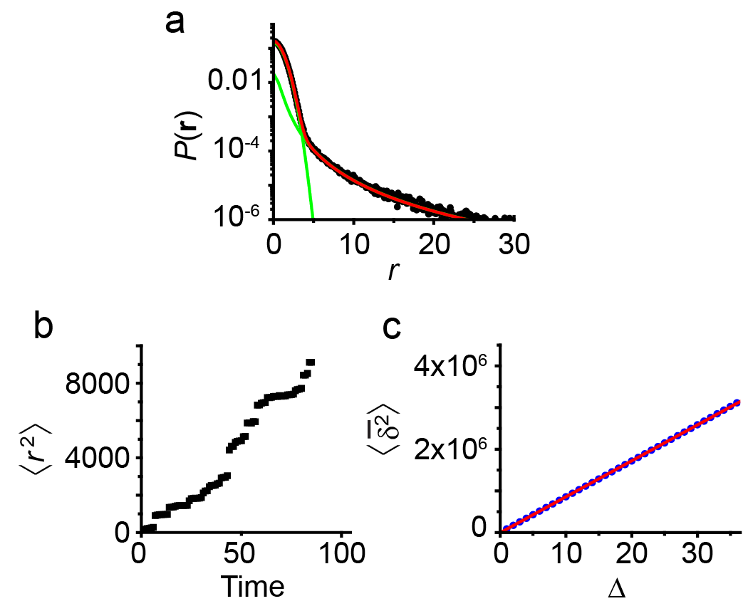
The distribution of displacements for the numerical simulations is shown in Figure 4a. As expected, there are two regimes: a central Gaussian part due to the two-dimensional diffusion on the membrane between bulk excursions, and a heavy tail that arises from the long distance behaviour of bulk excursions. The distributions for short times can again be modelled with a propagator that includes contributions from Gaussian surface diffusion and a Cauchy distribution due to bulk excursions. By fitting to equation (4), it is found (the value employed in the simulations is ) and .
MSD analysis
The dynamics of a particle with a Cauchy propagator are particularly interesting because the theoretical variance of the displacements diverges,
| (6) |
In practice, a diverging second moment implies that there is a non-negligible probability for the occurrence of extremely long jumps and this phenomenon has direct implications in the measured MSD. Figure 4b shows the ensemble-averaged MSD computed from the numerical simulations. The MSD increases in a superlinear fashion, i.e. by employing equation (1), we have , which implies the process is superdiffusive.
Let us now analyse the unexpected MSD behaviour, starting from the time-averaged MSD of individual trajectories. We can show that the time-averaged MSD is linear in lag time for any random walk with independent increments , such that when . From the definition of the time averaged MSD (equation (2)) froemberg2013random ,
| (7) | |||||
| (8) | |||||
| (9) |
where we have used the approximation that , we omitted the term because it is zero on average, and again we have used the parameter . Therefore we see that for symmetric random walks with independent increments, the time-averaged MSD is linear as observed in Figs. 3c and 4c.
Although the time-averaged MSD for individual trajectories is linear, the ensemble averaged MSD is not. We can understand the superdiffusive behaviour by assuming we can define the motion in terms of two independent processes , where is a two-dimensional Brownian motion and is a Lévy process with a probability density defined by equation (5). Then the MSD is . The first term is linear in time but the second term has a superdiffusive nature Bychuk1995 ; valiullin1997levy ; Chechkin2012 .
Discussion
The propagator for surface diffusion in the presence of bulk-mediated jumps (equation (4)) depends on the surface diffusion coefficient and the parameter that reflects the transition between the surface and the bulk phase. Namely, , where is the mean desorption time and is a dimensional factor. Bulk-mediated diffusion thus predicts , in agreement with the values we find for C2A and GST-C2A.
The surface motion of these membrane-targeting domains is well described by Lévy flights, a random walk where the step displacements have a heavy-tailed distribution. The heavy tail arises from the dissociation of molecules from the membrane, which then perform a three-dimensional random walk until they reach the surface again at another location. The process involves the first return to a surface and it converges to a power law according to the Sparre-Andersen theorem redner . This type of Lévy flight dynamics is fundamentally different from Lévy walks induced by molecular motors in the cytoplasm because periods of active motion require an energy input, typically in the form of ATP hydrolysis, while bulk excursions occur spontaneously.
One of the most interesting effects of the observed bulk-mediated diffusion statistics is that the ensemble-averaged MSD exhibit superdiffusive behaviour, whereas the temporal averages suggest normal diffusion. This nonergodic behaviour is similar to that of continuous time random walks (CTRW) where the sojourn time distribution between steps has a probability distribution that is heavy-tailed. Also in the CTRW, and , albeit the CTRW is subdiffusive with . The difference in the behaviour of temporal and ensemble averages is the key signature of weak ergodicity breaking in the process margolin2006nonergodicity .
To date, different groups have observed normal diffusion for membrane proteins in supported lipid bilayers, which appear to contradict our findings tamm1988lateral ; gambin2006lateral ; ramadurai2010influence ; ziemba2012assembly . There are several reasons for this apparent discrepancy. Single-particle tracking in lipid bilayers often focuses on time-averaged MSD, which does not show any non-linearity in lag time. Thus it would be reasonable to reach the conclusion that diffusion is not anomalous. Furthermore, anomalous diffusion in supported bilayers is known to develop as a result of packing and crowding. These mechanisms are modelled by a fractional Langevin equation, which is ergodic in nature, with anomalies that show up in the time averages. The distribution of displacements has also been previously reported as exhibiting Gaussian behaviour. Here we report on the motion of surface-bound membrane domains that exhibit desorption from the membrane within the experimental observation time. The behaviour of transmembrane proteins or lipids is very different because the free energy barrier for release from the membrane is too high to be observed within the constrains of experimental observations tamm1988lateral ; gambin2006lateral ; ramadurai2010influence . Previous works dealing with membrane-targeting domains such as C2 have generally been limited to short displacements in order to exclude the effect of long bulk-mediated jumps in diffusion measurements knight2010BJ ; ziemba2012assembly .
What are the biological implications of surface superdiffusion for peripheral membrane proteins? Search processes are ubiquitous in cell biology and it is feasible to assume that evolution has optimized search parameters. For signalling molecules delivered to the plasma membrane during a specific stimulus, the target molecule is often scarce in a sea of other lipids and proteins. Thus we can envision that if a molecule does not find its target in a given time, it becomes more efficient to start searching at a different location. Is it appropriate then to assume Lévy flights yield the optimal search for sparse targets when compared to Brownian motion? For one-dimensional intermittent processes that switch between Brownian motion and ballistic relocation phases, it has been shown that the search process is significantly more efficient when relocation times are power-law distributed, resulting in a Lévy walk lomholt2008levy . Notably, when Lévy dynamics are employed, the search is less sensitive to the target density lomholt2008levy . In general, the optimal strategy depends on the average target distance from the starting point Palyulin25022014 . However, blind searches inside a living cell are very different from a search in an unobstructed environment. Several aspects provide additional complexities in the plasma membrane, in particular krapf2015reviewCTM . Experimental measurements show that the plasma membrane is compartmentalized in a way that proteins and lipids have the tendency to remain transiently confined within small regions ritchie2003fence . Further, membrane proteins typically exhibit subdiffusion with anti-persistent increments where molecules drift towards the locations that they visited in the past. While this subdiffusive behaviour provides the opportunity for a thorough and compact search, it is definitely not the optimum situation to find sparse targets. A superdiffusive Lévy flight provides a mechanism to overcome the effects of anti-persistent correlated subdiffusive motion. Thus, we expect Lévy flight dynamics to often outperform a Brownian search.
The obstruction to the diffusion of membrane molecules has two different sources, both of them causing anti-persistent correlations in the random walk. On one hand, obstacles can be introduced by immobile transmembrane proteins which affect all lipids and membrane proteins. On the other hand, a more severe obstruction can be caused by cytoskeleton components that may not be in direct contact with the plasma membrane andrews2008actin . The effect of these barriers is not equal for all membrane proteins. Proteins that have large intracellular complexes are blocked much more efficiently than small molecules. In cases where a large signalling molecule adheres to the membrane via phospholipid-binding domains, bulk excursions allow for the exploration of larger areas. Otherwise, the molecule would remain confined for long times within cytoskeleton-formed corrals, even when no substrate target is found within this region.
In summary, we have observed the nonergodic, superdiffusive motion of membrane-targeting peptide domains in supported lipid bilayers. The motion is well-described by Lévy flights with jumps that have a heavy-tail distribution. The long jumps are caused by excursions into the liquid bulk. After dissociating from the membrane, the molecules diffuse in three dimensions until they reach the membrane again and bind at a new location. Diffusion in the liquid bulk is much faster than diffusion in the membrane, therefore we do not consider the delay time between dissociation and readsorption. The surface distances covered by jumps have a Cauchy distribution, which is responsible for the heavy tail in the superdiffusive Lévy flights. Model membranes provide an elegant system to study the effect of superdiffusive Lévy flights because they are not subjected to the interactions with other cell components that would mask its experimental observation. However, hopping was already observed on the surface of live cells Yasui2014 and we foresee these processes have broad physiological relevance in the surface diffusion of signalling molecules.
Methods
Imaging buffer
Imaging and rinsing during the preparation steps was performed in an imaging buffer consisting of 50 mM HEPES, 75 mM NaCl, 1 mM MgCl2, 2 mM tris(2-carboxyethyl)phosphine (TCEP), 200 M CaCl2. CaCl2 is necessary for C2 domain binding to the reconstituted membrane.
Preparation of phospholipid vesicles
Phospholipids were purchased from Avanti Polar Lipids (Alabaster, AL). Chloroform-suspended 18:1 (-Cis) PC (DOPC) and 18:1 PS (DOPS) were mixed at a ratio of 3:1. The phospholipid mixture was vacuum dried overnight and resuspended in imaging buffer to a final concentration of 3 mM followed by probe sonication to form sonicated unilamellar vesicles (SUVs).
Preparation of coveslips and supported lipid bilayers
Glass coverslips were cleaned by sonication in a detergent solution followed by soaking in 1M KOH. The coverslips were rinsed extensively in Milli-Q water and blown dry with a stream of nitrogen gas. Then, the coverslips were treated with an oxygen plasma. Immediately after the plasma cleaning, a perfusion chamber (CoverWell, Grace Bio-Labs) was adhered to the coverslip. In order to deposit the lipid bilayers a solution of SUVs (1.5-mM lipid) composed of phosphatidylcholine (PC) and phosphatidylserine (PS) at a 3:1 ratio in 1M NaCl and imaging buffer was introduced into the perfusion chamber and incubated for one hour at 4∘C. Refrigeration minimizes lipid oxidation. The surface was then rinsed with imaging buffer multiple times prior to addition of protein sample.
C2A and GST-C2A expression and purification
An expression plasmid containing the gene for a GST-ybbR-Synaptotagmin 7 (Syt7) C2A domain fusion protein was transformed into E. coli BL21-CodonPlus(DE3) competent cells. The ybbR segment provides a site for Sfp-catalysed fluorophore labelling ybbr . Cells were grown at 37∘C to an OD of 0.6 and then induced to express protein with 0.5 mM IPTG at room temperature for 6 hours. The harvested cells were lysed at 18,000 lb/in2 in a microfluidizer in a buffer containing 50 mM Tris pH 7.5, 400 mM NaCl and centrifuged at 17,000 rpm in a Sorval SS-34 rotor. The clarified lysate was loaded onto a 5-ml GSTrap FF column (GE Healthcare LifeSciences, Pittsburgh, PA) followed by gradient elution with 50 mM Tris, pH 8.0, 100 mM NaCl, and 10 mM glutathione. Fractions containing protein were pooled and diluted to reduce the salt to less than 0.1 M prior to loading onto a HiTrap Q HP column (GE Healthcare LifeSciences, Pittsburgh, PA) and eluting with a linear gradient to 1 M NaCl in 25 mM Tris, pH 8.5, 20(vol/vol) glycerol, and 0.02(wt/vol) NaN3. A portion of the construct was subjected to thrombin cleavage and then separated using a Superdex 200 gel filtration column (GE Healthcare LifeSciences, Pittsburgh, PA) equilibrated in 50 mM Tris, pH 7.5 and 100mM NaCl to yield a ybbr-Syt7 C2A construct.
Protein labeling
10 mM CoASH (New England Biolabs, Ipswich, MA) in 400 mM Tris, pH 7.5 was mixed with 10 mM ATTO-565 maleimide (ATTO-TEC, Siegen, Germany) in dimethylformamide and incubated at 30∘C overnight to form ATTO-565 CoA, then quenched with 5 mM DTT, 10 mM Tris pH 7.5. 10 M GST-ybbr-Syt7 C2A and ybbr-Syt7 C2A were labelled with the ATTO-565 via SFP synthase (4-phosphopantetheinyl transferase). Each reaction contained 50 mM tris 7.5, 10 mM MgCl2, 40 mM NaCl, 20 M ATTO-565 CoA and 1 M SFP synthase. Reactions were incubated at room temperature for 30 minutes, then placed at 4∘C overnight. Samples were dialysed against 1 L of 50 mM HEPES, pH 7.0, 75 mM NaCl, 4 mM MgCl2 and 5 glycerol overnight at 4∘C then concentrated to 10 M.
Imaging
All images were acquired using an objective-type total internal reflection fluorescence microscope (TIRFM). The microscope was home-built around an Olympus IX71 body weigel2011PNAS ; weigel2013pnas with a 561 nm laser line as excitation source. A back-illuminated electron-multiplied charge coupled device (EMCCD) camera (Andor iXon DU-888) liquid-cooled to -85∘C, with an electronic gain of 300 was used. In order to maintain constant focus during the whole imaging time we employed an autofocus system (CRISP, Applied Scientific Instrumentation, Eugene, OR) in combination with a piezoelectric stage (Z-100, Mad City Labs, Madison, WI). Videos were acquired at a frame rate of 20 frames/s.
Image processing and single-particle tracking
Images were acquired using Andor IQ 2.3 software and saved as 16-bit tiff files. Then the images were filtered using a Gaussian kernel with a standard deviation of 1.0 pixel in ImageJ. Single-particle tracking of Atto-C2 and Atto-GST-C2 was performed in MATLAB using the U-track algorithm developed by Jaqaman et al. jaqaman2008U-track under thorough manual inspection of detection and tracking.
References
- (1) Hurley JH (2006) Membrane binding domains. Biochim. Biophys. Acta 1761:805 – 811.
- (2) Lemmon MA (2008) Membrane recognition by phospholipid-binding domains. Nat. Rev. Mol. Cell Biol. 9:99–111.
- (3) Lemmon M, Ferguson K (2000) Signal-dependent membrane targeting by pleckstrin homology (PH) domains. Biochem. J. 350:1–18.
- (4) Cho W, Stahelin RV (2006) Membrane binding and subcellular targeting of C2 domains. Biochim. Biophys. Acta 1761:838 – 849.
- (5) Letunic I, Doerks T, Bork P (2012) SMART 7: recent updates to the protein domain annotation resource. Nucleic Acids Res. 40:D302–D305.
- (6) Knight JD, Falke JJ (2009) Single-molecule fluorescence studies of a PH domain: new insights into the membrane docking reaction. Biophys. J. 96:566–582.
- (7) Yasui M, Matsuoka S, Ueda M (2014) PTEN hopping on the cell membrane is regulated via a positively-charged C2 domain. PLoS Comput. Biol. 10:e1003817.
- (8) Barkai E, Garini Y, Metzler R (2012) Strange kinetics of single molecules in living cells. Phys. Today 65:29–35.
- (9) Höfling F, Franosch T (2013) Anomalous transport in the crowded world of biological cells. Rep. Prog. Phys. 76:046602.
- (10) Metzler R, Jeon JH, Cherstvy AG, Barkai E (2014) Anomalous diffusion models and their properties: non-stationarity, non-ergodicity, and ageing at the centenary of single particle tracking. Phys. Chem. Chem. Phys. 16:24128–24164.
- (11) Krapf D (2015) Mechanisms underlying anomalous diffusion in the plasma membrane. Curr. Top. Membr. 75:167–207.
- (12) Golding I, Cox EC (2006) Physical nature of bacterial cytoplasm. Phys. Rev. Lett. 96:098102.
- (13) Tolić-Norrelykke IM, Munteanu EL, Thon G, Oddershede L, Berg-Sorensen K (2004) Anomalous diffusion in living yeast cells. Phys. Rev. Lett. 93:078102.
- (14) Jeon JH et al. (2011) In vivo anomalous diffusion and weak ergodicity breaking of lipid granules. Phys. Rev. Lett. 106:048103.
- (15) Bronstein I et al. (2009) Transient anomalous diffusion of telomeres in the nucleus of mammalian cells. Phys. Rev. Lett. 103:018102.
- (16) Weigel AV, Tamkun MM, Krapf D (2013) Quantifying the dynamic interactions between a clathrin-coated pit and cargo molecules. Proc. Natl. Acad. Sci. U.S.A. 110:E4591–E4600.
- (17) Heinemann F, Vogel SK, Schwille P (2013) Lateral membrane diffusion modulated by a minimal actin cortex. Biophys. J. 104:1465–1475.
- (18) Torreno-Pina JA et al. (2014) Enhanced receptor–clathrin interactions induced by N-glycan–mediated membrane micropatterning. Proc. Natl. Acad. Sci. U.S.A. 111:11037–11042.
- (19) Banks DS, Fradin C (2005) Anomalous diffusion of proteins due to molecular crowding. Biophys. J. 89:2960–2971.
- (20) Szymanski J, Weiss M (2009) Elucidating the origin of anomalous diffusion in crowded fluids. Phys. Rev. Lett. 103:038102.
- (21) Horton MR, Höfling F, Rädler JO, Franosch T (2010) Development of anomalous diffusion among crowding proteins. Soft Matter 6:2648–2656.
- (22) Jeon JH, Monne HMS, Javanainen M, Metzler R (2012) Anomalous diffusion of phospholipids and cholesterols in a lipid bilayer and its origins. Phys. Rev. Lett. 109:188103.
- (23) Bursac P et al. (2007) Cytoskeleton dynamics: fluctuations within the network. Biochem. Biophys. Res. Comm. 355:324–330.
- (24) Kahana A, Kenan G, Feingold M, Elbaum M, Granek R (2008) Active transport on disordered microtubule networks: The generalized random velocity model. Phys. Rev. E 78:051912.
- (25) Bruno L, Levi V, Brunstein M, Despósito M (2009) Transition to superdiffusive behavior in intracellular actin-based transport mediated by molecular motors. Phys. Rev. E 80:011912.
- (26) Akimoto T (2012) Distributional response to biases in deterministic superdiffusion. Phys. Rev. Lett. 108:164101.
- (27) Skaug MJ, Mabry J, Schwartz DK (2013) Intermittent molecular hopping at the solid-liquid interface. Phys. Rev. Lett. 110:256101.
- (28) Yu C, Guan J, Chen K, Bae SC, Granick S (2013) Single-molecule observation of long jumps in polymer adsorption. ACS Nano 7:9735–9742.
- (29) Bouchaud JP (1992) Weak ergodicity breaking and aging in disordered systems. J. Physique I 2:1705–1713.
- (30) Bel G, Barkai E (2005) Weak ergodicity breaking in the continuous-time random walk. Phys. Rev. Lett. 94:240602.
- (31) Krapf D (2013) Nonergodicity in nanoscale electrodes. Phys. Chem. Chem. Phys. 15:459–465.
- (32) Weigel AV, Simon B, Tamkun MM, Krapf D (2011) Ergodic and nonergodic processes coexist in the plasma membrane as observed by single-molecule tracking. Proc. Natl. Acad. Sci. U.S.A. 108:6438–6443.
- (33) Manzo C et al. (2015) Weak ergodicity breaking of receptor motion in living cells stemming from random diffusivity. Phys. Rev. X 5:011021.
- (34) Tabei SA et al. (2013) Intracellular transport of insulin granules is a subordinated random walk. Proc. Natl. Acad. Sci. U.S.A. 110:4911–4916.
- (35) Brokmann X et al. (2003) Statistical aging and nonergodicity in the fluorescence of single nanocrystals. Phys. Rev. Lett. 90:120601.
- (36) Stefani FD, Hoogenboom JP, Barkai E (2009) Beyond quantum jumps: blinking nanoscale light emitters. Phys. Today 62:34–39.
- (37) Sugita S, Shin OH, Han W, Lao Y, Südhof TC (2002) Synaptotagmins form a hierarchy of exocytotic Ca2+ sensors with distinct Ca2+ affinities. EMBO J. 21:270–280.
- (38) Ziemba BP, Falke JJ (2013) Lateral diffusion of peripheral membrane proteins on supported lipid bilayers is controlled by the additive frictional drags of (1) bound lipids and (2) protein domains penetrating into the bilayer hydrocarbon core. Chem. Phys. Lipids 172:67–77.
- (39) Knight JD, Lerner MG, Marcano-Velázquez JG, Pastor RW, Falke JJ (2010) Single molecule diffusion of membrane-bound proteins: window into lipid contacts and bilayer dynamics. Biophys. J. 99:2879–2887.
- (40) Bychuk OV, O’Shaughnessy B (1995) Anomalous diffusion at liquid surfaces. Phys. Rev. Lett. 74:1795.
- (41) Redner S (2001) A guide to first-passage processes. (Cambridge University Press).
- (42) Chechkin AV, Zaid IM, Lomholt MA, Sokolov IM, Metzler R (2012) Bulk-mediated diffusion on a planar surface: Full solution. Phys. Rev. E 86:041101.
- (43) Froemberg D, Barkai E (2013) Random time averaged diffusivities for lévy walks. Eur. Phys. J. 86:1–13.
- (44) Valiullin R, Kimmich R, Fatkullin N (1997) Lévy walks of strong adsorbates on surfaces: Computer simulation and spin-lattice relaxation. Phys. Rev. E 56:4371.
- (45) Margolin G, Barkai E (2006) Nonergodicity of a time series obeying Lévy statistics. J. Statist. Phys. 122:137–167.
- (46) Tamm LK (1988) Lateral diffusion and fluorescence microscope studies on a monoclonal antibody specifically bound to supported phospholipid bilayers. Biochem. 27:1450–1457.
- (47) Gambin Y et al. (2006) Lateral mobility of proteins in liquid membranes revisited. Proc. Natl. Acad. Sci. U.S.A. 103:2098–2102.
- (48) Ramadurai S et al. (2010) Influence of hydrophobic mismatch and amino acid composition on the lateral diffusion of transmembrane peptides. Biophys. J. 99:1447–1454.
- (49) Ziemba BP, Knight JD, Falke JJ (2012) Assembly of membrane-bound protein complexes: detection and analysis by single molecule diffusion. Biochem. 51:1638–1647.
- (50) Lomholt MA, Tal K, Metzler R, Joseph K (2008) Lévy strategies in intermittent search processes are advantageous. Proc. Natl. Acad. Sci. U. S. A. 105:11055–11059.
- (51) Palyulin VV, Chechkin AV, Metzler R (2014) Lévy flights do not always optimize random blind search for sparse targets. Proc. Natl. Acad. Sci. U.S.A. 111:2931–2936.
- (52) Ritchie K, Iino R, Fujiwara T, Murase K, Kusumi A (2003) The fence and picket structure of the plasma membrane of live cells as revealed by single molecule techniques (Review). Mol. Membr. Biol. 20:13–18.
- (53) Andrews NL et al. (2008) Actin restricts FcRI diffusion and facilitates antigen-induced receptor immobilization. Nat. Cell Biol. 10:955–963.
- (54) Yin J, Lin AJ, Golan DE, Walsh CT (2006) Site-specific protein labeling by Sfp phosphopantetheinyl transferase. Nat. Protocols 1:280.
- (55) Jaqaman K et al. (2008) Robust single-particle tracking in live-cell time-lapse sequences. Nat. Methods 5:695–702.
Acknowledgments
We thank Jeff Knight for kindly supplying the plasmids for C2A and C2A-GST and for useful discussions. We also thank Kassi Prochazka for help in the initial part of the project and for her help in producing Figure 1. This work was supported by the National Science Foundation under grant 1401432 (to DK) and by the National Institutes of Health under grant R21AI111588 (to OBP).
Author contributions statement
G.C., O.B.P., and D.K. conceived the experiments; O.B.P. and D.K.supervised the project; G.C., K.N. and B.W.S. conducted the experiments; D.K. designed the analytical model and performed numerical simulations; K.N. and D.K. analysed the results; D.K. wrote the first draft; G.C., K.N., O.B.P.,and D.K. reviewed the manuscript.
Competing financial interests
The authors declare no competing financial interests.