Spectroscopic determination of the atomic f-electron symmetry underlying hidden order in URu2Si2
Abstract
The low temperature hidden order state of URu2Si2 has long been a subject of intense speculation, and is thought to represent an as yet undetermined many-body quantum state not realized by other known materials. Here, X-ray absorption spectroscopy (XAS) and high resolution resonant inelastic X-ray scattering (RIXS) are used to observe electronic excitation spectra of URu2Si2, as a means to identify the degrees of freedom available to constitute the hidden order wavefunction. Excitations are shown to have symmetries that derive from a correlated atomic multiplet basis that is modified by itinerancy. The features, amplitude and temperature dependence of linear dichroism are in agreement with ground states that closely resemble the doublet crystal field state of uranium.
The low temperature hidden order (HO) phase of URu2Si2 has been a mystery for more than 25 years, and is widely anticipated to represent a novel many-body quantum state. When cooling through THO=17.5K the material undergoes a second order phase transition, with a large loss of entropy that cannot be immediately explained by observed changes in the electronic structure entropyChange ; entropyUnexplained ; AmitsukaRXS ; WalkerRXS . Pinpointing the microscopic cause of this entropy change is challenging because basic properties of the atomic scale wavefunction are not decisively known. Experiments differ on whether the uranium valence state is closer to () inelastNeutron1 ; inelastNeutron2 or () EELS2010 . Proposed models have considered a wide range of local firstCEFmodel ; Gamma5doublet1 ; Gamma5doublet2 ; Gamma5doublet3 ; Gamma3early ; local2 ; local3 ; Gamma3review ; KotliarNatPhys ; FlouquetSimpleAnswer ; local6 ; HastaticNature ; HastaticReview and itinerant nonlocal1 ; nonlocal2 ; mydoshBigTheoryPRB ; nonlocal3 ; nonlocal3p5 ; nonlocal4 ; nonlocal5 ; nematicRank5 ; DasSOdensityWave low energy state bases for electrons, and explored many exciting possibilities for the “hidden” quantum state. Here, high resolution (35meV) resonant inelastic X-ray scattering (RIXS) and X-ray absorption spectroscopy (XAS) are used to measure fundamental excitations created by resonance with the uranium 5d core level (O-edge), to identify what electronic degrees of freedom are relevant for effective models of hidden order, and what degrees of freedom are energetically gapped out.
Measurements were performed on both a pristine crystalline surface cleaved in ultra high vacuum, and a cleaved surface that was oxidized by exposure to air at room temperature, promoting U4+ valence. The dominant spectral features observed from both surfaces are shown to derive from the excitations of a freestanding atom, revealing that atomically correlated Hund’s rule interactions play a key role in determining the electronic degrees of freedom that can contribute to the hidden order state. However, some low energy excitations of the pristine surface are found to be extremely short lived, implying that the symmetries they represent are not strictly eliminated from the hidden order ground state. Linear dichroism in the XAS spectrum is consistent with the crystalline electric field (CEF) doublet state , but inconsistent with CEF singlet ground states that have been predicted as the basis of hidden order (e.g. , , ).
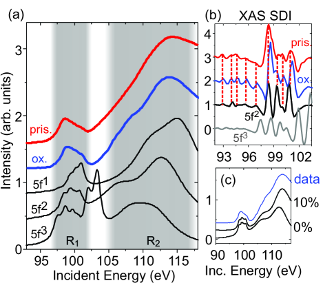
Measuring XAS at the O-edge reveals a fingerprint of how the valence electronic structure projects onto coherent core hole states deGrootInvisible ; 5fdichroismME . Colored curves in Fig. 1(a) show XAS measured on the pristine and air exposed samples within the hidden order state (T=10K). Intensity is divided between two principle regions labeled (97-102eV) and (104-118eV), split mostly by spin exchange interactions. Comparing the XAS measurements with black curves representing atomic multiplet (AM) calculations for isolated , , and uranium atoms shows reasonable correspondence with the multiplet structure, but other than the and features themselves, there are no prominent well resolved peaks that would provide a basis for detailed comparison. The simulation is relatively incompatible, because its intensity maximum at 103eV coincides with a local minimum of the experimental curve. Most analysis of XAS in this paper will focus on the region, because the Fano effect identified in Ref. AugerFano significantly influences total electron yield (TEY) XAS measurements on shorter-lived resonance states in the region (see Fig. 1(c)).
The technique of second derivative imaging (SDI) is used in Fig. 1(b) to more sharply resolve component features within the XAS spectra. The SDI spectrum of the air exposed sample reveals nine features near (see blue curve), including highly reproducible SM fine structure below the energy region. These peaks have a one to one correspondence with features in the calculation, as might be expected for an oxidized surface with U4+ nominal valence. More remarkably, features of the pristine sample also have a one-to-one correspondence with SDI local maxima seen from the air exposed sample (see red drop-lines), although the feature at 98.5 eV appears to be split into two components. SDI features of the pristine surface are broader and slightly lower in energy than those of the air-cleaved surface.
The correspondence of XAS with a multiplet strongly suggests that the atomic scale electronic degrees of freedom available for incorporation in the hidden order ground state come largely from the low energy states of a manifold. However, it is also possible that states may be underrepresented in the XAS spectrum if they are relatively itinerant and persist on the core hole site for too short a time to yield sharp excitation features. Technical concerns for relating O-edge XAS to valence estimates from other techniques are discussed in the online Supplemental Material (SM) SM .
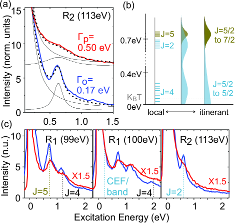
Performing RIXS at the same incident photon energies reveals low energy excitations that are left behind when the O-edge core hole decays, and provides much greater bulk sensitivity than XAS SM . The RIXS scattering process involves the short lived excitation of a core electron to the valence level, followed by another electronic transition that fills the core hole and leaves behind a low energy excitation ButorinReview ; KotaniRIXSreview ; AmentRIXSReview ; WrayFrontiers . Depending on how correlated or itinerant the electronic system is, electronic excitations on uranium may resemble band transitions between itinerant single electron states, which have J=5/2 or 7/2 angular momentum, or may be dominated by excitations that change the coherent relationship between locally entangled electrons (see diagram in Fig. 3(b)). Regardless of the entangled or itinerant nature of low energy electrons, the angular momentum coupling between a core hole and the valence electrons has an extremely large 15eV energy scale, leading to coherent resonance states with strong entanglement between valence electrons and the core hole, and qualitatively different spectra from previous studies of and core hole resonance EELS2010 ; M5M4 ; SM .
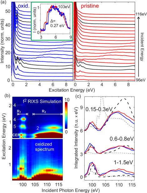
The incident energy dependence of RIXS spectra for the pristine and air exposed samples is shown in Fig. 2(a), with scattering intensity plotted in normalized units of the Si L-edge fluorescence amplitude. Both profiles show the same qualitative features, but there are numerous quantitative differences. In each case, almost all intensity at energy loss is found in a broad, linearly dispersive feature termed an emission line, which resonates at incident photon energies SM . Corresponding features of the emission line are shifted by between the pristine and air exposed samples (see Fig. 2(a) inset). Emission lines disperse with a slope of 1, and are thought to approximately represent the density of single particle electronic states below the Fermi level that can transition to fill the core hole JinghuaCKalpha ; JinghuaReview ; HancockRIXSmetal . In this picture, oxidation lowers the energy of ligand band structure (shifting features to larger energy loss) by reducing the surrounding charge density. No feature at the leading edge of the emission line is seen to be lost as a consequence of oxidation, suggesting that oxidation has little effect on the occupied electronic symmetries.
The relatively sharp air exposed RIXS spectrum is compared with a AM model in Fig. 2(b), and again establishes a one to one correspondence of features, with details very similar to the RIXS spectrum of uranium oxide (UO2) ButorinReview . The total angular momentum (J) quantum number of atomic multiplet excitations is labeled on the simulation. The CEF excitations will manifest as band excitations in an itinerant picture, and have therefore been set to 0.2 eV to correspond with the approximate energy scale expected for low energy uranium band transitions (between J=5/2 bands). The prominent 0.6 eV excitation visible at has J=2 angular momentum, and is unrelated to the nearly degenerate J=5 feature seen at . Comparing the incident energy dependence of the three lowest energy features shows that each can be qualitatively explained by a atomic multiplet model (thick dashed line in Fig. 2(c)), and has no distinct correspondence with a model (thin dashed line).
Excitations from 0.15 to 0.3 eV can account for most of the resonant intensity that the model attributes to CEF excitations (see Fig. 2(c), top curve). These CEF transitions are created by changing the coherent relationship between two electrons in J=5/2 single particle states on the same atom, and in an itinerant picture the electrons will rapidly delocalize into excitations between the J=5/2 bands that intersect the Fermi level. RIXS curves show no sharp features in this energy range (Fig. 3(c)), and neutron scattering does not reveal CEF excitations ButchNeutron , so one may conclude that CEF transitions are observed in the measurement, but are too short lived to be considered stable collective modes. The intensity at may be best interpreted as a combination of coherent band transitions and more complex dynamical modes triggered by a local change in the orientation of spin-orbit coupled angular momentum.
Other RIXS excitations of the pristine sample generally appear to be 5-10 higher in energy than those of the air exposed sample. A fitting of the atomic energetics (spin orbit coupling and 2-particle Coulomb interactions) is presented in the SM, identifying very standard degrees of renormalization (5-20) relative to first principles Hartree-Fock values. Although these parameters are slightly optimized to match experimental excitation energies, qualitative features of the calculated XAS and RIXS spectra are essentially parameter independent.
In distinguishing between itinerant and local pictures of uranium physics, it is useful to focus on the J=5 and J=2 excitations found close to 0.7eV. In an itinerant picture, the J=5 mode appears as a gapped inter-J (J=5/2 to 7/2) band excitation, while the J=2 excitation melts into the gapless J=5/2 to J=5/2 intra-J excitation continuum (see Fig. 3(b)). Both of these excitations are seen clearly in the air exposed sample, but the J=2 feature seen from the pristine sample is roughly three times broader (see Fig. 3(a)), with a fitted inverse lifetime of 0.5 eV that is comparable to the mode energy. The J=5 excitation has a similarly sharp line shape for each sample (Fig. 3(c), left panels), consistent with the picture that the J=5 mode is viable in an itinerant picture, but the J=2 mode decays as rapidly as electrons can delocalize away from the scattering site. The existence of a mostly-gapped J=2 mode in our spectra from URu2Si2 implies that electrons reside long enough on the same atom to enter coherent states composed primarily (but not exclusively) of the ground state manifold obtained from Hund’s rule energetics for uranium, which has the angular momentum quantum number J=4.
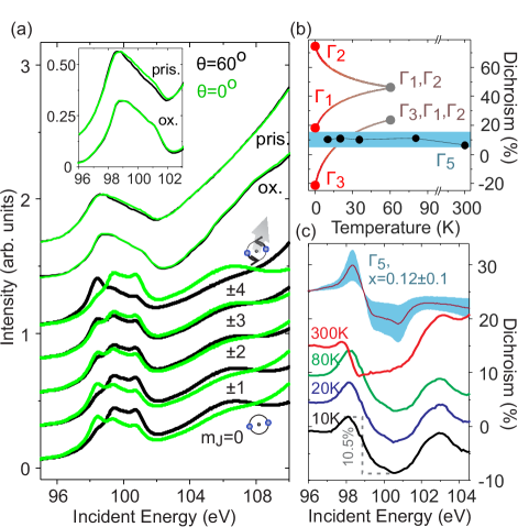
Provided that this is the case, an immediate next question is what superposition of J=4 moment states ( states) is occupied, and if this superposition changes as a function of temperature. A sizable fraction of the URu2Si2 literature has invoked CEF energy levels, which are coherent superpositions, as a key component in the low energy framework for understanding hidden order and the temperature dependence of susceptibility above hidden order. In scattering data, the occupation of states for systems can be directly evaluated from linear dichroism in O-edge X-ray absorption spectroscopy 5fdichroismME , as seen from the simulated XAS curves in Fig. 4(a) (thick curves). Dichroic contrast flips sign for wavefunction components with and , providing a qualitative metric to differentiate between models with a =2 ground state Gamma3early ; Gamma3review and ground states dominated by =3 or 4 components local3 ; firstCEFmodel ; Gamma3review ; Gamma5doublet1 ; Gamma5doublet2 ; Gamma5doublet3 ; KotliarNatPhys .
Measuring X-ray absorption from the pristine sample with contrasting polarization conditions yields spectra that differ by 5 of the maximum intensity (Fig. 4(c)). Spectra from the air exposed sample show no reproducible dichroism other than a slight difference in the intensity tail from , and will not be discussed in further detail. The dichroic difference spectrum of the pristine sample has a fluctuating pattern over , with two peaks near 98 eV and 102-103 eV, and a prominent valley from 99-101 eV. All of these features are universally present in simulations with ground state symmetry dominated by a basis (for example, the top curve in Fig. 4(c)), and appear with opposite sign in models with a ground state dominated by . Raising temperature yields no significant change across the T=17.5K hidden order transition, and the peak-to-valley difference in the dichroic spectrum keeps roughly the same amplitude (11) up to 80K, as plotted in Fig. 4(b). At room temperature, the dichroic spectrum is significantly distorted and has reduced amplitude, but retains qualitatively similar features.
A J=4 multiplet in the tetragonal crystal structure of URu2Si2 has 5 singlet states, of which three have been proposed by different studies as the hidden order ground state. These states are termed the (=0,4 components), (=4) and (=2), and a gap of roughly is required between the ground state and the next excited state to explain the sharp rise in magnetic susceptibility as temperature is raised across the hidden order transition entropyChange . Temperature dependence of dichroism as these states are thermalized into their magnetically polarizable manifolds is outlined in Fig. 4(b), using the mixing angle obtained numerically in Ref. KotliarNatPhys . The lack of temperature dependence in our data (black points in Fig. 4(b)) is qualitatively incompatible with these CEF hidden order pictures.
A last class of crystal field based models assigns the ground state as a doublet with symmetry (=3,1 components). The atomic ground state is degenerate and magnetically polarizable, and therefore does not directly explain hidden order, but provides a basis for pictures that incorporate interatomic dynamics such as the recent proposal of exotic hastatic order HastaticNature ; HastaticReview . The XAS dichroism of a ground state basis depends on the mixing parameter x=, where is the number operator for 2-electron configurations. A value of x=0.120.1 yields the simulation plotted at the top of 4(c), and is close to the value of x=0.2 considered in Ref. Gamma5doublet1 . The range of dichroic amplitude modeled for with x=0.120.1 is shaded in turquoise in Fig. 4(c), and encompasses the data points from pristine URu2Si2. Because the basis is magnetically polarizable, the suppression of magnetic susceptibility near the hidden order transition must be attributed to interatomic many-body effects, and one does not expect low temperature thermal activation of other CEF states.
In conclusion, these measurements show sharp excitation spectra of URu2Si2, which are definitively linked to the excitation modes of uranium through comparison with spectra from an oxidized surface. Crystal field excitation intensity, which has been elusive in inelastic neutron scattering, is finally observed, but found not to represent long-lived stable collective excitations. The large energy loss width of crystal field scattering intensity suggests that electrons are sufficiently itinerant to make the simplest rendering of CEF models untenable. However, itinerancy is not quite sufficient to dominate over the energy gap between the J=4 and J=2 atomic states that together give an almost complete basis for superpositions of the J=5/2 valence band electrons. The preserved energy gap between these modes implies that one should look for the principle components of hidden order in the basis of the J=4 atomic ground state. Linear dichroism is observed in XAS spectra, and analyzed to rule out CEF models that would directly explain hidden order via a singlet uranium ground state. The features, amplitude and temperature of XAS dichroism are found to be highly consistent with a doublet state that has formed the basis for intriguing many-body models and is compatible with experimental features such as nematicity okazakiNematic ; HastaticReview and large z-axis susceptibility. More generally, a non-singlet atomic ground state would enable Kondo-lattice physics within the hidden order phase, consistent with the observation of Kondo resonance features by point contact measurements KondoLatticeNature ; Kondo30K .
Acknowledgements: We are grateful for discussions with K. Wohlfeld. The Advanced Light Source is supported by the Director, Office of Science, Office of Basic Energy Sciences, of the U.S. Department of Energy under Contract No. DE-AC02-05CH11231. Crystal growth and characterization at UCSD was supported by the U.S. DOE under Grant No. DE-FG02-04-ER46105.
References
- (1) [
- (2) T. T. M. Palstra et al., Phys. Rev. Lett. 55 2727 (1985); M. B. Maple et al., Phys. Rev. Lett. 56, 185 (1986); W. J. Schlabitz, et al., Z. Phys. B 62, 171 (1986); R. A. Fisher et al., Physica B (Amsterdam) 163, 419 (1990).
- (3) L. P. Gor’kov and A. Sokol, Phys. Rev. Lett. 69, 2586 (1992); A. P. Ramirez et al., Phys. Rev. Lett. 68, 2680 (1992); M. B. Walker et al. Phys. Rev. Lett. 71, 2630 (1993).
- (4) H. Amitsuka et al. J. Phys. Conf. Ser. 200, 012007 (2010).
- (5) H. C. Walker et al., Phys. Rev. B 83, 193102 (2011).
- (6) C. Broholm et al., Phys. Rev B. 43, 12809 (1991).
- (7) J.-G. Park, K. A. McEwen, and M. J. Bull, Phys. Rev. B 66, 094502 (2002).
- (8) J. R. Jeffries, K. T. Moore, N. P. Butch, M. B. Maple, Phys. Rev. B 82, 033103 (2010).
- (9) G. J. Nieuwenhuys, Phys. Rev. B 35, 5260 (1987).
- (10) H. Amitsuka and T. Sakakibara, J. Phys. Soc. Jpn. 63, 736 (1994).
- (11) H. Amitsuka et al., Phys. Rev. Lett. 83, 5114 (1999).
- (12) Fusayoshi J. Ohkawa and Hirofumi Shimizu, J. Phys.: Condens. Matter 11, L519 (1999).
- (13) P. Santini and G. Amoretti, Phys. Rev. Lett. 73, 1027 (1994).
- (14) P. Chandra et al. Nature 417, 831 (2002).
- (15) A. Kiss and P. Fazekas, Phys. Rev. B 71, 054415 (2005).
- (16) Katsurou Hanzawa and Naoto Watanabe, J. Phys.: Condens. Matter 17, L419-L424 (2005).
- (17) Kristjan Haule and Gabriel Kotliar, Nat. Phys. 5, 796 (2009).
- (18) Hisatomo Harima, Kazumasa Miyake, and Jacques Flouquet, J. Phys. Soc. Jpn. 79, 033705 (2010).
- (19) H. Kusunose and H. Harima, J. Phys. Soc. Jpn 80, 084702 (2011).
- (20) Premala Chandra, Piers Coleman, and Rebecca Flint, Nature 493, 621 (2013).
- (21) Rebecca Flint, Premala Chandra, and Piers Coleman, J. Phys. Soc. Jpn. 83, 061003 (2014).
- (22) H. Ikeda and Y. Ohashi, Phys. Rev. Lett. 81, 3723 (1998).
- (23) C. M. Varma and L. Zhu, Phys. Rev. Lett. 96, 036405 (2006).
- (24) P. M. Oppeneer et al. Phys. Rev. B 82, 205103 (2010).
- (25) S. Elgazzar et al. Nature Materials 8, 337 (2009).
- (26) F. Cricchio, F. Bultmark, O. Granäs, and L. Nordström, Phys. Rev. Lett. 103, 107202 (2009).
- (27) Y. Dubi and A. V. Balatsky, Phys. Rev. Lett. 106, 086401 (2011).
- (28) S. Fujimoto, Phys. Rev. Lett. 106, 196407 (2011).
- (29) Hiroaki Ikeda et al., Nat. Phys. 8, 528 (2012).
- (30) Tanmoy Das, Sci. Rep. 2, 596 (2012).
- (31) K. O. Kvashnina and F. M. F. de Groot, J. of El. Spect. Rel. Phenom. 194, 88-93 (2014).
- (32) H. Ogasawara, A. Kotani, and B.T. Thole, Phys. Rev. B 44 2169 (1991); Gerrit van der Laan and B. T. Thole, Phys. Rev. B 53 14458 (1996).
- (33) H. Ogasawara and A. Kotani, J. Phys. Soc. Jpn. 64, 1394 (1995).
- (34) See Supplemental Material, which includes Refs. ARPES1 ; ARPES2 ; MengResARPES ; XrayPath ; SawatzkyShallowCore ; KotaniWrayIdea ; MagnusonSIAM .
- (35) F. L. Boariu, C. Bareille, H. Schwab, A. Nuber, P. Lejay, T. Durakiewicz, F. Reinert, and A.F. Santander-Syro, Phys. Rev. Lett. 110, 156404 (2013).
- (36) S. Chatterjee, J. Trinckauf, T. Hanke, D. E. Shai, J. W. Harter, T. J. Williams, G. M. Luke, K. M. Shen, and J. Geck, Phys. Rev. Lett. 110, 186401 (2013).
- (37) J.-Q. Meng et al., Phys. Rev. Lett. 111, 127002 (2013).
- (38) B. L. Henke, E. M. Gullikson, J. C. Davis, Atomic Data and Nuclear Data Tables 54, 181 (1993).
- (39) Subhra Sen Gupta et al., Phys. Rev. B 84, 075134 (2011).
- (40) K. Okada, A. Kotani, H. Ogasawara, Y. Seino, and B. T. Thole, Phys. Rev. B 47, 6203 (1993); H. Ogasawara, A. Kotani, and B. T. Thole, Phys. Rev. B 50, 12332 (1994); L. A. Wray et al., Phys. Rev. B 86, 195130 (2012).
- (41) M. Magnuson, S. M. Butorin, A. Agui, and J. Nordgren, J. Phys. Cond. Mat. 14, 3669 (2002).
- (42) Sergei M. Butorin, J. El. Spect. and Rel. Phenom. 110-111, 213-233 (2000).
- (43) A. Kotani, Eur. Phys. J. B 47, 3 (2005).
- (44) L. J. P Ament, M. van Veenendaal, T. P. Devereaux, J. P. Hill, and J. van den Brink, Rev. Mod. Phys. 83, 705 (2011).
- (45) L. A. Wray et al., Frontiers in Phys. 3, 32 (2015).
- (46) A. Yaouanc et al., Phys. Rev. B 58, 8793 (1998).
- (47) Jinghua Guo and Joseph Nordgren, J. El. Spect. Re. Phenom. 110-111, 105 (2000).
- (48) Jinghua Guo, Int. J. Nano. 1, 193 (2004).
- (49) J. N. Hancock et al., Phys. Rev. B 82, 020513(R) (2010).
- (50) Nicholas P. Butch, Michael E. Manley, Jason R. Jeffries, Marc Janoschek, Kevin Huang, M. Brian Maple, Ayman H. Said, Bogdan M. Leu, and Jeffrey W. Lynn, Phys. Rev. B 91, 035128 (2015).
- (51) R. Okazaki et al., Science 331, 439 (2011).
- (52) W. K. Park et al., Phys. Rev. Lett. 108, 246403 (2012).
- (53) A. R. Schmidt et al., Nature 465, 570 (2010).