Ultrafast near infrared photoinduced absorption in a multiferroic single crystal of bismuth ferrite
Abstract
We studied the ultrafast third-order optical nonlinearity in a single crystal of multiferroic bismuth ferrite (BiFeO3) in the near-infrared range of 0.5–1.0 eV, where the material is fundamentally transparent, at room temperature. With pump pulses at 1.55 eV, which is off-resonant to the strong inter-band charge transfer (CT) transition, we observed instantaneous transient absorption with pencil-like temporal profile originating from the two-photon CT transition from the oxygen 2 to the iron 3 levels. In contrast, under pumping with 3.10-eV photons, the pencil-like absorption change was not observed but decay profiles showed longer time constants. Although the two-photon absorption coefficient is estimated to be 1.5 cm/GW, which is ten (hundred) times smaller than that of two(one)-dimensional cuprates, it is larger than those of common semiconductors such as ZnSe and GaAs at the optical communication wavelength.
pacs:
42.70.Nq, 75.50.Ee, 78.20.Ci, 79.20.Ws, 78.47.jbI Introduction
A lot of efforts have been devoted for seeking materials for all optical switching, where large nonlinearity, quick response time, and operability at room temperature are demanded. Transition metal oxides of Mott Hubbard insulators are known to show ultrafast photoinduced phenomena reflecting broadband optical absorption. Especially in an one dimensional cuprate of Sr2CuO3, gigantic photoinduced absorption with an ultrafast decay constant of 1 ps occurs owing to the two-photon absorption process ogasawara ; ashida ; ashida2 . Since the wavelength range of probe pulses for the process includes the optical communication wavelength, such kind of materials are hopeful candidates for ultrafast all optical switches. So far, one and two dimensional cuprates have been well studied, however, there are not so many reports on other transition metal oxides especially three dimensional ones.
Bismuth ferrite (BiFeO3) is one of the multiferroics which possesses both ferroelectricity (FE) and antiferromagnetism (1100 K, 640 K), this material is studied from viewpoints of both fundamental physics and applications zhao ; lebeugle . Optical pump-probe measurements under excitation with above bandgap (2.6–2.8 eV) photons have been performed in films and single crystals of BiFeO3, where the authors discussed various photoinduced phenomena such as coherent oscillations due to magnon and phonon ruello ; doig ; chen , and spectral modulations in connection with the transport property yamada ; sheu . As a nonlinear medium, BiFeO3 has been studied in terms of the second-order nonlinearity in studies such as terahertz emission through optical rectification talbayev1 and ultrafast modulation of spontaneous polarization takahashi , and second harmonic generation spectroscopy kumar1 ; ramirez . However, there has been almost no study on the third-order nonlinearity especially around the optical communication wavelength in the near infrared region.
BiFeO3 is also one of the Mott insulators with ultrabroadband optical response continuously ranging from the far-infrared (terahertz) to the ultraviolet regions, reflecting strong interactions not only between electrons but also between electrons and lattice, spin, orbital degrees of freedoms, so that the material makes us expect strong and ultrafast nonlinear optical responses in the near-infrared below-gap region as ever reported in low-dimensional cuprates ogasawara ; ashida . Furthermore, BiFeO3 has the aspect of a ligand system, in which - transitions play important role in the optical responses in the visible and the near infrared regions, which makes the physics of the material more interesting.
As for the sample preparation, the fabrication of a single crystal of BiFeO3 has long been difficult. Recently, single crystals grown by the flux method have been utilized in many studies lobo ; talbayev1 ; cazayous ; singh ; rovillain ; xu ; talbayev2 ; sheu ; pisarev , however, the obtained size of the crystals has been limited. In 2011, Ito et al. realized the fabrication of large single crystals of BiFeO3 by using a modified floating zone method with laser diodes for heating ito . Besides the large size (4 mm in diameter), the leakage current under high bias voltage is extremely low. Hence, we think this crystal will enable us to extract only the intrinsic optical property of the material by making full use of the interaction length. Thus, in the present study, we optically pump the large single crystal of BiFeO3 with femtosecond pulses and probe the transient absorption change mainly in the near infrared region, to explore unknown ultrafast nonlinear response of BiFeO3.
II Experimental
A large single crystal of BiFeO3 with a diameter of 4 mm was grown with the floating zone method utilizing laser diodes as heat sources ito . By controlling the oxygen pressure during growth, oxygen deficiency is reduced a lot so that the leakage current flowing in the crystal under high bias-voltage is extremely low. Also the crystal does not show any magnetic hysteresis in the - curve which is usually observed due to the weak parasitic ferromagnetismin in crystals grown by the flux method.
The unit cell of BiFeO3 has the rhombohedral () structure, which is essentially a cubic elongated along the [111] direction. The ferroelectric polarization is along this axis. BiFeO3 is a G-type antiferromagnet in which all the nearest neighbor spin couplings, including both inter- and intra-plane ones, are antiferromagnetic. Due to the magnetic order, BiFeO3 forms the spiral spin structure with a period of 62 nm along the three equivalent wavevectors of [10-1], [01-1], and [1-10] directions. BiFeO3 is also a charge transfer (CT) type insulator with a bandgap energy of 2.62.8 eV, which corresponds to the lowest inter-band transition from the oxygen 2 to the iron 3 state. This broad CT band can be further decomposed into several peak structures centered at 2.5, 2.9, 3.2, 4.0, 4.5, and 6.1 eV pisarev . This CT band with strong oscillator strengths brings about strong absorption with coefficients of the order of 105 cm-1 xu . BiFeO3 also shows relatively weak (100 cm-1) - absorptions with broad peaks centered at 1.4 and 1.9 eV, which are also split into two by taking the spin degrees of freedom into account ramirez .
Pump-probe measurements were carried out with a titanium sapphire regenerative amplification system (800 nm, 35 fs, 1 kHz) as a light source. A (100)-oriented specimen of BiFeO3 with a thickness of 170 m was pumped with the fundamental (800 nm, 1.55 eV) or the second harmonic pulses (400 nm, 3.10 eV), and the transient transmission change was probed with signal or idler pulses from an optical parametric amplifier. The duration of the pump pulse was measeued to be 60 fs, and the cross correlation width of the pump and probe pulses was 100–110 fs, both at the sample. The penetration depths given by the inverse of the absorption coefficients xu are 33 m at 1.55 eV, and 0.49 m at 3.10 eV, respectively. The near-infrared transmission was measured with a combination of a monochromator and a cooled HgCdTe detector. The beam sizes of the pump and probe pulses were 1.35 mm and 130 m in diameter, respectively. First we pumped the specimen with 3.1-eV pulses and measured the transient optical conductivity with air-plasma based terahertz time domain spectroscopy. As a result, we observed no Drude-like response but observed an oscillation of transmission resembling a monocyclic terahertz pulse profile (not shown). This result is consistent with the high resistivity confirmed with the dc measurements ito and the oscillation profile seems to be caused by the generation of terahertz pulses due to the modulation of polarization by ultraviolet pulses takahashi . Since the electrons transport property is determined by oxygen deficiencies, the absence of the transient Drude response proves the suppression of them. Such high quality of the crystal will enable us to extract intrinsic property without artifacts.
III Results
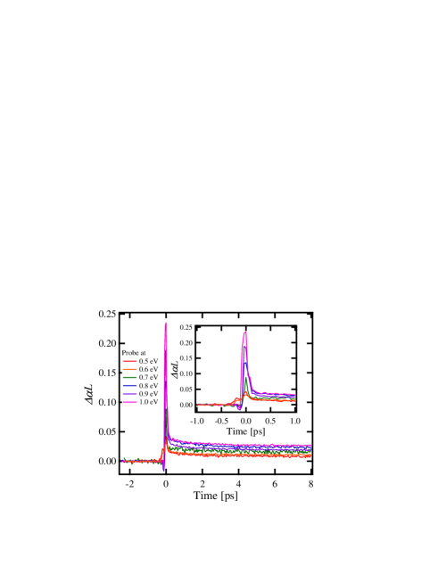
Figure 1 shows the time evolutions of the photoinduced absorption change at each probe photon energy (0.5–1.0 eV). The pump photon energy is fixed at 1.55 eV. At all probe photon energies, we can see steep increase of absorption and its ultrafast recovery. The amount of absorption change increases with the probe photon energy. The inset shows the expanded profiles around the time origin. Here the temporal profiles, especially for probe-photon energies greater than 0.8 eV, characteristically have pencil-like structures with a duration of 100–150 fs. This is close to the cross correlation width of the pump and probe pulses (100–110 fs). The decay profiles were well fitted by the expression assuming two exponential functions and a constant as,
| (1) |
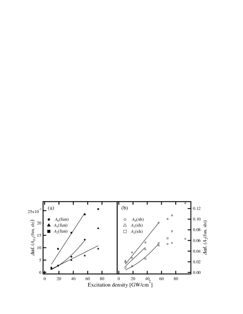
The constant component () certainly has a decay period beyond 1 ns, however, it did not exceed 1 ms, which is the separation period of the laser source. The values of and are fitted to be 50–60 fs and 1–3 ps, respectively. In the inset of Fig. 2 (a), we present how the transient absorption profiles probed at 0.9 eV are decomposed into three components. Importantly, the dotted curve showing the cross-correlation profiles of the pump and the probe pulses coincides with the dominant part of the transient absorption change. They appear to be essentially independent of the probe photon energy (wavelength) within the accuracy of the measurements. In Fig. 3 (a) we present the dependence of the magnitudes of the three components on the pump photon density. All the amplitudes of the three components increased with the probe-photon energy. More closely, and were proportional to the excitation density, while showed the quadratic dependence. We also examined the polarization dependence and found the temporal profile did not depend on the polarization of the pump and probe pulses, although the magnitude became smaller when the polarizations of two pulses were crossed.

To elucidate the physical mechanism, we changed the excitation photon energy to 3.10 eV and measured the transient absorption signal at the probe photon energy of 0.9 eV. The profile is shown in Fig. 2 (b). The decay curve was also decomposed into three parts with time constants of 140–170 fs, 1.9–2.6 ps, and nanoseconds (which can be regarded as constant). Hereafter, we denote the fitting results as (fun) or (sh) to distinguish those pumped by the fundamental (1.55 eV) and second harmonic (3.10 eV) pulses. The inset shows the profiles of each decomposed decay component. Apparently, the decay time of the fastest component for pumping with 3.10-eV pulses ((sh)) is longer than that with 1.55-eV pulses ((fun)), where the magnitude of (sh) is relatively smaller than (fun). The profile under pumping with 3.10-eV pulses is sharper at the top and does not have the pencil-like structure, which appeared under pumping with 1.55-eV photons, although the duration of 3.10-eV pulses is expected to be longer than that of the 1.55-eV ones due to the material dispersion in the nonlinear crystal. We also examined the dependence on the excitation density as presented with closed markers in Fig. 3 (b). (sh) was quadratically proportional to the excitation density up to 60 GW/cm2, approximately, while (sh) and (sh) showed linear dependence and the slopes become gentle around 40 and 55 GW/cm2, respectively. From these dependences and the absence of the pencil-like structure, the origin of photoinduced absorption under pumping with the second harmonic (3.10 eV) pulses is different from that with fundamental (1.55 eV) pulses.
IV Discussions
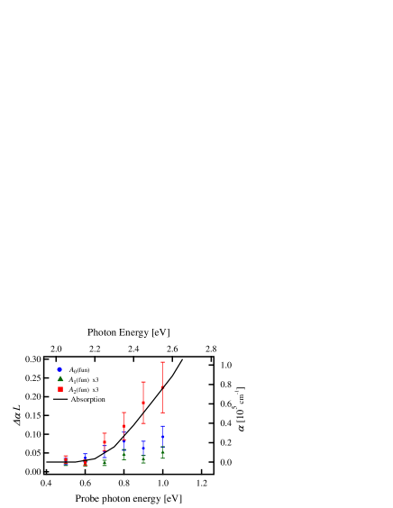
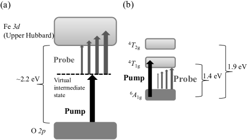
Now let us elucidate the microscopic origin of the present photoinduced phenomena. First, we focus on the pencil-like top structure in the transient absorption profile and the origin of the fastest decay component (fun). Ogasawara et al. observed a similar structure in the photoinduced transmission change profiles of a quasi-one-dimensional cuprate of Sr2CuO3, which they attributed to the two-photon excitation process from the oxygen to the even-parity band of Cu which lies at a higher frequency than the one-photon CT band ogasawara ; ashida ; ashida2 . We plot the amplitude of the fastest decay component as a function of the probe photon energy as shown in Fig. 4. By overlaying the absorption spectrum in such a manner that the horizontal axis (top) is shifted by the pump photon energy of 1.55 eV towards higher energy, the magnitude of the fastest component (fun) significantly rises up at 0.7 eV, where the linear absorption with the corresponding photon energy (2.2–2.3 eV) also starts to increase. From this and the pencil-like structure, we conclude that the two-photon absorption process from the O 2 and the Fe 3 level gives the fastest decay component as schematically shown in Fig. 5 (a). Note the unit cell of BiFeO3 lacks inversion symmetry at temperatures below 1100 K, so that the two-photon transition to the one-photon allowed level is allowed.
Let us further examine the probe photon energy (wavelength) dependence. While (fun) significantly increases with the probe photon energy, the increase of (fun) is minor. According to the result of the pump-probe measurements in the ultraviolet range, the total lifetime of the CT excited states is in the order of nanoseconds sheu . In the present case, one pump photon cannot create the CT excited state, however, the two-photon process can realize it. Hence, we consider the (fun) component comes from the lifetime of the two-photon excited CT states. We can attribute the gradual increase of (fun) with the probe photon-energy to the dependence of the oscillator strength of the CT transition on the photon energy. The pump photon energy of 1.55 eV is also close to the broadband - transition from to , so that the (fun) component with the decay constant of 1–3 ps probably corresponds to the lifetime of the - excited states, and the transition to the higher - levels or upper Hubbard band induces the transient absorption as schematically shown in Fig. 5 (b). The relatively small amplitude (fun) is due to the smaller oscillator strength compared with that of the CT transition by nearly one hundred times. As for the channel of the decay, we can think of some process involving the spin system, because the - transitions are known to be accompanied by magnon excitation in the form of sidebands ramirez .
Next let us discuss the photoinduced phenomena under pumping with 3.10-eV photons. The time evolution basically resembles the ones reported by Sheu et al. sheu , who pumped a single crystal of BiFeO3 with 3.1-eV photons and measured the transient reflection change also at 3.1 eV. From the quadratic dependence of (sh) on the excitation density, some cooperative interaction is certainly involved in the decay process. Similarly to the case in the previous report, the relaxation from the CT excited states to the bottom of the band through electron-electron or electron-phonon scatterings is highly possible as the origin. As already written, (sh) slightly decreases with the excitation density, which seems to support this speculation. Furthermore, while (fun) shows the quadratic dependence up to the excitation density of 56 GW/cm2, (sh) shows the linear dependence only up to 40 GW/cm2 and then the slope becomes gentle. In the case of excitation with fundamental (1.55 eV) pulses, some cooperative interaction seemingly works between the two-photon excited CT states. This is the origin of parabolic dependence of on the excitation density. In contrast, (sh) reflects the one-photon excited CT states, it can easily be saturated at the relatively weak excitation density as we observed in Fig. 3(b).
Now let us discuss the two-photon absorption coefficient of in BiFeO3. Using the transient transmission change of –0.11 at the probe photon energy of 1.0 eV (1.24 m), is estimated to be 1.5 cm/GW. The in the one-dimensional cuprate of Sr2CuO3 is 160 cm/GW at the peak of the two-photon bandogasawara , while that of the two-dimensional Sr2CuO2Cl2 is 12 cm/GW. This dimensionality dependence of the optical nonlinearity in materials relating to high cuprate superconductors was closely examined by Ashida et al., in which the in the two-dimensional cuprates was typically ten times smaller than that of the one-dimensional ones. The mechanism was explained using the cluster modelashida2 . Intuitively, to limit the coherent vibrational oscillations of electrons with large amplitudes in one direction can enhance the optical nonlinearity. While in the case of two or three dimensional materials, motion of electrons spreads into multiple directions even if electrons and lattice are vibrated in one direction by linearly polarized optical pulses. Thus the low-dimensional cuprates may be more beneficial in terms of application to optical device. However, the value is still larger than those of common semiconductors such as ZnSe and GaAs at the optical communication wavelength hutchings . BiFeO3 also has the strong aspect as a multiferroic, multifunctional responses relating to the magneto-optic effect can be expected in low temperature or polarization sensitive measurements. To explore such phenomena is our future work.
Finally, let us give a brief comment on the relationship between the present results and the optical anisotropy. BiFeO3 shows uniaxial birefringence with an extraordinary axis along the [111] direction. However, we observed no significant polarization dependence in the results of the pump probe spectroscopy. We can understand the reason considering the three dimensional chemical bonding structure in BiFeO3; in the present phenomenon, the CT excitation and - transition play important roles, while absorption due to phonons and magnons shows significant polarization dependence. At low temperatures, the coherence of elementary excitation will grow up enough to defeat dephasing factors and we will observe the transient anisotropic optical response originate from coherent phonon and magnon.
V Conclusions
In conclusion we demonstrated the ultrafast photoinduced absorption spectroscopy in a single crystal of BiFeO3 using sub-100-fs optical pulses in the near-infrared and ultraviolet ranges. With pump pulses at 1.55 eV, we observed sharp decrease in transmission which remained constant around the minimum for approximately 100 fs, it decayed with a time constant as short as 50–60 fs. Examining the dependences of the signal intensity on pump fluence and probe photon energy, we conclude the dominant transient absorption originates from the two-photon absorption process from the oxygen 2 to the iron 3 level. The decay profile also has two other components with constants of 1–3 ps, and nanoseconds which can be fitted as a constant in the present observation range. We attribute them as some relaxation process involving spins, and the total lifetime of the CT excited electrons via the two-photon process. The two-photon absorption coefficient is estimated to be 1.5 cm/GW, which is ten (hundred) times smaller than that of two(one)-dimensional cuprates. This result shows such ultrafast response is common in strongly correlated systems and may reflect the dimension dependence of the nonlinearity. The present value is still larger than those of common semiconductors such as ZnSe and GaAs at the optical communication wavelength. With pump pulses at 3.10 eV, the pencil like two-photon absorption did not appear, but the profiles had longer time components of 140–170 fs, 1.9–2.6 ps, and nanoseconds. We attribute them to the collective electron-electron and electron-phonon scattering in the CT band, some decay process involving spin or band to band relaxation, and the total lifetime of the CT excited levels, respectively.
References
- (1) T. Ogasawara, M. Ashida, N. Motoyama, H. Eisaki, S. Uchida, Y. Tokura, H. Ghosh, A. Shukla, S. Mazumdar, and M. Kuwata-Gonokami: Phys. Rev. Lett. 85 (2000) 2204.
- (2) M. Ashida, T. Ogasawara, Y. Tokura, S. Uchida, S. Mazumdar and M. Kuwata-Gonokami: Appl. Phys. Lett. 78 (2008) 2831.
- (3) M. Ashida, Y. Taguchi, Y. Tokura, R.T. Clay, S. Mazumdar, Yu. P. Svirko and M. Kuwata-Gonokami: Europhys. Lett. 58 (2002) 455.
- (4) T. Zhao, A. Scholl, F. Zavaliche, K. Lee, M. Barry, A. Doran, M. P. Cruz, Y. H. Chu, C. Ederer, N. A. Spaldin, R. R. Das, D. M. Kim, S. H. Baek, C. B. Eom and R. Ramesh: Nat. Mater. 5 (2006) 823.
- (5) D. Lebeugle, D. Colson, A. Forget, M. Viret, A. M. Bataille, and A. Gukasov: Phys. Rev. Lett. 100 (2008) 227602.
- (6) K.I. Doig, F. Aguesse, A.K. Axelsson, N. M. Alford, S. Nawaz, V.R. Palkar, S.P.P. Jones, R.D. Johnson, R.A. Synowicki, and J. Lloyd-Hughes: Phys. Rev. B 88 (2013) 094425.
- (7) L.Y. Chen, J.C. Yang, C.W. Luo, C.W. Laing, K.H. Wu, J.-Y. Lin, T.M. Uen, J.Y. Juang, Y.H. Chu and T. Kobayashi: Appl. Phys. Lett. 101 (2012) 041902.
- (8) P. Ruello, T. Pezeril, S. Avanesyan, G. Vaudel, V. Gusev1, I.C. Infante and B. Dkhil: Appl. Phys. Lett. 100 (2012) 212906.
- (9) Y. Yamada, T. Nakamura,S. Yasui, H. Funakubo, and Y. Kanemitsu: Phys. Rev. B 89 (2014) 035133.
- (10) Y. M. Sheu, S. A. Trugman, Y.-S. Park, S. Lee, H. T. Yi, S.-W. Cheong, Q. X. Jia, A. J. Taylor, and R. P. Prasankumar: Appl. Phys. Lett. 100 (2012) 242904.
- (11) D. Talbayev, S. Lee, S.-W. Cheong, and A. J. Taylor: Appl. Phys. Lett. 93 (2008) 212906.
- (12) K. Takahashi, N. Kida, and M. Tonouchi: Phys. Rev. Lett. 96 (2006) 117402.
- (13) A. Kumar, R.C. Rai, N.J. Podraza, S. Denev, M. Ramirez, Y-H. Chu, L.W. Martin, J. Ihlefeld, T. Heeg, J. Schubert, D.G. Schlom, J. Orenstein, R. Ramesh, R. W. Collins, J.L. Musfeldt and V. Gopalan: Appl. Phys. Lett. 92 (2008) 121915.
- (14) M.O. Ramirez, A.Kumar, S.A. Denev, N.J. Podraza, X.S. Xu, R.C. Rai, Y.H. Chu, J. Seidel, L.W. Martin, S.-Y. Yang, E. Saiz, J.F. Ihlefeld, S. Lee, J. Klug, S.W. Cheong, M.J. Bedzyk, O. Auciello, D.G. Schlom, R. Ramesh, J. Orenstein, J.L. Musfeldt, and V. Gopalan: Phys. Rev. B 79 (2009) 224106.
- (15) R. P. S. M. Lobo, R. L. Moreira, D. Lebeugle, and D. Colson: Phys. Rev. B 76 (2007) 172105.
- (16) M. Cazayous, Y. Gallais, A. Sacuto, R. de Sousa, D. Lebeugle, and D. Colson: Phys. Rev. Lett. 101 (2008) 037601.
- (17) M.K. Singh, R.S. Katiyar, and J.F. Scott: J. Phys.: Condens. Matter 20 (2008) 252203.
- (18) P. Rovillain, M. Cazayous, Y. Gallais, A. Sacuto, R. P. S. M. Lobo, D. Lebeugle, and D. Colson: Phys. Rev. B 79 (2009) 180411(R).
- (19) X.S. Xu, T.V. Brinzari, S. Lee, Y.H. Chu, L.W. Martin, A. Kumar, S. McGill, R.C. Rai, R. Ramesh, V. Gopalan, S.W. Cheong, and J.L. Musfeldt: Phys. Rev. B 79 (2009) 134425.
- (20) D. Talbayev, S. A. Trugman, S. Lee, H. T. Yi, S.-W. Cheong, and A. J. Taylor: Phys. Rev. B 83 (2011) 094403 .
- (21) R.V. Pisarev, A.S. Moskvin, A.M. Kalashnikova, and Th. Rasing: Phys. Rev. B 79 (2009) 235128.
- (22) T. Ito, T. Ushiyama, Y. Yanagisawa, R. Kumai, and Y. Tomioka: Crystal Growth and Design 11 (2011) 5139.
- (23) D.C. Hutchings and E.W. Van Stryland, J. Opt. Soc. B 11, 2065-2074 (1992).