MAKING A SWIM CENTER PATTERN GENERATOR OUT OF LATENT PARABOLIC BURSTERS
Abstract
We study the rhythmogenesis of oscillatory patterns emerging in network motifs composed of inhibitory coupled tonic spiking neurons represented by the Plant model of R15 nerve cells. Such motifs are argued to be used as building blocks for a larger central pattern generator network controlling swim locomotion of sea slug Melibe leonina.
keywords:
Plant model, parabolic bursting, half-center oscillation, central pattern generator, swim locomotion, see slug1 Introduction
A plethora of vital rhythmic motor behaviors, such as heartbeat, respiratory functions and locomotion are produced and governed by neural networks called central pattern generators (CPGs) Selverston [1985]; Bal et al. [1988]; Marder & Calabrese [1996]; Frost & Katz [1996]; Kristan et al. [2005]; Katz & Hooper [2007]. A CPG is a microcircuit of interneurons whose mutually synergetic interactions autonomously generate an array of multi-phase bursting rhythms underlying motor behaviors. There is a growing consensus in the community of neurophysiologists and computational researchers that some basic structural and functional elements must be shared by CPGs in invertebrate and vertebrate animals. As such, we should first understand these elements, find the universal principles, and develop efficient mathematical and computational tools for plausible and phenomenological models of CPG networks. Pairing experimental studies and modeling studies has proven to be key to unlocking insights into operational and dynamical principles of CPGs Gillner & Wallen [1985]; Kopell & Ermentrout [2004]; Matsuoka [1987]; Kopell [1988]; Canavier et al. [1994]; Skinner et al. [1994a]; Dror et al. [1999]; Prinz et al. [2003a]. Although various circuits and models of specific CPGs have been developed, it still remains unclear what makes the CPG dynamics so robust and flexible Best et al. [2005]; Belykh & Shilnikov [2008]; Sherwood et al. [2010]; Koch et al. [2011]; Calabrese et al. [2011]; Marder [2012]. It is also unclear what mechanisms a multi-functional motor system can use to generate polyrhythmic outcomes to govern several behaviors Kristan [2008]; Briggman & Kristan [2008]; Wojcik et al. [2014]. Our goal is to gain insight into the fundamental and universal rules governing pattern formation in complex networks of neurons. To achieve this goal, we should identify the rules underlying the emergence of cooperative rhythms in simple CPG networks.
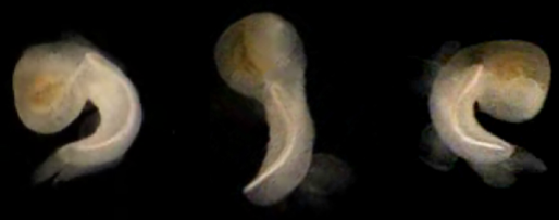
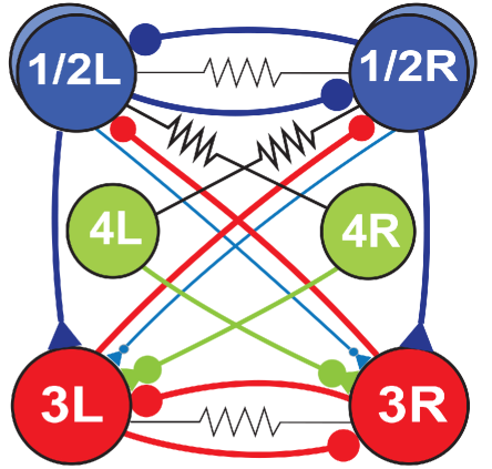
Recently, a great deal of computational studies have been focused on a range of 3-cell motifs of bursting neurons coupled by chemical (inhibitory and excitatory) and electrical synapses to disclose the role of coupling in generating sets of coexisting rhythmic outcomes, see Shilnikov et al. [2008]; Wojcik et al. [2011, 2014]; Schwabedal et al. [2014, 2015]; Collens et al. [2015] and references therein. These network structures reflect the known physiological details of various CPG networks in real animals. Next, we would like to explore dynamics and stability of some identified CPG circuits constituted by 4-cells Jalil et al. [2013]. Examples of such sub-networks can be found in the cerebral crustacean stomatogastric ganglion (STG) Selverston [1985]; Prinz et al. [2003b, 2004]; Marder [2012], as well as in the swim CPGs of the sea slugs – Melibe leonina (depicted during swimming in Fig. 1(a)) and Dendronotus iris Newcomb et al. [2012]; Sakurai et al. [2011]; Sakurai & Katz [2011]. Our greater goal is to create dynamical foundations for the onset, morphogenesis and structural robustness of rhythmic activity patterns produced by swim CPGs in these animals. A pilot mathematical model of the Melibe swim CPG will be discussed in this paper. The circuitry shown in Fig. 2(a) depicts only some core elements identified in the biological CPG; its detailed diagram can be found in Sakurai et al. [2014].
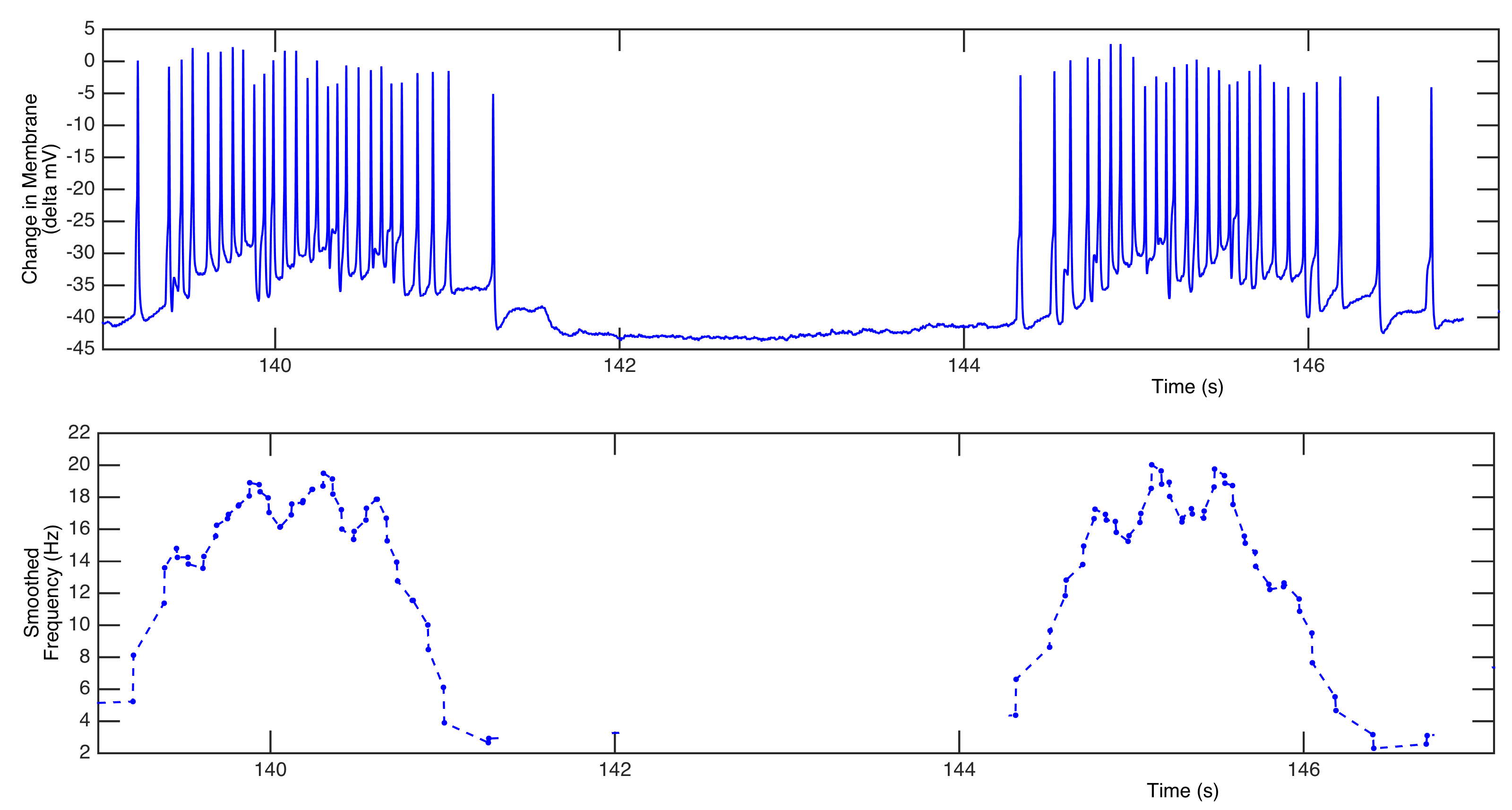
Being inspired by experimental studies of voltage activity recorded from the swim CPGs of the sea slugs Melibe leonina and Dendronotus iris, we would like to develop an assembly line for CPG constructors made of coupled biophysically plausible models. Our first simplifying assumption is that CPGs are made of universal building blocks – half center oscillations (HCOs) Hill et al. [2003]. Loosely speaking, a HCO is treated as a pair of interneurons interacting with each other through reciprocally inhibitory synapses and exhibiting anti-phase bursting. The interneurons of a HCO can be endogenous bursters, tonic spiking or quiescent ones, which exhibit alternating bursting only when they inhibit each other. Theoretical studies Wang & Rinzel [1985] have indicated that formation of an anti-phase bursting rhythm is always based on slow subsystem dynamics. There are three basic mechanisms to generate alternating bursting in the HCO: release, escape, and post-inhibitory rebound (PIR). The first mechanism is typical for endogenously bursting neurons Jalil et al. [2010, 2012]. The other two mechanisms underlie network bursting in HCOs comprised of neurons, which are hyperpolarized quiescent in isolation Perkel & Mulloney [1974]; Skinner et al. [1994b]; Angstadt et al. [2005]; Kopell & Ermentrout [2002]. Our second assumption is that the swim CPG interneurons are intrinsic tonic spikers that become network bursters only when externally driven or coupled by inhibitory synapses, as recent experimental studies suggest Sakurai et al. [2014]. The third assumption is that network bursting in the Melibe swim CPG is parabolic, i.e. the spike frequency within a burst increases at the middle, and decreases at the ends, as one can observe from Fig. 3. This observation indicates the type of neuronal models to be employed to describe network cores. Our model of choice for parabolic bursting is the Plant model Plant & Kim [1975, 1976]; Plant [1981]. The Plant model has been developed to accurately describe the voltage dynamics of the R15 neuron in a mollusk Aplysia Californica, which has turned out to be an endogenous burster Levitan & Levitan [1988]. Most dynamical properties of the R15 neuron have been modeled and studied in detail Canavier et al. [1991]; Bertran [1993]; Butera et al. [1995]; Butera [1998]; Sieling & Butera [2011]; Ji et al. [2013].
2 Methods: the Plant model of parabolic bursting
The conductance based Plant model Plant [1981] for the R15 neuron Sieling & Butera [2011] located in the abdominal ganglion of a slug Aplysia Californica is given by the following set of ordinary differential equations derived within the framework of the Hodgkin-Huxley formalism to describe the dynamics of the fast inward sodium [Na], outward potassium [K], slow TTX-resistant calcium [Ca] and an outward calcium sensitive potassium [KCa] currents:
| (1) |
The last three currents are the generic ohmic leak , external constant and synaptic currents flowing from a pre-synaptic neuron. The full details of the representation of the currents employed in the model are given in the Appendix below.
There are two bifurcation parameters in the individual model. The first one is the constant external current, , which is set . Following Shilnikov [2012], the other bifurcation parameter, , is introduced in the slowest equation:
| (2) |
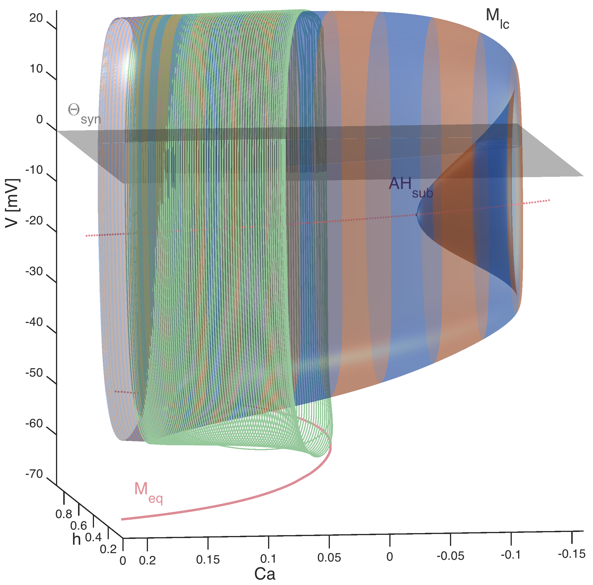
describing the concentration of the intracellular calcium in the Plant model. By construction, is a deviation from a mean value of the reversal potential evaluated experimentally for the calcium current in the R15 cells. As such, this makes a bifurcation parameter. Secondly its variations are not supposed to alter the topology of the slow motion manifolds in the 5D phase space, which are called tonic spiking and quiescent in the mathematical neuroscience context, as they are made of, respectively, round periodic orbits and equilibrium states [of the slow subsystem] of the model (Fig. 5).
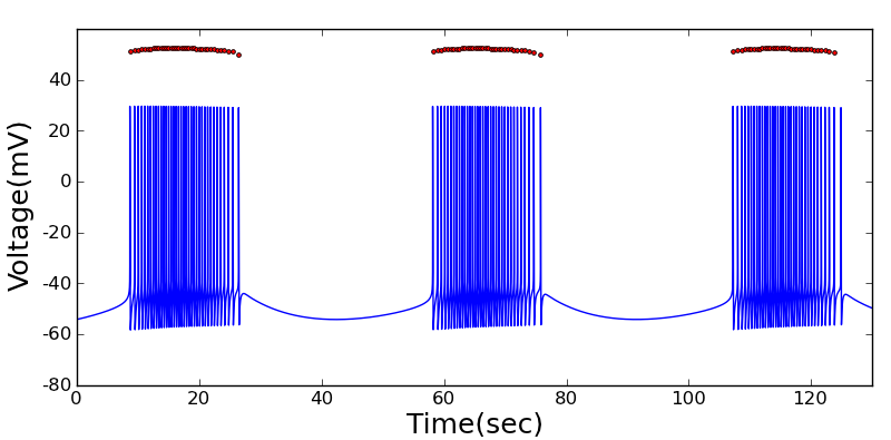
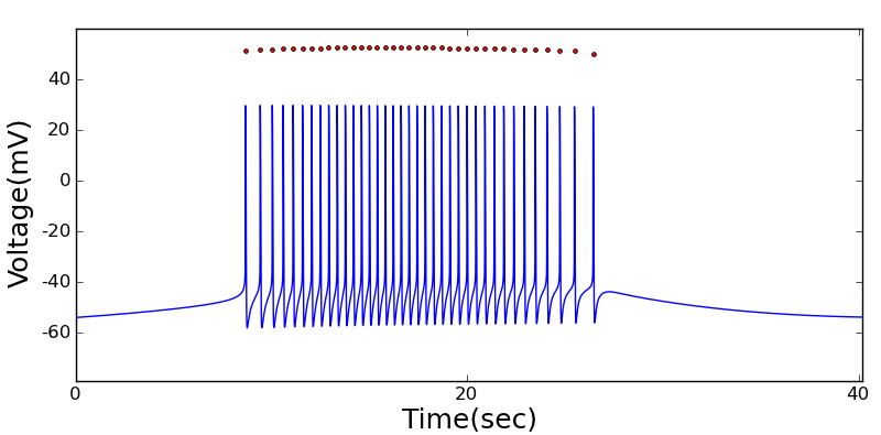
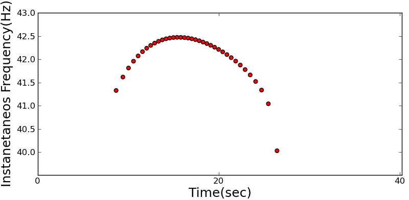
At , the neuron is an endogenous burster, see Fig. 4. According to Rinzel & Lee [1987], this type of bursting is termed parabolic. The reason for this term is that the spike frequency within bursts is maximized in the middle of bursts and minimized at the beginning and the end (see Fig. 4c). The parabolic structure of a burst is due to the calcium-activated potassium current. Its magnitude is determined by the intracellular calcium concentration. As the intracellular calcium concentration increases, the calcium dependent potassium current gets activated, which causes an increase of the inward potassium current. As the membrane potential increases over a threshold value, the intracellular calcium concentration decreases, as well as the inward potassium current (see Eq. (20) in the Appendix). The parabolic distribution of spikes within bursts is shown in Fig. 4. The instant frequency value is calculated by the reciprocal of each inter-spike interval. Panels b and c of Fig. 4 clearly disclose the parabolic inter-spike structure of bursts.
It was shown in Rinzel & Lee [1987] that the mechanism underlying a transition between quiescent and tonic spiking of bursting in the Plant model is due to a homoclinic bifurcation of a saddle-node equilibrium state Shilnikov [1963]; Afraimovich et al. [2014]. This bifurcation occurs in the fast 3D -subspace of the model and is modulated by the 2D slow dynamics in the -variables, which are determined by slow oscillations of the intracellular calcium concentration Plant & Kim [1975, 1976]. The unfolding of this codimension-one bifurcation includes an onset of a stable equilibrium, which is associated with a hyperpolarized phase of bursting, and on the other end, an emergent stable periodic orbit that is associated with tonic spiking phase of bursting. The period of this stable orbit decreases, as it moves further away from the saddle-node equilibrium mediated by decreasing calcium concentration. The period of the tonic spiking orbit grows with no upper bound as it approaches the homoclinic loop of the saddle-node Shilnikov et al. [1998,2001].
Variations of change the duty cycle of bursting, which is a ratio of the active tonic spiking phase of bursting to its period. Decreasing reduces the inactive, quiescent phase of bursting, i.e. increases its duty cycle. Zero duty cycle is associated with the homoclinic saddle-node bifurcation that makes the neuron hyperpolarized quiescent. This corresponds to an emergence of stable equilibrium state for all dynamical variables of the model (1). In other words, decreasing makes the active phase longer, so that below a threshold the neuron switches to tonic spiking activity. Tonic spiking activity is associated with the emergence of a stable periodic orbit in the fast -subspace, while the -variables of the slow subspace converge to a stable equilibrium state. As such, bursting occurs in the Plant and similar models due to relaxation of periodic oscillations in the 2D -subspace, which slowly modulates fast tonic spiking oscillations in the variables. The relaxation limit cycles emerge from one and collapse into the other equilibrium state in the -plane through Andronov-Hopf bifurcations, which can be sub- or super-critical. At the transitions between bursting and tonic spiking, and bursting and hyperpolarized quiescence, the neuron can produce chaotic dynamics, which are basically due to the membrane potential oscillatory perturbations of plain canards at the folds of the relaxation cycle.
3 Endogenous and network bursting. Inhibitory and excitatory drives
A half-center oscillator is a network of two neurons coupled by reciprocally inhibitory synapses that robustly produces bursting in alternation, or anti-phase bursting. Such a network can be multistable, i.e. produce other bursting rhythms as well, such as synchronous bursting Jalil et al. [2010] and rhythmic outcomes with slightly shifted phase lags between the endogenously bursting neurons Jalil et al. [2012].
In this study, the synaptic current is modeled through the fast threshold modulation (FTM) approach Kopell & Somers [1993]. The synapses are assumed to be fast and non-delayed, which is true for the swim CPG in both sea slugs under consideration. The synaptic current is given by
| (3) |
where is the maximal conductance of the current, which is used as a bifurcation parameter of the networked model; and are the voltages on the post-synaptic (driven) and pre-synaptic (driving) neurons; is the synaptic reversal potential. To make excitatory, we set , while in the inhibitory case we set . In Eq. (3), the second term is a Boltzmann coupling function that quickly, (), turns the synaptic current on and off as soon the voltage, , of the (driving) pre-synaptic cell(s) raises above and falls below the synaptic threshold, here (Fig. 5).
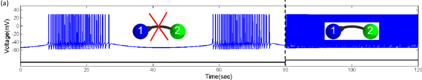
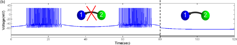
To model the constant synaptic drive onto the post-synaptic neuron, we assume that . This allows us to calibrate the state of the post-synaptic neuron, and to determine the drive threshold that separates the qualitatively distinct states of the individual and networked neurons. This statement is illustrated in Fig. 6 by simulating responses of the endogenous parabolic burster to network perturbation. Figure 6(a) shows, with a properly adjusted excitatory drive, that the endogenous burster switches into tonic spiking activity. On the other hand, bursting in the networked neuron can be halted when it receives a sufficient inhibitory drive from the pre-synaptic neuron of the network (Figure 6(b)). Eliminating either drive makes the post-synaptic neuron return to its natural state, i.e. these experiments de-facto prove that the neuron is mono-stable for the given parameter values.
A HCO, in the canonical Brown definition Graham-Brown [1911], is a pair of neurons bursting in anti-phase when they are networked by inhibitory synapses. In isolation, such neurons are not endogenous bursters but tonic spikers instead, or remain quiescent Marder & Calabrese [1996]. There are multiple mechanisms underlying such anti-phase bursting, or, more accurately, anti-phase oscillations in HCOs and CPGs made of relaxation oscillators Kopell & Ermentrout [2002]; Daun et al. [2009]. The list includes the well studied mechanisms of post-inhibitory rebound and escape for quiescent neurons Perkel & Mulloney [1974]; Wang & Rinzel [1985]; Skinner et al. [1994b]; Destexhe et al. [1994]; Matveev et al. [2007], as well as less-known mechanisms of HCOs constituted by intrinsically spiking neurons. Such networks utilizing the Plant models are discussed below.
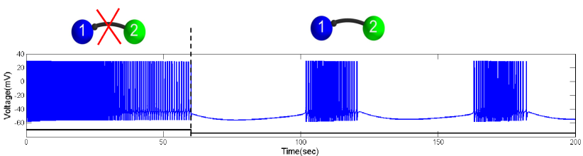
To construct such a HCO with relatively weak inhibitory coupling, the Plant model must be first set into the tonic spiking mode. This is done by setting the bifurcation parameter, , see Fig. 7. Next, we consider a unidirectional network where the tonic spiking neuron 1 starts receiving, an inhibitory drive of from the post-synaptic neuron 2 at . The inhibitory drive is sufficient to shift the post-inhibitory neuron over the bifurcation transition back into bursting activity. The minimal inhibitory drive must be increased proportionally to make the targeted neuron a network burster whenever it stays further away from the bifurcation transition between tonic spiking and bursting in isolation.
4 Forming a half-center oscillator
In this section, we discuss the dynamics of half-center oscillators made of two tonically spiking Plant neurons reciprocally coupled by inhibitory synapses. As before, we describe such synapses within the framework of the fast threshold modulation (FTM) paradigm using Eq. (3) to match the shape and magnitude of inhibitory postsynaptic potentials (IPSPs) in the post-synaptic neurons. IPSPs are the indicators of the type and the strength of synapses in the network.
We perform simulations in a fashion that is analogous to the dynamic clamp technique used in neurophysiological experiments. The approach involves the dynamic block, restoration and modulation of synaptic connections during simulation. These modeling perturbations should closely resemble the experimental techniques of drug-induced synaptic blockade, modulation, wash-out, etc. Restoring the chemical synapses during a simulation makes the HCO regain network bursting activity with specific phase characteristics. Depending on the coupling strength as well as the way the tonically spiking neurons are clamped, the network bursting may change phase-locked states, i.e. be potentially multi-stable. Experimental observations also suggest specific constraints on the range of coupling strengths of the reciprocal inhibition, such that the networks stably and generically achieve the desired phase-locking.
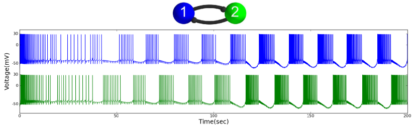
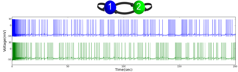
Figure 8 demonstrates the stages of anti-phase bursting formation in the HCO. The uncoupled neurons are initiated in tonic spiking mode. After turning on the reciprocally inhibitory synapses , the HCO quickly transitions to the regime of robust anti-phase bursting. Turning off the synapses restores the native tonic spiking activity in both neurons. Turning on the reciprocal synapses makes the HCO regain the network bursting. Note that the length of transients from tonic spiking to network bursting depends on the strength of the synaptic coupling for the fixed parameters of the individual Plant neurons. By comparing the magnitude of IPSPs in the voltage traces represented in Figs. 8 and 9, one can conclude that the coupling in the later case is weaker. This is why, the onset of network bursting in the HCO is less pronounced.
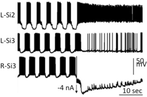
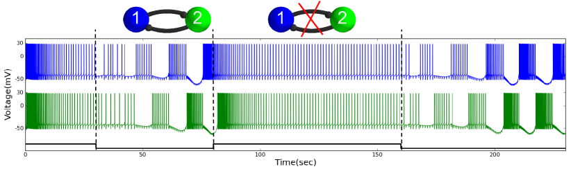
Our modeling studies agree well with experimental recordings from the identified interneurons in the Melibe swim CPG which suggests that the observed bursting is due to synergetic interactions of interneurons of the network Sakurai et al. [2014]. One can see from Fig. 1(b) that network bursting in the biological HCO formed by two Si3 interneurons of the Melibe swim CPG is seized as soon as the right one, Si3R, receives a negative current pulse that makes it hyperpolarized quiescent, while its left bursting counterpart, Si3L, turns into tonic spiking activity instead. Moreover, one can deduct from the wiring diagram of the CPG depicted in Fig. 2(a) and the analysis of voltage traces represented in Fig. 1(b) that the interneuron Si2L becomes a tonic-spiker as soon as the pre-synaptic interneuron Si3R stops inhibiting it (compare with Fig. 8.) This further supports the assertion that the swim CPG is made of intrinsically tonic spiking interneurons.
To test the robustness of network anti-phase bursting to perturbation and to calibrate the necessary influx of reciprocal inhibition generated by the Plant neurons, we consider a HCO with excitatory autapses. The objective here is to determine an equivalent amount of excitatory drive to be projected onto the post-inhibitory network burster to cancel out the inhibitory drive and shift it back to the initial tonic-spiking mode.
An autapse is a synapse of a neuron onto itself, where the axon of the neuron ends on its own dendrite. After their discovery Van der Loos & Glaser [1972] autapses have been observed in a range of nervous systems. The autapses are arguably to be responsible for tuning of neural networks. This particular configuration of the HCO depicted in Fig. 10 is formally motivated by the swim CPG circuitry, see Fig. 2(a). One can see from it that the interneurons of the bottom HCO receive excitatory drives from the top interneurons forming the top HCO. We would like to find the threshold over which the neurons no longer form a stably bursting HCO. This would allow us to calibrate and quantify the relative strengths of the mixed synaptic connections in the swim CPG models.
In this HCO configuration, each neuron inhibits its counterpart and self-excites through the autapse. Both autapses are introduced to the model using the FTM approach with . In this experiment, the conductance values for inhibitory synapses are set at . This is sufficient for the HCO to generate robust anti-phase bursting as seen in Fig. 9. Next, we add the autapses along with inhibition and gradually increase . We found that increasing proportionally increase the delay. At , the network stops exhibiting anti-phase bursting. We note that unlike a permanent excitatory drive from pre-synaptic neurons, an introduction of the excitatory autapse, acting only when the self-driving neuron is above the synaptic threshold, is effectively perturbation equivalent for the calibration purpose.
5 Assembly line of a Melibe swim CPG
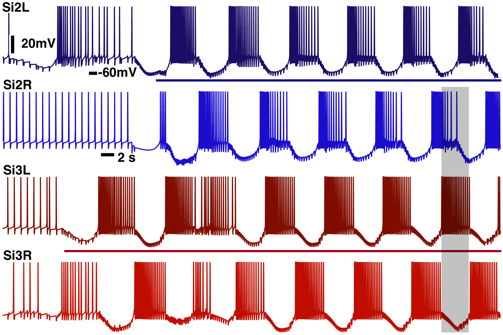
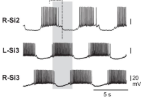
In this final section, we put together a pilot model of the Melibe swim CPG according to a circuitry based on identified interneurons and synapses; its wiring diagram is sketched in Fig. 2(a). This network model is made of the two HCOs constituted by tonic spiking Plant neurons. We would like to find out whether this sample CPG model can already produce phase lags similar to those between bursting interneurons in the biological CPG. For the sake of simplicity, we do not include Si4R/L interneurons in the model and we also omit electrical synapses. It is known from experimental studies Sakurai et al. [2014] that blocking chemical, inhibitory and excitatory synapses between the interneurons may be sufficient to break down the motor pattern by the network. Figure 2(b) points out that the interneurons of either HCO burst in anti-phase and there is the characteristic 3/4 phase lag between the burst initiation in the neurons Si2L and Si3L, as well as between Si2R and Si3L. This phase lag is repeatedly observed in both adult and juvenile animals.
As before, we use the Plant neurons initiated in the tonic spiking mode, relatively close to the transition to bursting. Initial conditions of the neurons are randomized. After letting the neurons settled down to tonic spiking activity, the network connections are turned on. As Fig. 11 shows, with the reciprocal inhibition being first turned on, the bottom interneurons Si3L and Si3R become anti-phase network bursters, and so do Si2R and Si2L as soon as the reciprocal inhibition between them is turned them on, too. At this stage, the CPG model is formed by two uncoupled HCOs. A few seconds later, they become coupled by simultaneous turning on the unidirectional cross-lateral inhibition from Si3R(L) projected onto Si2L(R), and bi-lateral excitation from Si2R(L) down onto Si3R(L). One can see from this figure that all four interneurons of the CPG model exhibit network bursting with the desired phase lags. These are 0.5 (half period) between the interneurons of each HCO, and 3/4 (a fraction of the network period) between the HCOs, or between the corresponding reference interneurons Si2L and Si3L. We note that such a phase shift was reported in a similar Melibe swim CPG constituted by endogenous bursters; that model also incorporated electrical synapses Jalil et al. [2013]. There is a great room for improvement of CPG network models to include other identified interneurons and to incorporate additional electrical synapses to find out whether additions of new elements can stabilize or desynchronize the desired bursting pattern as it was done using the Poincaré return maps for endogenous bursters Wojcik et al. [2014]. Of our special interest are various problems concerning structural stability of the network, and its robustness (Lyapunov stability) for bursting outcomes subjected to perturbations by pulses of the external current, as well as reductions to return maps between burst initiations in constituent neurons. These questions are beyond the scopes of the given examination and will be addressed in full detail in our forthcoming publications soon. The question about a possible linking of the characteristic phase lag and the Melibe leonina lateral swim style is the paramount one among them.
6 Summary
We have discussed a basic procedure for building network bursting CPGs made of intrinsically tonic spiking neurons. As a model for such networks, we have employed the biophysically plausible Plant model that was originally proposed to describe endogenous bursting R15-cells in the Aplysia mollusk. Such bursting was intracellularly recorded, and identified as parabolic, from the known interneurons in the swim CPGs of two sea slugs: Melibe leonina and Dendronotus iris. There is experimental evidence that bursting in these swim CPGs is due to synergetic interactions of all constituent neurons that are intrinsic tonic-spikers in isolation. To model the Melibe swim CPG, we have first examined dynamical and structural properties of the Plant model and its responses to perturbations. These perturbations include inhibitory and excitatory inputs from pre-synaptic neurons in the network. We have identified the transition boundary beyond which the bursting Plant model becomes a tonic-spiker and shifted it slightly over the threshold using an introduced bifurcation parameter. We have shown that the perturbed/calibrated Plant neuron, exhibiting intrinsically tonic spiking activity, becomes a network burster when it receives an inhibitory drive from a pre-synaptic neuron. By combining two such neurons, we have created a genuine half-center oscillator robustly producing anti-phase bursting dynamics. We have also considered a HCO configuration with two excitatory autapses to assess the robustness of anti-phase bursting with respect to excitatory perturbations. Finally, we have employed all necessary components to assemble a truncated model of the Melibe swim CPG with the characteristic 3/4-phase lags between the bursting onsets in the four constituent interneurons. In future studies, we plan to examine the dynamics of the CPG models with all synaptic connections, including electrical, as well as incorporating additional identified interneurons. We will also explore their structural stability, robustness and potential multi-stability of their bursting outcomes with various phase lags. An additional goal is to find out whether the motor pattern with the 3/4-phase lags will persist in networks with interneurons represented by other mathematical models including phenomenologically reduced ones. Potentially, these findings shall provide a systematic basis for comprehension of plausible biophysical mechanisms for the origination and regulation of rhythmic patterns generated by various CPGs. Our goal is to extend and generalize the dynamical principles disclosed in the considered networks for other neural systems besides locomotion, such as olfactory cellular networks.
7 Acknowledgments
A.S. also acknowledges the support from GSU Brain and Behaviors pilot grant, RFFI 11-01-00001, RSF grant 14-41-00044 at the Lobachevsky University of Nizhny Novgorod and the grant in the agreement of August 27, 2013 N 02.B.49.21.0003 between the Ministry of education and science of the Russian Federation and Lobachevsky State University of Nizhni Novgorod (Sections 2-4), as well as NSF BIO-DMS grant XXXXX. We thank A. Sakurai and P.S. Katz for sharing experimental data and suggestions, and the acting and associated members of the NEURDS (Neuro Dynamical Systems) lab at GSU: A. Kelley, K. Pusuluri, S. Pusuluri, T.Xing, M. Fen, F. Fen, J. Collens, D. Knapper, A. Noriega, P. Ozluk, B. Chung and E. Latash for helpful discussions, and R. Clewley for his guidance on the PyDSTool package Clewley et al. [2006] used in simulations.
8 Appendix: the conductance based Plant model
The model in this study is adopted from Plant [1981]. The dynamics of the membrane potential, , is governed by the following equation:
| (4) |
where is the membrane capacitance, is the current, is the current, is the current, is the activated current, is the leak current, is the synaptic current. The fast inward sodium current is given by
| (5) |
where the reversal potential and the maximum conductance value . The instantaneous activation variable is defined as
| (6) |
where
| (7) |
while the dynamics of inactivation variable is given by
| (8) |
where
| (9) |
with
| (10) |
where
| (11) |
The fast potassium current is given by the equation
| (12) |
where the reversal potential is and the maximum conductance value is .The dynamics of inactivation gating variable is described by
| (13) |
where
| (14) |
with
| (15) |
The TTX-resistant calcium current is given by
| (16) |
where the reversal potential is and the maximum conductance is . The dynamics of the slow activation variable is described by
| (17) |
where
| (18) |
The outward activated current is given by
| (19) |
where the reversal potential is . The dynamics of intracellular calcium concentration is governed by
| (20) |
where the reversal potential is , and the constant values are and . The leak current is given by
| (21) |
where the reversal potential and the maximum conductance value . The synaptic current is defined as
| (22) |
with the synaptic reversal potential for inhibitory synapses and for excitatory synapses and the synaptic threshold , and .
References
- Afraimovich et al. [2014] Afraimovich, V., Gonchenko, S., Lerman, L., Shilnikov, A. & Turaev, D. [2014] “Scientific heritage of L.P. Shilnikov. Part 1.” Regular and Chaotic Dynamics 19, 435–460.
- Angstadt et al. [2005] Angstadt, J., Grassmann, J., Theriault, K. & Levasseur, S. [2005] “Mechanisms of postinhibitory rebound and its modulation by serotonin in excitatory swim motor neurons of the medicinal leech,” Journal of Comparative Physiology A-Neuroethology, Sensory, Neural and Behavioral Physiology 191, 715–732, 10.1007/s00359-005-0628-6.
- Bal et al. [1988] Bal, T., Nagy, F. & Moulins, M. [1988] “The pyloric central pattern generator in crustacea: a set of conditional neural oscillators,” Journal of Comparative Physiology A 163, 715–727.
- Belykh & Shilnikov [2008] Belykh, I. & Shilnikov, A. [2008] “When weak inhibition synchronizes strongly desynchronizing networks of bursting neurons.” Phys Rev Lett 101, 078102.
- Bertran [1993] Bertran, R. [1993] “A computational study of the effects of serotonin on a molluscan burster neuron,” Biol. Cybern. 69, 257–267.
- Best et al. [2005] Best, J., Borisyuk, A., Rubin, J., Terman, D. & M., W. [2005] “The dynamic range of bursting in a model respiratory pacemaker network,” SIAM J. Appl. Dyn. Syst. 4, 1107–1139.
- Briggman & Kristan [2008] Briggman, K. L. & Kristan, W. B. [2008] “Multifunctional pattern-generating circuits.” Annu Rev Neurosci 31, 271–294, 10.1146/annurev.neuro.31.060407.125552, URL http://dx.doi.org/10.1146/annurev.neuro.31.060407.125552.
- Butera [1998] Butera, R. [1998] “Multirhythmic bursting,” Chaos 8, 274–284.
- Butera et al. [1995] Butera, R., Clark, J. W. J., Canavier, C. C., Baxter, D. A. & Byrne, J. H. [1995] “Analysis of the effects of modulatory agents on a modeled bursting neuron: Dynamic interactions between voltage and calcium dependent systems,” J Comput. Neuroscience 2, 19–44.
- Calabrese et al. [2011] Calabrese, RL., Norris, BJ., Wenning, A. & Wright, TM. [2011] “Coping with variability in small neuronal networks,” Integrative and Comparative Biology 51, 845–855, 10.1093/icb/icr074.
- Canavier et al. [1991] Canavier, C., Clark, J. & Byrne, J. [1991] “Simulation of the bursting activity of neuron R15 in Aplysia: Role of ionic currents, calcium balance, and modulatory transmitters” J Neurophysiology 66, 2107–2124.
- Canavier et al. [1994] Canavier, C., Baxter, DA., Clark, JW. & Byrne, JH. [1994] “Multiple modes of activity in a model neuron suggest a novel mechanism for the effects of neuromodulator” J Neurophysiology 72, 872–882.
- Clewley et al. [2006] Clewley, R., Sherwood, W., LaMar, M. & Guckenheimer, J. [2006] “Pydstool: an integrated simulation, modeling, and analysis package for dynamical systems.” Tech. rep., http://pydstool.sourceforge.net.
- Collens et al. [2015] Collens, J., Kelley, A., Alacam, D., Xing, T., D., K., Schwabedal, J. & Shilnikov, A. [2015] “Intrinsic mechanisms for pattern generation in three-node networks,” submitted .
- Daun et al. [2009] Daun, S., Rubin, J. E. & Rybak, I. A. [2009] “Control of oscillation periods and phase durations in half-center central pattern generators: a comparative mechanistic analysis,” Journal of Computational Neuroscience 27, 3–36, 10.1007/s10827-008-0124-4, URL http://dx.doi.org/10.1007/s10827-008-0124-4.
- Destexhe et al. [1994] Destexhe, A., Contreras, D., Sejnowski, T. & Steriade, M. [1994] “A Model of Spindle Rhythmicity in the Isolated Thalamic Reticular Nucleus,” Journal of Neurophysiology 72, 803–818.
- Dror et al. [1999] Dror, R. O., Canavier, C. C., Butera, R. J., Clark, J. W. & Byrne, J. H. [1999] “A mathematical criterion based on phase response curves for stability in a ring of coupled oscillators.” Biol Cybern 80, 11–23, 10.1007/s004220050501, URL http://dx.doi.org/10.1007/s004220050501.
- Frost & Katz [1996] Frost, W. N. & Katz, P. S. [1996] “Single neuron control over a complex motor program,” Proc.Nat.Acad. Sc. 93, 422–426.
- Gillner & Wallen [1985] Gillner, S. & Wallen, P. [1985] “Central pattern generators for locomotion, with special references to vertebrates,” Ann. Rev. Neurosci. 8, 233–261.
- Graham-Brown [1911] Graham-Brown, T. [1911] “The intrinsic factors in the act of progression in the mammal,” Lond B Biol Society 84, 308–319.
- Hill et al. [2003] Hill, A., Van Hooser, S. & Calabrese, R. [2003] “Half-center oscillators underlying rhythmic movements,” The Handbook of Brain Theory and Neural Networks, ed. Arbib, M. A. (The MIT Press).
- Jalil et al. [2013] Jalil, S., Allen, D., Youker, J. & Shilnikov, A. [2013] “Toward robust phase-locking in melibe swim central pattern generator models,” Chaos 23, 046105, http://dx.doi.org/10.1063/1.4825389, URL http://scitation.aip.org/content/aip/journal/chaos/23/4/10.1063/1.4825389.
- Jalil et al. [2010] Jalil, S., Belykh, I. & Shilnikov, A. [2010] “Fast reciprocal inhibition can synchronize bursting neurons,” Physical Review E 81, 10.1103/PhysRevE.81.045201.
- Jalil et al. [2012] Jalil, S., Belykh, I. & Shilnikov, A. [2012] “Spikes matter for phase-locked bursting in inhibitory neurons,” Physical Review E 85, 10.1103/PhysRevE.85.036214.
- Ji et al. [2013] Ji, L., Zhang, J., Lang, X. & Zhang, X. [2013] “Coupling and noise induced spiking-bursting transition in a parabolic bursting model,” Chaos: An Interdisciplinary Journal of Nonlinear Science 23, 013141, http://dx.doi.org/10.1063/1.4795281, URL http://scitation.aip.org/content/aip/journal/chaos/23/1/10.1063/1.4795281.
- Katz & Hooper [2007] Katz, P. S. & Hooper, S. [2007] “Invertebrate central pattern generators,” Invertebrate Neurobiology, eds. North, G. & R. Greenspan, R. (Cold Spring Harbor Laboratory Press, NY, New York).
- Koch et al. [2011] Koch, H., Garcia, A. J. & Ramirez, J.-M. [2011] “Network reconfiguration and neuronal plasticity in rhythm-generating networks,” Integrative and Comparative Biology 51, 856–868, 10.1093/icb/icr099.
- Kopell [1988] Kopell, N. [1988] “Toward a theory of modelling central pattern generators,” Neural Control of Rhythmic Movements in Vertebrates, eds. Cohen, A., Rossingol, S. & Grillner, S. (Wiley, New York).
- Kopell & Ermentrout [2004] Kopell, N. & Ermentrout, B. [2004] “Chemical and electrical synapses perform complementary roles in the synchronization of interneuronal networks,” Proc. Natl. Acad, Sci. 101, 15482–15487, 10.1073/pnas.0406343101.
- Kopell & Ermentrout [2002] Kopell, N. & Ermentrout, G. [2002] “Mechanisms of phase-locking and frequency control in pairs of coupled neural oscillators,” Handbook on Dynamical Systems 2, 3–54.
- Kopell & Somers [1993] Kopell, N. & Somers, D. [1993] “Rapid synchronization through fast threshold modulation,” Biol. Cybern. 68, 5.
- Kristan [2008] Kristan, W. [2008] “Neuronal decision-making circuits.” Curr Biol 18, R928–R932, 10.1016/j.cub.2008.07.081, URL http://dx.doi.org/10.1016/j.cub.2008.07.081.
- Kristan et al. [2005] Kristan, W., Calabrese, R. & Friesen, W. [2005] “Neuronal control of leech behavior,” Prog. Neurobiol. 76, 279.
- Levitan & Levitan [1988] Levitan, E. & Levitan, I. B. [1988] “Serotonin acting via cyclic amp enhances both the hyperpolarizing and depolarizing phases of bursting pacemaker activity in the aplysia neuron R15,” J Neuroscience 8, 1152–1161.
- Marder [2012] Marder, E. [2012] “Neuromodulation of neuronal circuits: back to the future,” Neuron 76, 1.
- Marder & Calabrese [1996] Marder, E. & Calabrese, R. [1996] “Principles of rhythmic motor pattern generation,” Physiological Reviews 76, 687–717.
- Matsuoka [1987] Matsuoka, K. [1987] “Mechanisms of frequency and pattern control in the neural rhythms generators,” Biol. Cybernetics 1, 1.
- Matveev et al. [2007] Matveev, V., Bose, A. & Nadim, F. [2007] “Capturing the bursting dynamics of a two-cell inhibitory network using a one-dimensional map,” Journal of Computational Neuroscience 23, 169–187, 10.1007/s10827-007-0026-x.
- Newcomb et al. [2012] Newcomb, J., Sakurai, A., Lillvis, J., Gunaratne, C. & Katz, P. S. [2012] “Homology and homoplasy of swimming behaviors and neural circuits in the nudipleura (mollusca, gastropoda, opistho-branchia),” Proc. Natl. Acad. Sci. 109, 10669–76.
- Perkel & Mulloney [1974] Perkel, D. & Mulloney, B. [1974] “Mechanism of postinhibitory rebound in molluscan neurons,” Science 185, 181–183, 10.1126/science.185.4146.181.
- Plant [1981] Plant, R. [1981] “Bifurcation and resonance in a model for bursting nerve cells,” J. Math. Biol 11, 15–32.
- Plant & Kim [1975] Plant, R. & Kim, M. [1975] “On the mechanism underlying bursting in the aplysia abdominal ganglion r15 cell,” Math. Biosci. 26, 357–375.
- Plant & Kim [1976] Plant, R. E. & Kim, M. [1976] “Mathematical description of a bursting pacemaker neuron by a modification of the hodgkin-huxley equations,” Biophys J 16, 227–244.
- Prinz et al. [2003a] Prinz, A., Billimoria, C. & Marder, E. [2003a] “Alternative to hand-tuning conductance-based models: construction and analysis of databases of model neurons,” J.Neurophysiol. 90, 3998–4015, URL PM:12944532.
- Prinz et al. [2004] Prinz, A. A., Bucher, D. & Marder, E. [2004] “Similar network activity from disparate circuit parameters,” Nature neuroscience 7, 1345–1352.
- Prinz et al. [2003b] Prinz, A. A., Thirumalai, V. & Marder, E. [2003b] “The functional consequences of changes in the strength and duration of synaptic inputs to oscillatory neurons,” The Journal of neuroscience 23, 943–954.
- Rinzel & Lee [1987] Rinzel, J. & Lee, Y. S. [1987] “Dissection of a model for neuronal parabolic bursting,” Journal of Mathematical Biology 25, 653–675.
- Sakurai et al. [2014] Sakurai, A., Gunaratne, C. A. & Katz, P. S. [2014] “Two interconnected kernels of reciprocally inhibitory interneurons underlie alternating left-right swim motor pattern generation in the mollusk melibe leonina,” Journal of Neurophysiology 112, 317–1328.
- Sakurai & Katz [2011] Sakurai, A. & Katz, P. S. [2011] “Distinct neural circuit architectures produce analogous rhythmic behaviors in related species,” Soc. Neurosci. Abstr. 37.918.04.
- Sakurai et al. [2011] Sakurai, A., Newcomb, J., Lillvis, J. & Katz, P. S. [2011] “Synaptic patterning of left-right alternation in a computational model of the rodent hindlimb central pattern generator,” Curr. Biol. 21, 1036.
- Schwabedal et al. [2015] Schwabedal, J., Knapper, D. & Shilnikov, A. [2015] “Qualitative and quantitative stability of polyrhythmic circuits,” submitted .
- Schwabedal et al. [2014] Schwabedal, J. T. C., Neiman, A. B. & Shilnikov, A. L. [2014] “Robust design of polyrhythmic neural circuits,” Phys. Rev. E 90, 022715, 10.1103/PhysRevE.90.022715, URL http://link.aps.org/doi/10.1103/PhysRevE.90.022715.
- Selverston [1985] Selverston, A. (ed.) [1985] Model Neural Networks and Behavior (Springer, Berlin).
- Sherwood et al. [2010] Sherwood, W., Harris-Warrick, R. & Guckenheimer, J. [2010] “Synaptic patterning of left-right alternation in a computational model of the rodent hindlimb central pattern generator,” J. Comput Neuroscience 30, 323.
- Shilnikov [2012] Shilnikov, A. [2012] “Complete dynamical analysis of a neuron model,” Nonlinear Dynamics 68, 305–328, 10.1007/s11071-011-0046-y.
- Shilnikov et al. [2008] Shilnikov, A., Gordon, R. & Belykh, I. [2008] “Polyrhythmic synchronization in bursting networking motifs,” Chaos 18, 10.1063/1.2959850.
- Shilnikov [1963] Shilnikov, L. [1963] “Some cases of generation of periodic motion from singular trajectories.” Math. USSR Sbornik 61, 443–466.
- Shilnikov et al. [1998,2001] Shilnikov, L., Shilnikov, A., Turaev, D. & Chua, L. [1998,2001] Methods of Qualitative Theory in Nonlinear Dynamics, Parts I and II (World Scientific Publ.), ISBN 9789810240721.
- Sieling & Butera [2011] Sieling, F. & Butera, R. [2011] “Aplysia R15 neuron,” Scholarpedia 6, 4181, URL doi:10.4249/scholarpedia.4181.
- Skinner et al. [1994a] Skinner, F., Kopell, N. & Marder, E. [1994a] “Mechanisms for oscillation and frequency control in networks of mutually inhibitory relaxation oscillators,” Comput. Neurosci. 1, 69.
- Skinner et al. [1994b] Skinner, F., Kopell, N. & Marder, E. [1994b] “Mechanisms for oscillation and frequency control in reciprocally inhibitory model neural networks,” Journal of Computational Neuroscience 1, 69–87.
- Van der Loos & Glaser [1972] Van der Loos, H. & Glaser, E. [1972] “Autapses in neocortex cerebri: synapses between a pyramidal cell’s axon and its own dendrites,” Brain Research 48.
- Wang & Rinzel [1985] Wang, X.-J. & Rinzel, J. [1985] “Alternating and synchronous rhythms in reciprocally inhibitory model neurons,” Ann. Rev. Neurosci. 8, 233–261.
- Wojcik et al. [2014] Wojcik, J., Clewley, R., Schwabedal, J. & Shilnikov, A. [2014] “Key bifurcations of bursting polyrhythms in 3-cell central pattern generators,” PLoS ONE 9.
- Wojcik et al. [2011] Wojcik, J., Clewley, R. & Shilnikov, A. [2011] “Order parameter for bursting polyrhythms in multifunctional central pattern generators,” Phys Rev E 83, 056209–6.
- Wojcik et al. [2014] Wojcik, J., Schwabedal, J., Clewley, R. & Shilnikov, A. [2014] “Key bifurcation of bursting polyrhythms in 3-cell central pattern generators,” PLoS ONE 9, e92918.