Topological effects of charge transfer in telomere G-quadruplex: Mechanism on telomerase activation and inhibition
Abstract
We explore charge transfer in the telomere G-Quadruplex (TG4) DNA theoretically by the nonequilibrium Green’s function method, and reveal the topological effect of charge transport in TG4 DNA. The consecutive TG4(CTG4) is semiconducting with energy gap. Charges transfers favorably in the consecutive TG4, but are trapped in the non-consecutive TG4 (NCTG4). The global conductance is inversely proportional to the local conductance for NCTG4. The topological structure transition from NCTG4 to CTG4 induces abruptly charge current, which provide a microscopic clue to understand the telomerase activated or inhibited by TG4. Our findings reveal the fundamental property of charge transfer in TG4 and its relationship with the topological structure of TG4.
keywords:
DNA; Polymers; organic compounds1 Introduction
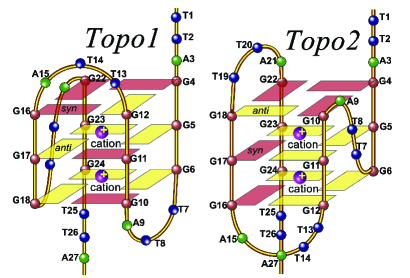
Charge transfer along double-stranded DNA has attracted much attention among biomedicine, chemistry and physics communities in the past decade.[1, 2] As a specialized DNA sequence, telomeres involve many essential physiological processes, DNA damage, cell replication, aging, genetic stability and cancer.[3, 4] Human telomeres consist of tandem repeats of the hexanucleotide 5-10kb in length 5’-3’ toward the chromosome end, terminating in a single-stranded 3’-overhang of 100-200 bases.[5] In normal cells, the telomere shortens with each cell replication. When a critical length is reached, cells undergo apoptosis.[6] Nevertheless, 80-90% of cancer cells preserve their telomere length by the activation of telomerase and thus become abnormal.[7] Interestingly, the G-rich sequence can fold into G-quadruplex (G4) (see in Fig.1), which is a secondary structure consisting of stacked G-tetrad planes connected by a network of Hoogsteen hydrogen bonds and stabilized by centre monovalent cations, such as and . For human telomeric DNA, the repeats of the telomere form the telomere G4 (TG4), where intramolecular G4 forms three-layer packet (Fig.1),[8] which can form several topological structures. It has been found that the formation of TG4 inhibits telomerase activity, which obstructs tumor immortal mechanism.[9] Hence, TG4 has attracted extensive studies as an attractive target for cancer therapeutic intervention.[3]
Moreover, because of DNA self-assembly properties, its paring specificity and conformational flexibility offer great potential for the rational design of DNA-based nanostructure and nano electronics.[10] Different topological structures of G4 DNA provide more possibilities for nano devices.
However, there are divergent opinons on charge transfer in the G-rich DNA sequences. Sugiyama et. al. believe that the G base is favor for charge transfer because the ionization potential of G base is lowest among the nucleobases.[11] The numerical study also shows G4 DNA favoring charge transport based on a simplified mono-G tight-binding G4 model.[12] In fact, charge transfer in G4 could be quite different from normal DNA due to the cation effect as a new hydrogen bond in G4 DNA. Barton’s group found experimentally that G4 has great trapping potency on charges by chemical florescence method.[13] Actually, the problem of charge transfer in G4 is still open because of the complicated topological structures of G4. The full understanding of charge transfer in G4 requires comparing different topological structures of G4 DNA. However, it is not easy to compare functions of different topological structures of G4 by biomedical methods. Physical method can provide a microscopic insight to understand the mechanism of the G4 functions in physiological process and charge transfer for nano devices.
In this paper we will study charge transfer in intramolecular TG4 by the effective tight-binding model with nonequilibrium Green’s function (NEGF) method. Investigating the current-voltage (I-V) characteristics of two predominant topological structures of TG4 in vivo, we find novel charge transfer properties of TG4 beyond the classical charge transfer law, which provide basic properties for designing G4-based nano devices and inspire a physical mechanism on the telomerase activation inhibited by TG4.[9]
2 General G4 model
The TG4 DNA can be viewed as comprising of four parallel stacks for conducting charge channels through the superposition of orbitals along DNA molecules, which is observed by the NMR spectroscopy and x-ray crystallography.[14] We consider the two ends of G4 DNA connected with two semi-infinite one-dimensional electrodes. The Hamiltonian of G4-DNA model can be written as
| (1) |
The charge channels in the backbone of G4 DNA is very small and the orbit channel dominates charge transfer in G4 DNA.[10] Thus, the effective tight-binding Hamiltonian of G4 DNA can be expressed as
where is the on-site energy of each bases,[11] where the second term is the ion effect in G4 packet, where is the distance between the ion and electrons, and is the effective dielectric constant.[15] The second term in Eq.(2) describes the electron hoppings between Gs in G4 packet. The is the nearest electron hopping parameters listed in table I.[11, 16] The notation 5’-XY-3’ indicates the direction along the DNA strand (see,Fig.1(b) [17]. The describes the hopping amplitude between Gs in G4 packet. The in Eq.(1) is the electrodes at the left and right ends of G4 DNA,
| (3) |
The in Eq.(1) is the couplings between the G4 DNA and electrodes,
| (4) |
The is the creation (annihilation) operator of electron or hole at k sites. The ( ) is the on-site energy (hopping parameter) in the electrode, respectively. To minimize the contact effect, we use the optimal injection condition and the strong coupling in the electrode ,[18] where denotes A, C, G, and T.
The on-site energy and the electron hopping parameter between nearest bases. 5’-XY-3’(eV) X/Y G C A T G 0.119 -0.295 -0.186 0.334 C 0.026 0.042 -0.008 -0.161 A -0.013 0.091 -0.038 -0.157 T 0.044 -0.066 -0.068 0.180 7.75 8.87 8.24 9.14
Since realistic DNA molecules are under physiological conditions, we consider DNA having water and ion environment. These water and ion environments of provides a stochastic electric field background to influence the structure as a stochastic perturbation. We introduce the describing these environment effects by the stochastic fluctuation of the structure. Thus, the has the same form to , but the parameters are and , where describes the stochastic strength and randomly.[19, 20]
It should be remarked that we introduce the electrode such that we can calculate the I-V characteristic, which can be compared directly with experiments. For the case of G4 DNA in vivo, suppose that the physiological environment provides an electric potential bias to G4 DNA. Thus, the situation of G4 DNA in vivo maps similarly to the case we consider.
Using NEGF Method, the current can be expressed in terms of [21]
| (5) |
where is the retard (advanced) Green’s Function; the describes the level-width function, where is the retard (advanced) self-energy of electrodes. The is the Fermi function of the left (right) electrodes.
3 TG4s and their classifications
In general, the telomere sequence can be folded to many types of TG4 DNA. The human telomere has been found to be folded to two topological structures (TP1 and TP2) in vivo shown in Fig.1.[15] The TG4 packet as an basic element can be constructed to different configurations of TG4 DNA, which can be classified into three classes, the consecutive mono(hybrid)-TG4(CM(H)TG4) DNA, and non-consecutive mono(hybrid) TG4 (NCM(H)TG4) DNA. Moreover, we may compare the TG4 packet with and without ion. For convenience, we label the TG4 DNA by or , where sn labels the telomere sequence ; the labels the TG4 topological structure and the last and labels ion and absence of ion. For example we define and when is folded to two TP1s and TP2s with ion. We label it by that is CHTG4. When is folded to one TP1 and two TP2 without ion. We label it as that is NCHTG4, where 0 labels the sequence of non-TG4 packet.
4 Topological effect of charge transfer
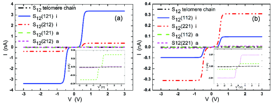
In order to capture the basic physics of the charge transfer in , we first turn off the environment effects . For CMTG4, , it can be folded to only one TG4 packet. We compare the I-V characteristic of the s4 telomere chain and two topological structures (TP1 and TP2) of TG4 with and without ion, namely , , , and , shown in Fig.2 (a). The s4 chain is of little conductance compared with CMTG4. The and show semiconducting with the energy gap 0.3eV. The currents saturate at around 1V due to only finite states for molecule systems. The saturated currents of and reach 6nA and 1nA respectively, which is much higher than that of , and (0.06nA). That means that the ion in TG4 enhances charge transfer. For longer telomere sequences, , and its CMTG4, the basic behaviors of the I-V characteristics are similar to that of the s4 case, but the energy gaps for the non-ion cases are very small. The saturated currents of is larger than that of , but reverse for the case, which can been seen in Fig.2 (b).

For CHTG4 the basic physical behavior of I-V characteristics is same to that of CMTG4. They are also semiconductor with energy gap and the saturated currents of the case with ion are much larger than that of the cases without ion. The saturated currents depend on the configurations of the topological structures of TG4 shown in Fig.3.
For NCHTG4 in Fig.4, the saturated currents of NCHTG4 with and without ion become very small and actually vanish for some cases, which is quite different from CMTG4 and CHTG4. The saturated current of the telomere chain is larger than that of NCM(H)TG4. It implies that TG4 in NCM(H)TG4 can trap charges, which is consistent with the experimental results.[13]
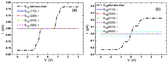
Charge transport in NCM(H)TG4 exhibits novel topological properties. In common sense of charge transport in classical and quantum systems, the global conductance of the system is proportional to the local conductance of the system. However, for NCH(H)TG4, the global conductance is inversely proportional to the local conductance. The conductance of TG4-DNA system may be regarded as to be proportional to the saturated current. We find the conductance from results in Figs.2 and 3. When the telomere chain forms NCMTG4, , the conductance becomes shown in Fig.4. It implies that the global conductance of NCM(H)TG4 is inversely proportional to the local conductance even though the local conductance . In other words, charge transfer in NCM(H)TG4 exhibits nonlinearity. The topology of TG4 induces the anomalous charge transport. We can understand these novel charge transfer properties in NCM(H)TG4 from quantum mechanics. Physically, mobile charges form Bloch wave in a periodic or quasi-periodic structures of CM(H)TG4, while mobile charges in NCM(H)TG4 are trapped due to Anderson’s localization effect in disorder chain. That is why charges transfer favorably in the CM(H)TG4. Actually, DNA sequence may be regarded as a quantum system. Charge transfer in DNA sequence has been found exhibiting some quantum nature.[2] The anomalous conductance in NCM(H)TG4 we find is a novel quantum property of charge transfer induced by topology of TG4 DNA.
5 Topological structure transition and mechanism on telomerase activation and inhibition
The charge transfer properties of TG4 DNA can give a physical clue to understand TG4 how to activate or inhibit telomerase. It has been found that cancer cells obtain their immortality by activating telomerase to preserve the telomere length.[7] The telomerase activation or inhibition is related to TG4 DNA.[7] The challenging problem is how to activate or inhibit the telomerase.[22, 23] Suppose that the ligands of the telomerase offer an electric bias on the telomere DNA to form a circuit inducing charge current. The topological structure transition from NCM(H)TG4 to CM(H)TG4 generates abruptly the charge current from (see Figs. 2 and 4). This current as a threshold current can activate or inhibit telomease. This scenario can provide a clue to understand TG4 blockading the cancer cell immortal pathways and eventually perishing tumors in the biomedical experimental observation.[9] This finding inspires a further challenging problem for biomedicine how to realize or control the topological structure transition between NCM(H)TG4 and CM(H)TG4.

6 Discussion
It should be remarked that the ratio of the saturated currents between CM(H)TG4 and NCM(H)TG4 has orders. The solvent leads only eV on-site energy fluctuation to DNA bases, which cannot lead to higher G4 structures.[20] We investigate the environment effects in Fig. 5 for two cases. When the environment fluctuation is small , the error bar of the ratio of the currents for the is larger than that of the , which implies that the current fluctuation depends on the detail configurations of the TG4 in the small environment fluctuation. When the environment fluctuation becomes larger, , the current fluctuation trends to be insensitive to the detail configurations of TG4. More importantly, we do see that the -order current difference induced by the topological structure transition from NCM to CM TG4s is robust against the environment fluctuation. Moreover, it has been found that two basic mechanisms of charge transfer in DNA chain, coherent tunneling (hopping), and thermal diffusive hopping.[24] The coherent tunneling or hopping occurs betwen pairs and the thermal hopping occurs in more than consecutive pairs.[24] For TG4, , the coherent hopping dominates the charge transfer due to only consecutive pairs in TG4. Thus, the coherent transport method is valid for TG4. In addition, the energy gap at Fermi energy is about for TG4, which is order higher than the influence of temperature fluctuation in room temperature, and the charge transfer in TG4 is not sensitive to temperature fluctuation because the coherent hopping dominates the charge transfer in TG4 instead of the thermal diffusive hopping. Namely, temperature fluctuation in room temperature is not sensitive for our results. Therefore, in room temperature all our conclusions are invariant.
7 Conclusion
In summary, we reveal novel charge transport properties in TG4 DNA. The CM(H)TG4 is semiconducting with energy gap. The conductances of different configurations of TG4 DNA follow the inequalites, , where means telomere chain. The conductances depend on the topological structures and configurations of TG4. The cation in TG4 enhances charge transfer. The TG4 packet in NCM(H)TG4 suppresses charge transfer, which agrees with the experimental results.[13] The topological structure transition between NCM(H)TG4 and CM(H)TG4 induces a threshold charge current to activate or inhibit telomease. Our findings not only reveal novel charge transfer properties in TG4 DNA that can offer many opportunities for DNA-based electronics, but also provide physical insights and hints to understand the telomerase activation inhibited by TG4.
The biomedical investigation found that tumor growth is related to the telomerase activation and inhibition. The telomerase activation and inhibition is related to TG4. [7] It implies strongly that states of TG4 could be related tumor growth even cancer. However, the biomedical investigation cannot find the working mechanism of these three factors, TG4, tumor growth and telomerase activation and inhibition. Our results provide an understanding of the working mechanism of the telomerase activation and inhibition. We found that the topological structure transition from NCM(H)TG4 to CM(H)TG4 generates abruptly the charge current from (6 order difference) when the ligands of the telomerase offer an electric bias on the telomere DNA. It strongly implies that this abrupt current as a threshold current can activate or inhibit telomease. Namely, if we can control the topological structure transition from NCM(H)TG4 to CM(H)TG4 we can control the tumor growth. This scenario can provide a hint to understand TG4 blockading the cancer cell immortal pathways and eventually perishing tumors in the biomedical experimental observation.[9] Consequently, the further challenging issue is how to tune the topological structure transition of TG4 by biomedical method, which is expected to further study.
These novel charge transfer properties in TG4 DNA give some fundamental relationships between topology, anomalous charge transport and biomedical function.
Acknowledgements
The authors acknowledge the financial supports of the projects from the Elite Student Program from National Education Department, and the Fundamental Research Funds for the Central Universities.
References
References
- [1] Hans Achim Wagenknecht, Charge transfer in DNA, Wiley-VCH Verlag GmbH &Co.KGaA, (2005).
- [2] Tapash Chakraborty, Charge Migration in DNA, Springer-Verlag, (2007).
- [3] J.L. Mergny, C. Helene, Nat. Med.,4, 1366 (1988) ; L.H. Hurley, Nat. Rev. Cancer,2, 188 (2002); S. Neidle, G. Parkinson, Nat. Rev. Drug Discov., 1, 383 (2002); A.G. Bodnar, et.al., Science,279, 349 (1998).
- [4] J.A. Hackett, D.M. Feldser, C.W. Greider, Cell, 106, 275 (2001).
- [5] E.H. Blackburn, Nature 53, 408 (2000); V.L. Makarov, Y. Hirose, J.P.,Cell 88, 657 (1997).
- [6] C.B. Harley, A.B. Futcher, C.W. Greider, Nature 345, 458 (1990).
- [7] N.W. Kim, M.A. Piatyszek, K.R. Prowse, C.B. Harley, M.D. West, P.L. Ho, G.M. Coviello, W.E. Wright, S.L. Weinrich, J.W. Shay, Science 266, 2011 (1994).
- [8] J. Dai, M. Carver, C. Punchihewa, R.A. Jones and D.Yang, Nucleic Acids Research 35, 4927 (2007).
- [9] A.M. Zahler, J.R. Williamson, T.R. Cech, D.M. Prescott, Nature 350, 718 (1991); S.Balasubramanian,S. Neidle, Curr. Opin. Chem. Biol. 13, 345 (2009).
- [10] G. Cuniberti, E. Macia A. Rodriguez, and R. A. Romer, Charge Migration in DNA edited by T.Chakraborty, Springer-Verlag, Berlin, (2007).
- [11] H. Sugiyama and I. Saito, J. Am. Chem. Soc. 118, 7063 (1996).
- [12] A.M. Guo, S.J. Xiong, Phy. Rev.B80, 035115 (2009).
- [13] S.Delaney, J.K. Barton, Biochemistry 42, 14159 (2003).
- [14] J. Feigon, Nature 356, 164 (1992); F. Aboul-ela, A. I. H. Murchie, and D. M. J. Lilley, Nature 360, 280 (1992).
- [15] S. Neidle, S. Balasubramanlian, Quadruplex Nucleic Acids , RSC Publishing, Cambridge, (2006).
- [16] K. Senthilkumar, et.al., J. Am. Chem. Soc. 127, 14894 (2005).
- [17] R.G. Endres, D.L.Cox, R.R.P.Songh, Rev. Mod. Phys. 76, 195 (2004).
- [18] E. Maciá, F. Triozon, and S. Roche, Phys. Rev. B71, 113106 (2006); X. F. Wang, T. Chakraborty, Phys. Rev. Lett. 97, 106602 (2006).
- [19] B.Giese, J.Amaudrut, A. Kőhler, M.Spormann, S.Wessely, Nature 412, 318 (2001); S. Roche, Phys. Rev. Lett. 91, (2003) 108101; S. Roche, D.Bicout, E. Maciá, E.Kats., Phys. Rev. Lett. 91, 228101 (2003).
- [20] P. Benjamin Woiczikowski, et. al., J. of Chem. and Phys. 133, 035103 (2010); R. Gutierrez, et. al., Phys. Rev. Lett. 102, 208102 (2009).
- [21] J.K.Viljas, J.C.Cuevas, F.Pauly, and M.Häfner, Phys. Rev. B72, 245415 (2005).
- [22] N.W. Kim, et. al., Science 266, 2011 (1994).
- [23] D. Hanahan, R.A. Weinberg, Cell 100, 57 (2000).
- [24] B. Giese, Acc. Chem. Res. 33, 631 (2000).