Rotational state microwave mixing for laser cooling of complex diatomic molecules
Abstract
We demonstrate the mixing of rotational states in the ground electronic state using microwave radiation to enhance optical cycling in the molecule yttrium (II) monoxide (YO). This mixing technique is used in conjunction with a frequency modulated and chirped continuous wave laser to slow longitudinally a cryogenic buffer gas beam of YO. We generate a measurable flux of YO below 10 m/s, directly loadable into a three-dimensional magneto-optical trap. This technique opens the door for laser cooling of molecules with more complex structure.
pacs:
37.10.Mn, 37.10.Pq, 37.10.VzThe field of cold and ultracold molecules is undergoing rapid growth with the development of new techniques and applications Carr et al. (2009). Cold polar molecules are continuing to improve tests of fundamental symmetries Baron et al., (2014); Hudson et al. (2011); Hunter et al. (2012); Zhuang et al. (2011); Isaev et al. (2010). The strong dipolar interaction present in polar molecules also yields a rich set of applications in novel quantum matter Büchler et al. (2007); Yao et al. (2013); Yan et al. (2013) and cold chemistry Ospelkaus et al. (2010); Bell and Softley (2009); Sawyer et al. (2011).
In many of these applications it is desirable to have a slow source of molecules to either enhance the interaction time or for loading into a trap. The magneto-association and coherent state transfer of ultracold alkali atoms Ni et al. (2008) can produce polar molecules near quantum degeneracy. Supersonic beams produce molecules with forward velocity 300 m/s Miller (1988), whereas cryogenic buffer gas beams have velocities of 50-200 m/s, depending on source configuration Hutzler et al. (2012) and with the lower velocity beams directly loadable into a trap Lu et al. (2014). Many techniques to further slow these beams have been developed. Stark van de Meerakker et al. (2006); Sawyer et al. (2008) and Zeeman Narevicius et al. (2008); Hogan et al. (2008) deceleration have been used to load conservative traps, which can be used for evaporation Stuhl et al. (2012). Centrifugal slowing Chervenkov et al. (2014) and opto-electric cooling was demonstrated with CH3F molecules Zeppenfeld et al. (2012). The rovibrational branching that prevents optical cycling transitions in molecules was addressed theoretically Di Rosa (2004); Stuhl et al. (2008) and optical Doppler cooling was subsequently demonstrated in the SrF system Shuman et al. (2010). Magneto-optical trapping was proposed for TiO Stuhl et al. (2008) and demonstrated in two dimensions for YO Hummon et al. (2013) and in three dimensions for SrF Barry et al. (2014); McCarron et al. .
While it is possible to load directly a buffer-gas cooled atomic beam into a three-dimensional (3D) magneto-optic trap (MOT) Hemmerling et al. (2014), for molecules with much lower photon scattering rates longitudinal optical cooling is necessary for loading into a 3D MOT. Typically photons must be scattered to slow molecules to within the capture velocities of a normal MOT. During the slowing process, the molecular transition used for optical cooling experiences a Doppler frequency shift of tens or hundreds of resonance linewidths, resulting in inefficient photon scattering for a monochromatic unchirped laser beam. To date, molecules that have been laser cooled all have the magnetic sub-levels in the ground electronic state continually remixed by the multi-leveled optical cycling process. Hence, the widely used atomic Zeeman slower Phillips and Metcalf (1982), which relies on the Zeeman shift of a single magnetic sublevel to compensate for the Doppler shift during deceleration is not applicable. To maintain a sufficiently large optical scattering rate throughout the slowing process, two techniques have been employed. Chirping the laser frequency was shown to reduce the velocity of a supersonic beam of CaF by 30 m/s Zhelyazkova et al. (2014). For SrF, broadband laser radiation was used to slow molecules to be loaded directly into a 3D MOT Barry et al. (2014).
The advent of optical cycling in molecules Stuhl et al. (2008); Shuman et al. (2010) is critical for the radiation pressure force to directly slow and cool molecular beams. However, despite the achievement of quasi-closed optical cycling in several molecular systems, preventing optical pumping into dark states still remains difficult for a large class of molecules. While vibrational dark states can be addressed with repump lasers Di Rosa (2004), rotational dark states often have parity selection rules that prevent direct optical repumping. There are several loss mechanisms that break the rotational closure in the current schemes for molecular optical cycling transitions, and they arise from additional decays via intermediate states or higher order transition moments. Vibrational states typically have lifetimes 1 s Vanhaecke and Dulieu (2007), and rovibrational decay is not yet a limiting factor for current techniques. Decays via magnetic dipole transitions are typically suppressed by a factor of compared to electric dipole transitions ( is the fine structure constant), though recent measurements in OH indicate that they can sometimes be a factor of 10 faster than expected Kirste et al. (2012). Decays through intermediate electronic states, present in for example YO Chalek and Gole (1976) and BaF Allouche et al. (1993), are suppressed by the relatively long transition wavelengths. For YO, the suppression factor is , however the decay is sufficient to cause population leakage to rotational dark states that reduce photon scattering. In this Letter, we report microwave mixing of rotational states to close these additional loss channels. This enables us to slow longitudinally a beam of YO emitting from a two-stage buffer gas cell to molecular velocities 10 m/s, which can be loaded into a 3D MOT.
The optical cycling transition in YO proceeds between the states as described in Ref. Hummon et al. (2013). In brief, highly diagonal Franck-Condon factors limit vibrational branching Bernard and Gravina (1983) such that a single repump laser limits branching loss to (Fig. 1(a)). Rotational branching is suppressed by a combination of parity and angular momentum selection rules Stuhl et al. (2008); Shuman et al. (2009) when driving the level, where is the rotation quantum number. Figure 1(b) shows the hyperfine structure of the states, where is the coupled nuclear and electronic spin, and is the total angular momentum. For the excited and states, is the total angular momentum excluding nuclear spin. For all three electronic states, represents the parity.
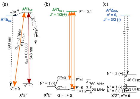
Figure 1(a) shows the additional loss channel where the , state decays to the , state. Decays to states of higher are forbidden due to angular momentum selection rules. This state has a radiative lifetime of 1 s Chalek and Gole (1976) and will rapidly decay back to the state. Since this is a three-photon process back to the ground state, parity selection rules will only allow decays to even rotational states (Fig. 1(c)). In the state, while is not a good quantum number, each state of can be expressed as a superposition of states with (for , J = 1/2). The angular momentum selection rule prevents decay to and thus leads to optical pumping into the states. The rotational constant, , for the state is 11.634 GHz. Microwave radiation tuned to and can mix the levels respectively with the level. This mixing effectively removes the rotational dark states; however it also lowers the maximum optical scattering rate as the number of states in involved in optical cycling increases by a factor of two.
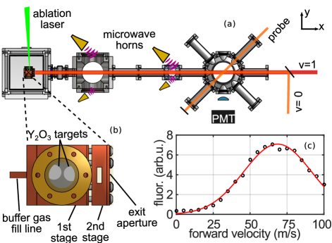
We employ this rotational state microwave mixing scheme for longitudinal slowing of a YO beam generated from a two-stage cryogenic buffer gas beam source Hutzler et al. (2012). We laser ablate (with a 10 Hz repetition rate) sintered Y2O3 pellets in a copper cell filled with helium buffer gas at 3.5 K (Fig. 2(a)). The hot YO thermalizes rotationally and translationally to the cold helium buffer gas and is hydrodynamically extracted through a 3 mm aperture into the second stage cell. By virtue of the 4.5 mm gap between the two stages and the larger exit aperture diameter of mm, the helium density is times smaller in the second stage than that of the first stage and the molecules experience only a few additional collisions before exiting the second stage cell. This slows the m/s beam from the first stage Hummon et al. (2013) to m/s (Fig. 2(c)). The YO beam then travels cm to the detection region, during which it may be illuminated by a 6 mm diameter, counter-propagating slowing beam. The nm laser has 70 mW of power and the nm laser has mW (Fig. 1(a)). The slowing laser has three frequency components to address the hyperfine manifold (Fig. 1(b)). To provide the rotational mixing, we use two pairs of microwave horns strategically located along the beam path, each pair producing 1 mW of either the 23 GHz or 46 GHz microwaves.
For velocity sensitive detection, we measure the fluorescence induced by a low intensity 4 mW/cm2, 614 nm beam aligned to the molecular beam. This probe laser has a saturation parameter of 0.5 and two frequency components separated by 780 MHz (Fig. 1(b)). This eliminates mechanical and optical pumping effects from the detection beam. The detection laser can be scanned over a range of 100 MHz at a rate of 1 kHz, corresponding to a velocity detection range of 100 m/s.
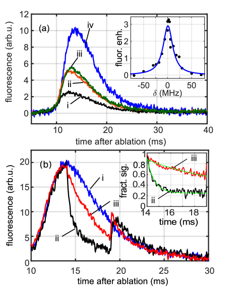
We study the effects of rotational mixing by observing the fluorescence induced by the slowing beam in Fig. 3(a). We apply the microwaves in four binary combinations. For case i, no microwaves are applied. In contrast, when microwave mixing is applied to both , (curve iv), there is a factor of four increase in fluorescence. Applying only the or mixing (Fig. 3(b) ii, iii respectively), we see a factor of two increase in fluorescence compared to the no mixing case. We estimate the vibrational lifetime of the state from molecular parameters calculated in Ref. Langhoff (1988) to be 600 ms, which is much longer than the optical pumping time scale. The branching ratios for magnetic dipole transitions from the state is 2/3(1/3) for the levels in the state, while electric dipole transitions from the state have equal branching ratios to the rotational levels, consistent with curves ii and iii. Hence, decays through the intermediate state are identified to be the dominant process.
To verify that the mixing microwaves are sufficiently strong to address all hyperfine states without the need of additional frequency sidebands, we simultaneously vary the frequency of the mixing microwave by and measure the corresponding enhancement in fluorescence (Fig. 3(a) inset). This enhancement follows a Lorentzian lineshape with a FWHM of MHz, which is larger than the hyperfine variations of 5 MHz among the three relevant rotational manifolds Childs et al. (1988); Brown and Carrington (2003). The mixing for higher vibrational states is suppressed because the rotational constant for is MHz smaller than that for . This results in a MHz detuning for the transition in . Hence, we do not suffer any further reduction in the maximum optical cycling rate.
Figure 3(b) shows a measurement of the branching loss from the state to the state. Again we measure the fluorescence induced by the high power slowing beam. For case i, the slowing and repumping light as well as microwaves are turned on at all times. For case ii, the repump laser is rapidly shut off in 10 s from ms to 19 ms after ablation. This leads to an abrupt decrease in the fluorescence as the YO molecules are rapidly pumped into the state and hence turn dark. To extract this pumping rate, we divide curve (ii) by curve (i) and fit an exponential with a constant background ratio of 0.25, resulting in a time constant of 620 s. Combining this with the calculated Franck-Condon factor Bernard and Gravina (1983), yields a photon scattering rate of s-1. This is smaller than the maximum possible scattering rate by a factor of 5 Hummon et al. (2013) and may be due to inefficient depletion due to highly Doppler detuned molecules, insufficient power in the wings of the slowing beam or an overestimate of the Franck-Condon factor. Curve iii shows the fluorescence when only the rotational mixing microwaves are turned off between ms. The decay due to pumping into and 2 dark states has a ms time constant (inset curve iii). By combining the ratio of the two loss rates and , we estimate the branching ratio from to to be . This branching ratio agrees with the value derived from the transition dipole moments calculated in Ref. Langhoff (1988) and is similar to the combined branching ratio to . At 19 ms after ablation, the vibration repump (curve ii) and mixing microwaves (curve iii) are turned back on and the fluorescence immediately recovers.
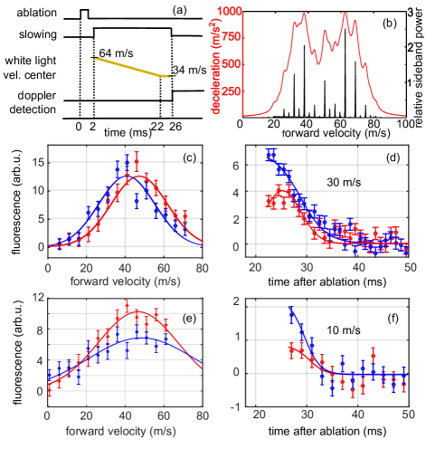
We estimate the capture velocity of a 3D MOT for YO with a laser beam diameter of 1 cm to be m/s. No detectable population of 10 m/s molecules exists in the buffer gas beam source. To slow some fraction of the molecules to below this capture velocity, we apply longitudinal radiation pressure slowing with protocols shown in Fig. 4(a). To increase the fraction of molecules addressed by the slowing beam, we spectrally broaden the light Zhu et al. (1991) via a resonant electro-optic modulator (EOM) with a modulation frequency of MHz and a phase modulation index of . This spectrally broadens each laser frequency to MHz, corresponding to a velocity spread of m/s. Polarization switching used for destabilizing Zeeman dark states Hummon et al. (2013) further adds MHz sidebands and thus we obtain frequency components spaced by the natural linewidth of the main cooling transition, ensuring that we have continuous deceleration across this velocity range (Fig. 4(b)).
As the range of the slowing force is smaller than the velocity spread of the YO beam itself, we additionally implement frequency chirping on the slowing light. In Fig. 4(c) and (d), the slowing sequence is designed to enhance molecules at m/s. The molecules propagate freely for the first 12 ms after ablation, then the slowing beam is turned on from 12-22 ms. During this time, the center of the broadband radiation is linearly swept from being resonant with m/s to m/s molecules. In panel (c), we see a molecular population enhancement between m/s (negligible for below m/s), and depletion for velocities greater than 40 m/s. In panel (d), we maintain the probe laser’s frequency to be resonant with the m/s molecule and we observe a clear enhancement in the number of molecules in this velocity class compared to the original beam. Due to off-resonant excitations, some faster molecules ( m/s) contribute to the detected signal at 27 ms for the unslowed case.
To produce even slower molecules, we increase the time during which the slowing laser is turned on and perform a frequency chirp to address lower velocity molecules. The slowing light is turned on 2 ms after ablation and is left on for the next 24 ms. During the first ms, the center frequency of the broadband slowing laser is linearly increased from being resonant with 64 m/s to 34 m/s molecules (Fig. 4(a)). Figure 4(e) shows the velocity distribution after the slowing sequence compared to the original velocity distribution. Slowing enhances the molecular population below 20 m/s in the detection region and depletes molecules with velocities higher than 20 m/s. Again, we tune the Doppler-sensitive probe laser to be resonant with the 10 m/s molecules and we see a clear enhancement in the number of molecules in this velocity class (Fig. 4(f)). Unslowed molecules leaving the cell with 10 m/s forward velocity will arrive 89 ms after the ablation, well after the detection time. Hence, we attribute the non-zero 10 m/s signal in the unslowed case to off-resonant excitations from fast molecules.
We have demonstrated enhanced optical cycling in YO via microwave mixing of rotational states in the ground electronic state. This enabled us to slow longitudinally the YO beam via radiation pressure, and hence produce molecules that can be loaded directly into a 3D MOT. To further close the vibrational branching to , an additional repump laser at nm can be used Hummon et al. (2013); Bernard and Gravina (1983). By turning off the microwave mixing for at an appropriate time, we can accumulate trapped cold YO in the ro-vibrational ground state. The state lifetime is 1 s, which is 10 times longer than that of . This opens up the possibility of a narrow line MOT Loftus et al. (2004); Collopy et al. with a Doppler temperature of K as compared to the K obtainable from the , to transition.
Acknowledgements.
We thank M. Petzold and M. Kuhnert for their contributions to the early stage of this work. We acknowledge funding support from ARO and AFOSR (MURI), Gordon and Betty Moore Foundation, NIST, and NSF.References
- Carr et al. (2009) L. D. Carr, D. DeMille, R. V. Krems, and J. Ye, New. J. Phys. 11, 055049 (2009).
- Baron et al., (2014) (The ACME Collaboration) J. Baron et al., Science 343, 269 (2014).
- Hudson et al. (2011) J. J. Hudson, D. M. Kara, I. J. Smallman, B. E. Sauer, M. R. Tarbutt, and E. A. Hinds, Nature 473, 493 (2011).
- Hunter et al. (2012) L. R. Hunter, S. K. Peck, A. S. Greenspon, S. S. Alam, and D. DeMille, Phys. Rev. A 85, 012511 (2012).
- Zhuang et al. (2011) X. Zhuang, A. Le, T. C. Steimle, N. E. Bulleid, I. J. Smallman, R. J. Hendricks, S. M. Skoff, J. J. Hudson, B. E. Sauer, E. A. Hinds, and M. R. Tarbutt, Phys. Chem. Chem. Phys. 13, 19013 (2011).
- Isaev et al. (2010) T. A. Isaev, S. Hoekstra, and R. Berger, Phys. Rev. A 82, 052521 (2010).
- Büchler et al. (2007) H. P. Büchler, E. Demler, M. Lukin, A. Micheli, N. Prokof’ev, G. Pupillo, and P. Zoller, Phys. Rev. Lett. 98, 060404 (2007).
- Yao et al. (2013) N. Y. Yao, A. V. Gorshkov, C. R. Laumann, A. M. Läuchli, J. Ye, and M. D. Lukin, Phys. Rev. Lett. 110, 185302 (2013).
- Yan et al. (2013) B. Yan, S. A. Moses, B. Gadway, J. P. Covey, K. R. A. Hazzard, A. M. Rey, D. S. Jin, and J. Ye, Nature 501, 521 (2013).
- Ospelkaus et al. (2010) S. Ospelkaus, K.-K. Ni, D. Wang, M. H. G. de Miranda, B. Neyenhuis, G. Quéméner, P. S. Julienne, J. L. Bohn, D. S. Jin, and J. Ye, Science 327, 853 (2010).
- Bell and Softley (2009) M. T. Bell and T. P. Softley, Mol. Phys. 107, 99 (2009).
- Sawyer et al. (2011) B. C. Sawyer, B. K. Stuhl, M. Yeo, T. V. Tscherbul, M. T. Hummon, Y. Xia, J. Klos, D. Patterson, J. M. Doyle, and J. Ye, Phys. Chem. Chem. Phys. 13, 19059 (2011).
- Ni et al. (2008) K. Ni, S. Ospelkaus, M. H. G. D. Miranda, A. Pe’er, B. Neyenhuis, J. J. Zirbel, S. Kotochigova, P. S. Julienne, D. S. Jin, and J. Ye, Science 322, 231 (2008).
- Miller (1988) D. R. Miller, Atomic and Molecular Beam Methods, edited by G. Scoles (Oxford University Press, 1988) p. 20.
- Hutzler et al. (2012) N. R. Hutzler, Hsin-I. Lu, and J. M. Doyle, Chem. Rev. 112, 4803 (2012).
- Lu et al. (2014) H. -I. Lu, I. Kozyryev, B. Hemmerling, J. Piskorski, and J. M. Doyle, Phys. Rev. Lett. 112, 113006 (2014).
- van de Meerakker et al. (2006) S. Y. T. van de Meerakker, N. Vanhaecke, and G. Meijer, Annu. Rev. Phys. Chem. 57, 159 (2006).
- Sawyer et al. (2008) B. C. Sawyer, B. K. Stuhl, D. Wang, M. Yeo, and J. Ye, Phys. Rev. Lett. 101, 203203 (2008).
- Narevicius et al. (2008) E. Narevicius, A. Libson, C. G. Parthey, I. Chavez, J. Narevicius, U. Even, and M. G. Raizen, Phys. Rev. Lett. 100, 093003 (2008).
- Hogan et al. (2008) S. D. Hogan, A. W. Wiederkehr, H. Schmutz, and F. Merkt, Phys. Rev. Lett. 101, 143001 (2008).
- Stuhl et al. (2012) B. K. Stuhl, M. T. Hummon, M. Yeo, G. Quéméner, J. L. Bohn, and J. Ye, Nature 492, 396 (2012).
- Chervenkov et al. (2014) S. Chervenkov, X. Wu, J. Bayerl, A. Rohlfes, T. Gantner, M. Zeppenfeld, and G. Rempe, Phys. Rev. Lett. 112, 013001 (2014).
- Zeppenfeld et al. (2012) M. Zeppenfeld, B. G. U. Englert, R. Glöckner, A. Prehn, M. Mielenz, C. Sommer, L. D. van Buuren, M. Motsch, and G. Rempe, Nature 491, 570 (2012).
- Di Rosa (2004) M. Di Rosa, Eur. Phys. J. D. 31, 395 (2004).
- Stuhl et al. (2008) B. K. Stuhl, B. C. Sawyer, D. Wang, and J. Ye, Phys. Rev. Lett. 101, 243002 (2008).
- Shuman et al. (2010) E. S. Shuman, J. F. Barry, and D. Demille, Nature 467, 820 (2010).
- Hummon et al. (2013) M. T. Hummon, M. Yeo, B. K. Stuhl, A. L. Collopy, Y. Xia, and J. Ye, Phys. Rev. Lett. 110, 143001 (2013).
- Barry et al. (2014) J. F. Barry, D. J. McCarron, E. B. Norrgard, M. H. Steinecker, and D. DeMille, Nature 512, 286 (2014).
- (29) D. J. McCarron, E. B. Norrgard, M. H. Steinecker, and D. DeMille, arXiv:1412.8220 .
- Hemmerling et al. (2014) B. Hemmerling, G. K. Drayna, E. Chae, A. Ravi, and J. M. Doyle, New J. Phys. 16, 063070 (2014).
- Phillips and Metcalf (1982) W. D. Phillips and H. Metcalf, Phys. Rev. Lett. 48, 596 (1982).
- Zhelyazkova et al. (2014) V. Zhelyazkova, A. Cournol, T. E. Wall, A. Matsushima, J. J. Hudson, E. A. Hinds, M. R. Tarbutt, and B. E. Sauer, Phys. Rev. A 89, 053416 (2014).
- Vanhaecke and Dulieu (2007) N. Vanhaecke and O. Dulieu, Mol. Phys. 105, 1723 (2007).
- Kirste et al. (2012) M. Kirste, X. Wang, G. Meijer, K. B. Gubbels, A. van der Avoird, G. C. Groenenboom, and S. Y. T. van de Meerakker, J. Chem. Phys. 137, 101102 (2012).
- Chalek and Gole (1976) C. L. Chalek and J. L. Gole, J. Chem. Phys. 65, 2845 (1976).
- Allouche et al. (1993) A. R. Allouche, G. Wannous, and M. Aubert-Frécon, Chem. Phys. 170, 11 (1993).
- Bernard and Gravina (1983) A. Bernard and R. Gravina, Astrophys. J. Suppl. Ser. 52, 443 (1983).
- Shuman et al. (2009) E. S. Shuman, J. F. Barry, D. R. Glenn, and D. DeMille, Phys. Rev. Lett. 103, 223001 (2009).
- Langhoff (1988) S. Langhoff, J. Chem. Phys. 89, 2160 (1988).
- Childs et al. (1988) W. J. Childs, O. Poulsen, and T. C. Steimle, J. Chem. Phys. 88, 598 (1988).
- Brown and Carrington (2003) J. M. Brown and A. Carrington, Rotational Spectroscopy of Diatomic Molecules (Cambridge University Press, 2003).
- Zhu et al. (1991) M. Zhu, C. W. Oates, and J. L. Hall, Phys. Rev. Lett. 67, 46 (1991).
- Loftus et al. (2004) T. H. Loftus, T. Ido, A. D. Ludlow, M. M. Boyd, and J. Ye, Phys. Rev. Lett. 93, 073003 (2004).
- (44) A. L. Collopy, M. H. Hummon, M. Yeo, B. Yan, and J. Ye, “Prospects for a narrow line MOT in YO,” arXiv:to be published (2015) .