Effect of dibucaine on phase behavior of ternary liposome
Abstract
We investigated the effect of Dibucaine hydrochloride (DCHCl), one of the local anesthetics, on phase behavior of ternary liposome composed of dioleoylphosphatidylcholine (DOPC), dipalmitoylphosphatidylcholine (DPPC), and cholesterol (Chol). The large DOPC/DPPC/Chol liposome, that is directly observable by optical microscope, is commonly known to be laterally separated into liquid-ordered (Lo) phase (raft-like domain) and liquid-disordered (Ld) phase under certain conditions and is useful for study of lipid-raft-like domains as a simple model system. In order to confirm the effect of DCHCl on a miscibility transition temperature, , of the ternary liposome, we observed the liposomes with three concentrations, 0, 0.05, and 0.2 mM, of DCHCl at various temperatures. In addition, we calculated the angle-averaged two-dimensional autocorrelation (2D-AC) functions in order to quantify the phase behavior. The results of these observations and calculations revealed that the DCHCl molecules induce the reduction of of the ternary liposome. Furthermore, we calculated the circularity of Lo domain in order to confirm the change in the line tension of the Lo/Ld phase boundary and revealed that the insertion of the DC molecules induces the reduction of line tension. In terms of the critical phenomena, we conclude that the insertion of the DC molecules induces the reduction of the of the ternary liposome due to reduction of line tension. This suggests that the DC molecules may disturb function of ion channels via affecting the lipid bilayers which surround ion channels.
pacs:
I Introduction
Because it is important to understand the physical mechanism of functional expression of anesthetics in the pharmacology, the effect of anesthetic molecules on biological membrane containing ion channels has been studied for many years. For instance, effect of chloroform and other general anesthetics on sodium current in a squid giant axon and interactions of chloroform and/or benzyl alcohol with lipid bilayer have been studied by using experimental and/or simulation methodsHaydon and Urban (1983); Ebihara et al. (1979); Reyes and Latorre (1979); Reigada (2011, 2013).
Recently, local anesthetics have especially been paid attention and studied. A lot of studies have reported that local anesthetics affect the sodium current across the cell membrane Muroi and Chanda (2009); Hanck et al. (2009); Yamagishi et al. (2009); Arcisio-Miranda et al. (2010); Sheets et al. (2011); Lee et al. (2012); Bant et al. (2013). In addition, interaction between anesthetics and lipid bilayer which surrounds the ion channels has also been studied because it is suggested that the perturbation of the lipids due to invasion of the anesthetics disturbs the function of ion channelsHata et al. (2000, 2001); Matsuki et al. (2001); Takeda et al. (2009); Tsuchiya et al. (2010a). Furthermore, the effect on lipid-raft domains, which are mainly composed of saturated lipids, cholesterol and membrane proteins such as ion channels, and are dispersed in the cell membranes (believed to be a kind of phase separation)Simons and Ikonen (1997) should be investigated in order to clarify the detailed mechanism of local anesthetic function because the lipid rafts are believed to play important roles in the various cell functionsSimons and Ikonen (1997). Therefore, the phase-separated liposome with the raft-like domains has been used as a simple model system of cell membrane for studies of interactions between local anesthetics and lipid bilayersTsuchiya et al. (2010b); Bandeiras et al. (2013). However, the detailed physicochemical mechanism of functional expression of local anesthesia is still unclear.
Large phase-separated liposome, that is observable by optical microscope, is useful for the direct investigations of the raft-like domains. The multi-component liposome is commonly known to be separated into two or three phases due to difference of thermodynamical properties of their lipids, and the phase-separated domains of large liposome are observable by fluorescence microscopy with a dye which is only incorporated into a certain phaseBaumgart et al. (2003); Veatch and Keller (2003a); Diguet et al. (2012); Morita et al. (2012); Shimokawa et al. (2010); Himeno et al. (2014). Indeed, large phase-separated liposome has been used for many studies of the lipid membranes, and the physical properties of the lipid membrane have been revealedBaumgart et al. (2003); Diguet et al. (2012); Morita et al. (2012); Shimokawa et al. (2010); Himeno et al. (2014); Cicuta et al. (2007); Sakuma et al. (2013); Chen and Santore (2014).
In the similar cases to the present study, the large liposome and cell-derived giant plasma membrane vesicle (GPMV) which is similar to the multi-component liposome are also used as a model system. For example, it has been reported that vitamin E, Triton-X 100, and benzyl alcohol affect the phase morphology of large multi-component liposomesMuddana et al. (2012). Gray et al. have also reported that the general anesthetics including alcohols reduce the miscibility transition temperature, , of GPMVsGray et al. (2013). However, a direct observation of the influence of local anesthetics on the phase behavior of large liposome has not been reported.
In the present study, we investigated the influence of dibucaine hydrochloride (DCHCl), one of the commonly used local anesthetics, on phase behavior of liposome composed of dioleoylphosphatidylcholine (DOPC), dipalmitoylphosphatidylcholine (DPPC), and cholesterol (Chol). The DOPC/DPPC/Chol liposome is one of the famous systems and is known to be laterally separated into liquid-ordered (Lo) and liquid-disordered (Ld) phases under certain conditionsVeatch and Keller (2003b). The Lo phase, mainly composed of DPPC and Chol, represents the raft-like domain, and the Ld phase is DOPC-rich regionVeatch and Keller (2003b). We calculated angle-averaged two-dimensional autocorrelation (2D-AC) function of images of liposomes with three concentrations of DCHCl at various temperatures in order to estimate the change in due to the effect of DC. The results indicate that the DC molecules reduce the of ternary liposome.
The 2D-AC and/or angle-averaged 2D-AC functions have been widely used in the various fields such as biologyHwang et al. (1998) and physicsFacsko et al. (1999); Geringer et al. (2009). In addition, the angle-averaged 2D-AC analysis was performed in order to clarify the correlation length of GPMV surface in the similar study to this articleVeatch et al. (2008). However, to our knowledge, this is the first report of quantitatively demonstrating the change in of ternary liposome due to addition of local anesthetics by calculating the angle-averaged 2D-AC function of membrane domain patterns.
II Materials and methods
II.1 Materials
Dibucaine hydrochloride (DCHCl), DOPC (chain melting temperature, )Koynova and Caffrey (1998), DPPC ()Koynova and Caffrey (1998), Chol and chloroform were purchased from Wako Pure Chemical Industries, Ltd. (Japan). The chemical structure of DC is described in Figure 1. Methanol was purchased from Showa Chemical Industry Co., Ltd. (Japan). Rhodamine B 1,2-dihexadecanoyl-sn-glycero-3-phosphoethanolamine (rhodamine DHPE, ), fluorescent phospholipid, was obtained from Invitrogen (U.S.A.). Ultra-pure water was obtained using a WT101UV AUTOPURE (Yamato Scientific Co., Ltd., Japan).

II.2 Preparation of Ternary Liposome
We prepared liposomes using the natural swelling methodsLasic (1988); Yoshida et al. (2014), The lipids of DOPC/DPPC/Chol = 50/25/25 (mol%) with 0.5 mol% of rhodamine DHPE were dissolved in mixture solvent of chloroform and methanol with a volume ratio of 2:1. Then, we removed the organic solvents through an air flow and placing the sample in an aspirated desiccator for more than 8 hours in order to make a dry lipid film. Finally, liposomes formed through hydration of the lipid film with the ultra-pure water at 37 for more than 24 hours. The lipid concentration was 0.2 mM.
II.3 Observation
40 L of liposome suspension and 40 L of DCHCl aqueous solution were gently mixed, and the solution was incubated at room temperature (RT, 21.7 0.4 ) for more than 10 minutes in order to wait for sufficient dispersion of DCHCl molecules into the lipid membrane. In this study, we used 0, 0.1, and 0.4 mM of DCHCl aqueous solutions. The final concentration of lipids was 0.1 mM with 0, 0.05, or 0.2 mM of DCHCl. Then, 5 L of the mixed solution was placed between two cover glasses and was sealed with vacuum grease. The sample was placed on a copper plate, and the temperature of the plate was controlled using a peltier device (deviation ) with monitoring the plate temperature using a thermistor probe placed in the copper plate (under the sample). We put a thin layer of thermal grease between the sample cell and the copper plate in order to keep the high thermal conduction from the plate to the sample. The sample placed on the plate was incubated for more than 10 minutes in order to archive the thermal equilibrium state before observation and was observed using a fluorescence microscope BX40 (Olympus, Japan) through a objective lens of 40 at various temperature. We recorded the microscopic images using a digital camera DP73 (Olympus, Japan). Excitation-light irradiation ( nm) was applied using a Hg lamp through a WIG filter set (Olympus, Japan).
II.4 Analysis of microscopic images
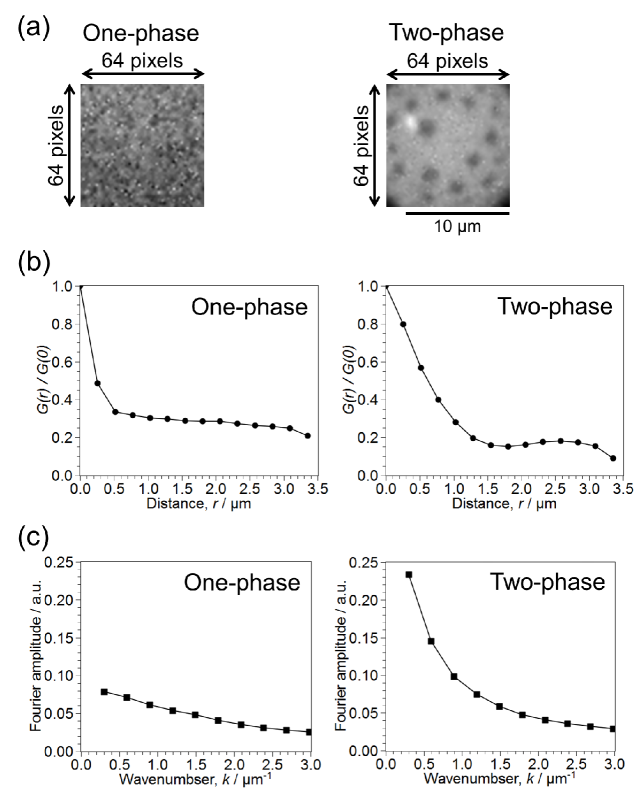
We calculated the angle-averaged 2D-AC using a macro program of the ImageJSchneider et al. (2012), First, fluorescence microscopic images of liposome surface were cut into 64 64 pixels and were converted into gray scale (16 bit). Then, we calculated two-dimensional fast Fourier transform (2D-FFT) using brightnesses of each pixel of images and inverse transform of their power spectrum in order to derive 2D-AC function. Further, the 2D-AC function was averaged for all angles. Finally, we calculated the FFT of curves, where is a angle-averaged 2D-AC as a function of radial position, , in the real space. Typical results of the analysis of one- and two-phase images are described in the Appendix A.
II.5 Calculation of Lo-domain circularity
The circularity was also calculated using ImageJSchneider et al. (2012). We transformed the microscopic images to binary images in order to obtain the phase boundary of Lo/Ld. The circularity, dimensionless parameter, of the Lo domains was defined as
| (1) |
where the is the ratio of the circumference of a circle to its diameter, is a area of Lo domain, and is a perimeter of Lo domain. The circularity of 1 corresponds to perfect circle. We calculated the circularity in order to estimate the change in line tension of phase boundary in the case of .
III Results and discussion
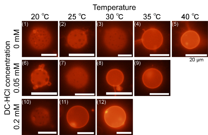
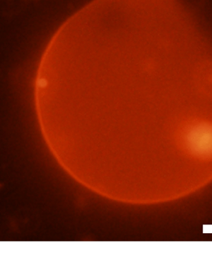
We observed the DOPC/DPPC/Chol liposomes with 0, 0.05, and 0.2 mM of DCHCl at , 25, 30, 35, and in order to confirm the influence of DCHCl on phase behavior of the ternary liposomes. Figure 2 shows the typical microscopic images at each condition. Since rhodamine DHPE is mainly localized in Ld phase of the phase-separated systemsChiantia et al. (2006); Shimokawa et al. (2010), the dark region corresponds to DPPC- and Chol-rich domain (Lo, raft-like domain) while the bright region corresponds to DOPC-rich domain (Ld). Image 1 described in the Figure 2 shows the phase-separated liposome with scattered Lo domains. The number of phase-separated liposome was reduced with increasing temperature (images 1-5). In the case of 0 mM and at , we observed many one-phase liposomes. There is no phase-separated liposome at , corresponding to image 5. Next, we also observed the ternary liposomes at various temperature in the case of 0.05 mM DCHCl. We observed the phase separation in the most of liposomes with 0.05 mM DCHCl at , corresponding to image 6. There are several phase-separated liposomes with smaller Lo domains as shown in the image 7 and a few liposomes with large domains as shown in Figure 8 described in Appendix B at 25 . We observed a few phase-separated liposomes with smaller Lo domains at (image 8) , and most of liposomes dose not have the Lo domains at 35 (image 9). Further, in the case of 0.2 mM, the number of phase-separated liposomes was also reduced with increasing temperature. At 20 , phase-separated liposomes are majority, however, there are several liposomes which have unclear phase boundaries. At 25 and above, most of liposomes have one phase without raft-like domains, corresponding to images 11 and 12. We especially focus on the images at 25 with the three DCHCl concentrations, corresponding to images 2, 7, and 11. The Lo domains in the liposome surfaces became smaller with increasing DCHCl concentration. These results indicate that DC molecule have an ability to change the phase behavior of DOPC/DPPC/Chol liposomes. It is considered that the effect of HCl is ignorable because the lipids used in this study is electrically neutral (not charged lipids)Shimokawa et al. (2010); Himeno et al. (2014).
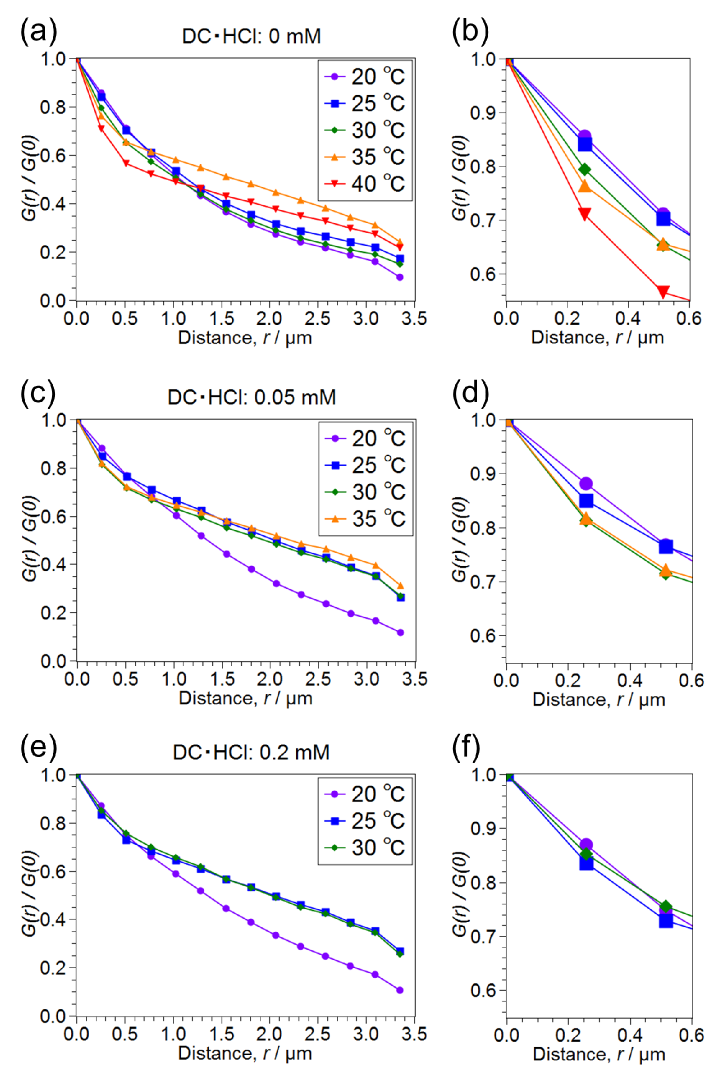
In order to quantify the phase behavior, we calculated the 2D-AC functions of domain pattern in the ternary liposomes. Figures 3(a, c, and e) show mean values of the normalized angle-averaged 2D-AC, , as a function of radial position, , at 0, 0.05, and 0.2 mM, respectively, and Figures 3(b, d, and f) show the enlarged view of the short distance region of the angle-averaged 2D-AC functions, corresponding to Figures 3(a, c, and e), respectively. The numbers of the observed liposomes are not less than 10 at each condition. Value of the short distance region (especially m) of the decreases with increasing temperature. It is considered that the correlation of the short distance region dependents on the ratio of the phase-separated liposomes. The correlation of the phase-separated liposome tends to have higher value due to Lo domains than that of the one-phase liposome as shown in Figure S1. The results indicate that the ratio of the phase-separated liposomes is reduced with increasing temperature. In the case of the 0.2 mM, dispersion of the values at m is smaller than that of two other concentrations because the differences of ratio of phase-separated liposomes between three temperatures seem to be smaller than that of 0 and 0.05 mM.
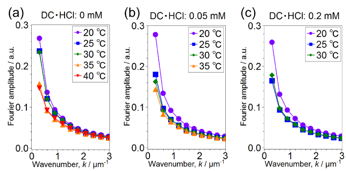
Since we need further informations in order to quantitatively confirm the effect of DCHCl molecules on miscibility transition temperature, , FFT of the curves of individual liposomes was calculated. Figures 4(a, b, and c) show the Fourier spectrum of the curves at 0, 0.05, and 0.2 mM, respectively (). Fourier amplitude of phase-separated liposome at wavenumber and m-1 tends to have larger value due to Lo domains than that of one-phase one as shown in Figure S1, where the wavenumber and m-1 correspond to wavelength and m, respectively. Therefore, the values of the Fourier amplitudes depend on the rate of phase-separated liposomes. The amplitudes at and m-1 are reduced with increasing temperature at all concentrations. In addition, largest distance point of Fourier amplitudes corresponds from 30 to 35 at 0 mM (Figure 4(a)), while the points correspond from 20 to 25 at 0.05 and 0.2 mM (Figures 4(b and c)). This indicates that the DCHCl molecules reduce the because it is considered that such largest distance point roughly corresponds to .
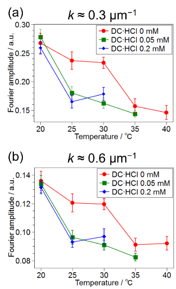
Furthermore, relation between Fourier amplitude and temperature was shown in Figure 5. The result indicates that the is reduced with increasing DCHCl concentration. The downwardly shifts more than 5 from 0 mM to 0.05 and 0.2 mM. The difference of Fourier amplitude between 0.05 and 0.2 mM at 25 is larger at m-1 than at m-1. This indicates that the amount of the liposomes with Lo domains corresponded to m-1 is larger at 0.05 mM than at 0.2 mM, and that the amount of the liposomes with Lo domains corresponded to m-1 at 0.05 mM is similar to at 0.2 mM. In other words, the ratio of liposomes having the large domains is higher at 0.05 mM than at 0.2 mM at 25 . This suggests that the difference of the DCHCl concentration influences the phase behavior. To our knowledge, this is the first report of quantitatively showing the change in of DOPC/DPPC/Chol liposome due to effect of local anesthetic molecule by 2D-AC analysis.
In this study, we demonstrated the change in of DOPC/DPPC/Chol liposome due to effect of DCHCl molecules. We discuss the cause of change in by calculating a circularity of the Lo domains. Figure 6 shows the circularity, , at . The circularity is reduced with increasing the DCHCl concentration. This suggests that the line tension of the Lo/Ld phase boundary is reduced with the DCHCl concentration because the domain circularity is related to line tension of phase boundaryGarcía-Sáez et al. (2007). In other words, the DC molecule induces the reduction of line tension of phase boundary. It was previously shown that the DC molecules has an ability to invade the lipid bilayersWeizenmann et al. (2012), which led us to consider this possibility in the case of DOPC/DPPC/Chol systems, and the effect of HCl is ignorable as described above. In addition, line-tension change induced by incorporation of other molecules was reported by several groupsAkimov et al. (2009); Hutchison et al. (2012). Therefore, it is considered that the insertion of the DC molecules into bilayers induces the reduction of the line tension.
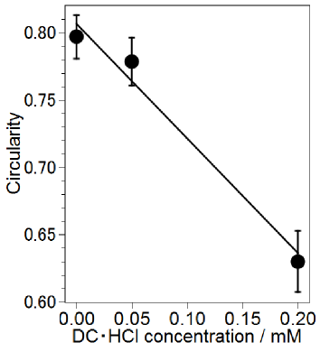
Previously, line tension of the phase boundary was expressed as
| (2) |
where the is a line tension of the Lo/Ld phase boundary, and the is a critical exponentBaumgart et al. (2003); Honerkamp-Smith et al. (2008). In terms of this equation, the miscibility transition temperature, , is reduced with the decrease in the line tension, , when the temperature of the system, , is constant. Therefore, it is considered that the insertion of the DC molecules into bilayers induces the reduction of the (Figure 5) through the reduction of the line tension.
A similar studies to the present work suggested that the general anesthetics disturb the phase separation of liposome and GPMV (reduction of ) by using the neutron diffraction and/or X-ray diffraction methodsWeinrich et al. (2012); Weinrich and Worcester (2013) and the direct observationGray et al. (2013). It is believed that the behavior in these previous studies is also induced by insertion of the anesthetic molecules to bilayers. Actually, Hamada et al. have reported that photoisomerization of the azobenzene derivative reversibly switches the phase pattern due to change in lateral line tensionHamada et al. (2011).
IV Conclusion
In summary, we have investigated the influence of DCHCl molecules, one of the local anesthetics, on phase behavior of DOPC/DPPC/Chol (mol%) liposome. In order to confirm the effect of DCHCl molecules, the ternary liposomes with 0, 0.05, and 0.2 mM of DCHCl have been observed at various temperatures. As a result, we have clarified the DC molecules has an ability to change the miscibility transition temperature, of the ternary liposome by calculating the angle-averaged 2D-AC function. Furthermore, we calculated a circularity of the Lo domains at in order to estimate the change in line tension of the Lo/Ld phase boundary and revealed that insertion of the DC molecules into bilayers induces the reduction of the via decrease in line tension. This suggests that the DC molecules disturb the function of the ion channels (anesthetic function of DC) through affecting the lipid bilayers which surround the ion channels. Since these physicochemical findings hint at the mechanism of the anesthetic function, this study may play an important role in the pharmacology.
V Acknowledgements
The authors thank Dr. Yasuhiro Fujii (Ritsumeikan Univ.), Prof. Tsutomu Hamada (JAIST), Dr. Rina Kagawa (M.D., Univ. Tokyo), and Prof. Miho Yanagisawa (Tokyo Univ. Agr. Tech.) for their significant advises.
Appendix A Appendix A: Two-dimensional aoutocorrelation (2D-AC) analysis
Appendix B Appendix B: Microscopic image of other domain pattern
References
- Haydon and Urban (1983) D. A. Haydon and B. W. Urban, J. Physiol. 341, 429 (1983).
- Ebihara et al. (1979) L. Ebihara, J. E. Hall, R. C. MacDonald, T. J. McIntosh, and S. A. Simon, Biophys. J. 28, 185 (1979).
- Reyes and Latorre (1979) J. Reyes and R. Latorre, Biophys. J. 28, 259 (1979).
- Reigada (2011) R. Reigada, J. Phys. Chem. B 115, 2527 (2011).
- Reigada (2013) R. Reigada, PLoS ONE 8, e52631 (2013).
- Muroi and Chanda (2009) Y. Muroi and B. Chanda, J. Gen. Physiol. 133, 1 (2009), http://jgp.rupress.org/content/133/1/1.full.pdf+html .
- Hanck et al. (2009) D. A. Hanck, E. Nikitina, M. M. McNulty, H. A. Fozzard, G. M. Lipkind, and M. F. Sheets, Circ. Res. 105, 492 (2009), http://circres.ahajournals.org/content/105/5/492.full.pdf+html .
- Yamagishi et al. (2009) T. Yamagishi, W. Xiong, A. Kondratiev, P. Vélez, A. Méndez-Fitzwilliam, J. R. Balser, E. Marbán, and G. F. Tomaselli, Mol. Pharm. 76, 861 (2009), http://molpharm.aspetjournals.org/content/76/4/861.full.pdf+html .
- Arcisio-Miranda et al. (2010) M. Arcisio-Miranda, Y. Muroi, S. Chowdhury, and B. Chanda, J. Gen. Physiol. 136, 541 (2010), http://jgp.rupress.org/content/136/5/541.full.pdf+html .
- Sheets et al. (2011) M. Sheets, T. Chen, and D. A. Hanck, Eur. J. Physiol. 461, 91 (2011).
- Lee et al. (2012) S. Lee, S. J. Goodchild, and C. A. Ahern, J. Gen. Physiol. 139, 507 (2012), http://jgp.rupress.org/content/139/6/507.full.pdf+html .
- Bant et al. (2013) J. S. Bant, T. K. Aman, and I. M. Raman, J. Neuro. 33, 4976 (2013), http://www.jneurosci.org/content/33/11/4976.full.pdf+html .
- Hata et al. (2000) T. Hata, H. Matsuki, and S. Kaneshina, Coll. Surf. B: Biointer. 18, 41 (2000).
- Hata et al. (2001) T. Hata, T. Sakamoto, H. Matsuki, and S. Kaneshina, Coll. Surf. B: Biointer. 22, 77 (2001).
- Matsuki et al. (2001) H. Matsuki, T. Hata, M. Yamanaka, and S. Kaneshina, Coll. Surf. B: Biointer. 22, 69 (2001).
- Takeda et al. (2009) K. Takeda, Y. Sano, S. Ichikawa, Y. Hirata, H. Matsuki, and S. Kaneshina, J. Oleo Sci. 58, 369 (2009).
- Tsuchiya et al. (2010a) H. Tsuchiya, T. Ueno, M. Mizogami, and K. Takakura, Chem. Biol. Interact. 183, 19 (2010a).
- Simons and Ikonen (1997) K. Simons and E. Ikonen, Nature 387, 569 (1997).
- Tsuchiya et al. (2010b) H. Tsuchiya, T. Ueno, M. Mizogami, and K. Takakura, J. Anesth. 24, 639 (2010b).
- Bandeiras et al. (2013) C. Bandeiras, A. P. Serro, K. Luzyanin, A. Fernandes, and B. Saramago, Eur. J. Pharm. Sci. 48, 153 (2013).
- Baumgart et al. (2003) T. Baumgart, S. T. Hess, and W. W. Webb, Nature 425, 821 (2003).
- Veatch and Keller (2003a) S. L. Veatch and S. L. Keller, Biophys. J. 85, 3074 (2003a).
- Diguet et al. (2012) A. Diguet, M. Yanagisawa, Y.-J. Liu, E. Brun, S. Abadie, S. Rudiuk, and D. Baigl, J. Am. Chem. Soc. 134, 4898 (2012), http://pubs.acs.org/doi/pdf/10.1021/ja211664f .
- Morita et al. (2012) M. Morita, T. Hamada, Y. Tendo, T. Hata, M. C. Vestergaard, and M. Takagi, Soft Matter 8, 2816 (2012).
- Shimokawa et al. (2010) N. Shimokawa, M. Hishida, H. Seto, and K. Yoshikawa, Chem. Phys. Lett. 496, 59 (2010).
- Himeno et al. (2014) H. Himeno, N. Shimokawa, S. Komura, D. Andelman, T. Hamada, and M. Takagi, Soft Matter 10, 7959 (2014).
- Cicuta et al. (2007) P. Cicuta, S. L. Keller, and S. L. Veatch, J. Phys. Chem. B 111, 3328 (2007), http://dx.doi.org/10.1021/jp0702088 .
- Sakuma et al. (2013) Y. Sakuma, T. Taniguchi, T. Kawakatsu, and M. Imai, Biophys. J. 105, 2074 (2013).
- Chen and Santore (2014) D. Chen and M. M. Santore, Langmuir 30, 9484 (2014), http://dx.doi.org/10.1021/la502089t .
- Muddana et al. (2012) H. S. Muddana, H. H. Chiang, and P. J. Butler, Biophys. J. 102, 489 (2012).
- Gray et al. (2013) E. Gray, J. Karslake, B. Machta, and S. Veatch, Biophys. J. 105, 2751 (2013).
- Veatch and Keller (2003b) S. L. Veatch and S. L. Keller, Biophys. J. 85, 3074 (2003b).
- Hwang et al. (1998) J. Hwang, L. A. Gheber, L. Margolis, and M. Edidin, Biophys J. 74, 2184 (1998).
- Facsko et al. (1999) S. Facsko, T. Dekorsy, C. Koerdt, C. Trappe, H. Kurz, A. Vogt, and H. L. Hartnagel, Science 285, 1551 (1999), http://www.sciencemag.org/content/285/5433/1551.full.pdf .
- Geringer et al. (2009) V. Geringer, M. Liebmann, T. Echtermeyer, S. Runte, M. Schmidt, R. Rückamp, M. C. Lemme, and M. Morgenstern, Phys. Rev. Lett. 102, 076102 (2009).
- Veatch et al. (2008) S. L. Veatch, P. Cicuta, P. Sengupta, A. Honerkamp-Smith, D. Holowka, and B. Baird, ACS Chem. Biol. 3, 287 (2008), http://dx.doi.org/10.1021/cb800012x .
- Koynova and Caffrey (1998) R. Koynova and M. Caffrey, Biochim. Biophys. Acta - Biomembr. 1376, 91 (1998).
- Lasic (1988) D. D. Lasic, Biochem. J. 256, 1 (1988), http://www.biochemj.org/bj/256/0001/2560001.pdf .
- Yoshida et al. (2014) K. Yoshida, Y. Fujii, and I. Nishio, J. Phys. Chem. B 118, 4115 (2014), http://dx.doi.org/10.1021/jp412710f .
- Schneider et al. (2012) C. A. Schneider, W. S. Rasband, and K. W. Eliceiri, Nat. Methods 9, 671 (2012).
- Chiantia et al. (2006) S. Chiantia, N. Kahya, J. Ries, and P. Schwille, Biophys. J. 90, 4500 (2006).
- García-Sáez et al. (2007) A. J. García-Sáez, S. Chiantia, and P. Schwille, J. Biol. Chem. 282, 33537 (2007).
- Weizenmann et al. (2012) N. Weizenmann, D. Huster, and H. A. Scheidt, Biochim. Biophys. Acta - Biomembr. 1818, 3010 (2012).
- Akimov et al. (2009) S. Akimov, E. Hlaponin, P. Bashkirov, I. Boldyrev, I. Mikhalyov, W. Telford, and I. Molotkovskaya, Biochemistry (Moscow) Supplement Series A: Membrane and Cell Biology 3, 216 (2009).
- Hutchison et al. (2012) J. B. Hutchison, R. M. Weis, and A. D. Dinsmore, Langmuir 28, 5176 (2012), http://dx.doi.org/10.1021/la204225a .
- Honerkamp-Smith et al. (2008) A. R. Honerkamp-Smith, P. Cicuta, M. D. Collins, S. L. Veatch, M. den Nijs, M. Schick, and S. L. Keller, Biophys. J. 95, 236 (2008).
- Weinrich et al. (2012) M. Weinrich, H. Nanda, D. L. Worcester, C. F. Majkrzak, B. B. Maranville, and S. M. Bezrukov, Langmuir 28, 4723 (2012), http://pubs.acs.org/doi/pdf/10.1021/la204317k .
- Weinrich and Worcester (2013) M. Weinrich and D. L. Worcester, J. Phys. Chem. B 117, 16141 (2013), http://pubs.acs.org/doi/pdf/10.1021/jp411261g .
- Hamada et al. (2011) T. Hamada, R. Sugimoto, T. Nagasaki, and M. Takagi, Soft Matter 7, 220 (2011).