Accepted for publication in the Proceedings of the National Academy of Sciences of the United States of America (2014). \urlwww.pnas.org/cgi/doi/10.1073/pnas.1322966111 \issuedateIssue Date \issuenumberIssue Number
Renormalization of myoglobin-ligand binding energetics by quantum many-body effects
Abstract
We carry out a first-principles atomistic study of the electronic mechanisms of ligand binding and discrimination in the myoglobin protein. Electronic correlation effects are taken into account using one of the most advanced methods currently available, namely a linear-scaling density functional theory (DFT) approach wherein the treatment of localized iron electrons is further refined using dynamical mean-field theory (DMFT). This combination of methods explicitly accounts for dynamical and multi-reference quantum physics, such as valence and spin fluctuations, of the electrons, whilst treating a significant proportion of the protein (more than 1000 atoms) with density functional theory. The computed electronic structure of the myoglobin complexes and the nature of the Fe–O2 bonding are validated against experimental spectroscopic observables. We elucidate and solve a long standing problem related to the quantum-mechanical description of the respiration process, namely that DFT calculations predict a strong imbalance between O2 and CO binding, favoring the latter to an unphysically large extent. We show that the explicit inclusion of many body-effects induced by the Hund’s coupling mechanism results in the correct prediction of similar binding energies for oxy- and carbonmonoxymyoglobin.
keywords:
metalloprotein — strong correlation — optical absorption — quantum-mechanical simulation — natural bond orbitals1 Significance Statement
Heme-based metalloproteins play a central role in respiration by transporting and storing oxygen, a function that is inhibited by carbon monoxide. Density-functional theory has been unable to provide a complete description of the binding of these ligands to heme s central iron atom, predicting an unrealistically high relative affinity for carbon monoxide. Here, we solve this problem using dynamical mean-field theory in combination with linear-scaling density-functional theory, thus allowing for a simultaneous description of crucial quantum entanglement and protein discrimination effects in the ground-state of the oxygen-heme complex. By simulating the binding process within a 1,000-atom quantum-mechanical model of the myoglobin metalloprotein, we obtain a significantly improved description of its spectroscopic and energetic observables.
2 Introduction
The ability of metalloporphyrins to bind small ligands is of great interest in the field of biochemistry. One such example is the heme molecule, which reversibly binds diatomic ligands, such as oxygen (O2) and carbon monoxide (CO), and plays a crucial role in human respiration. Heme is employed in myoglobin (Mb) and hemoglobin (Hb) proteins to store and transport O2 in vertebrates. The heme group of Mb is packed within a predominantly -helical secondary structure and is coordinated by a histidine residue (known as the proximal histidine) as the fifth ligand of the heme’s central Fe ion.
Despite intensive studies [1, 2, 3, 4, 5, 6], the nature of the bonding of O2 to the iron binding site of the heme molecule remains poorly understood, mainly due to the strong electronic correlation effects associated with its localized Fe electrons. It is known that these electrons are energetically well-aligned with the acceptor orbitals of CO and O2, and that the molecules’ bound conformations seek to maximize intermolecular orbital overlap [7, 8, 9]. In the case of MbO2, the short Fe–O bond (1.81 Å [9]) implies that -bonding is supplemented, to some extent, by -bonding [8]. Indeed, calculations employing the ab initio complete active space self-consistent method, in combination with a molecular mechanics force field to describe the protein (CASSCF/MM) [4], have identified a weak -bonding mechanism in the Fe–O2 bond that gives rise to an antiferromagnetic (open-shell singlet) state.
However, recent Fe L-edge X-ray absorption spectroscopy measurements on small biomimetic heme models lack the signature low-energy peak that is characteristic of the hole, formed by metal-to-ligand charge transfer into the ligand orbitals [10]. Although these spectroscopic results are more consistent with a strong Fe–O interaction, some uncertainty remains about whether the same bonding picture holds in MbO2, since the experiment was performed on a small model system (Fe(pfp)1-MeImO2), which, in particular, neglects the distal histidine (His 64) that hydrogen bonds directly with O2 in the protein.
Furthermore, while the diamagnetic nature of MbO2 is well-established, there is little experimental evidence that directly addresses the extent of the charge transfer from Fe to O2. Both CASSCF/MM [4] and L-edge X-ray absorption spectroscopy [10] suggest strong -donation from O2 into the d orbital of iron (ligand-to-metal back charge transfer), which is hypothesized to limit the charge on the O2 molecule to around e [4]. However, the stretching frequency of the O–O bond in MbO2 has been shown to be close to that of the free O ion [11, 8], which motivates further study.
The energetics of diatomic ligand binding to the Mb protein are expected to depend strongly on the electronic structure of the heme site, and in particular on its orbital polarization. Specifically, Mb reduces the heme group’s natural preference for CO binding: based on experimental equilibrium association constants, the binding free energy of CO, relative to O2, is reduced from around 5.9 kcal/mol in a non-polar solvent to 1.9 kcal/mol in the protein environment [12]. The combination of the strong electronic correlation centered at the Fe binding site and long-ranged interactions between the protein and the charged O2 molecule make computational modeling of the energetics of these complexes extremely challenging.
Typically, such studies calculate the protein effect [13, 14, 15], or relative spin state energies [16], or focus on small model systems [6]. However, most approaches applied to large system sizes did not include a proper treatment of electronic correlations. The effect of electronic correlations in the iron states was investigated by some of us [13], where it was included, to an extent, in ab initio simulations of ligand discrimination in myoglobin via a DFT+ treatment. DFT+ has been shown to be an efficient method for correcting self-interaction errors in the approximate DFT description of transition-metal chemistry [19], and when combined with linear-scaling approaches [17, 18], it allows us to tackle such systems comprising thousands of atoms. It was found, in the case of Mb [13], that the protein discrimination effect is dominated by polar interactions between O2 and the distal protein residue His 64. However, a problem in the DFT+ calculations is that a strong residual energetic imbalance that favors CO over O2 binding was observed [13], suggesting that approaches beyond static DFT+ are called for in order to obtain a proper description of myoglobin.
Recent progress has been made in the study of strongly-correlated electrons by means of dynamical mean-field theory (DMFT) [20], a sophisticated method that includes quantum dynamical effects, and takes into account both valence and spin fluctuations. DMFT is routinely used to describe materials, and recently has also been extended to nanoscopic systems [23, 24]. DMFT also explicitly includes the Hund’s exchange coupling typically, although not always, neglected in DFT and DFT+ studies. DMFT was recently combined with linear-scaling DFT [21] to produce a linear-scaling DFT+DMFT approach [22]. By means of the latter, we have pointed out that strong correlation effects in heme are controlled by the Hund’s coupling , and not the Hubbard repulsion alone [25], suggesting that subtle quantum many body effects are missing in the DFT+ treatment of myoglobin [13]. However, the computational model included just the heme group and diatomic ligands, neglecting entirely the protein environment and proximal histidine ligand, thereby strongly overestimating the binding energy of CO relative to O2.
In the present work, we bridge state-of-the-art DMFT many-body calculations with large scale DFT calculations. We perform simulations of realistic models of the MbO2 and MbCO complexes, comprising 1007 atoms, using linear-scaling DFT+DMFT. We thereby treat the electrostatic, steric, and hydrogen-bonding effects due to protein materials, together with the multi-reference, finite-temperature and explicit Hund’s exchange coupling effects associated with the iron binding site, in single, self-consistent calculations for the first time. We systematically investigate how the Hund’s coupling alters the electronic structure at the heme site and, at the same time, corrects the long-simulated, unphysical imbalance between CO and O2 binding affinities.
3 Results
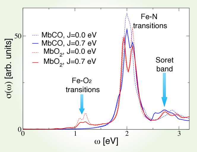
We first discuss our results for the porphyrin-plane component of the optical absorption spectra of ligated myoglobin, computed using DFT+DMFT (Fig. 1), where a realistic value of the Hund’s coupling eV is considered (results obtained for eV are shown for comparison). The absorption spectrum of this protein has been reported experimentally to be qualitatively dependent on its ligation state, in that a peak is present in the infrared region in the MbO2 case but not for MbCO [26]. Our theoretical absorption spectrum is in good agreement with the experimental data obtained from sperm whale MbO2 single crystals [27], and reproduces an MbO2 infrared absorption band at eV, observed experimentally at eV [28]. Our calculations associate this feature with a charge transfer band generated by the hybridization of the Fe atom and the O2 molecule, in particular transitions of occupied porphyrin and iron orbital states into empty O2 () orbitals. In MbCO, due to the strong covalent bond, the porphyrin and hybridized orbitals are at a lower energy and, hence, there is no contribution to the infrared spectrum. The double peak structure in the optical transition obtained at eV and 2.2 eV is also very close to experiments, where they are obtained respectively at 2.1 and 2.3 eV [28, 27]. We attribute this feature to the porphyrin Q band ( to absorptions) and to corresponding charge transfer excitations. We find a broad Soret band centered theoretically at eV, close to the experimental peak obtained at 2.95 eV [28, 27]. For MbO2, the spectrum at eV is qualitatively similar. However, Fig. 1 also reveals that a non-zero is required to recover the experimentally observed double-peak structure of the MbCO Q band [27]. Analysis of the spectral weight below the Fermi level in MbCO reveals the source of this splitting. For eV, the orbital character of the HOMO is almost degenerate between the three orbitals. However, for eV, we observe a splitting of the spectral weight of the dxy and the dxz,yz orbitals of eV, thus recovering the expected splitting of the charge transfer Q band in the optical absorption spectrum.
In order to further understand the nature of the bonding in the MbO2 complex, we have found it instructive to transform the atomic basis functions, used to expand the DFT+DMFT density-matrix (that is, the frequency-integrated Green’s function), into a set of natural bond orbitals (NBOs) [29, 30, 31]. The transformation is constructed such that the resulting orbitals may be categorized into localized Lewis-type bonding and lone pair orbitals, as well as their anti-bonding and Rydberg counterparts, thus allowing a chemically-intuitive population analysis to be applied to the DFT+DMFT many-body wave-function. Fig. 2 shows the - and -bonding many-body natural bond orbitals of the MbO2 complex. We find, in particular, that an O2 NBO (Fig. 2a), which has an occupancy of e, and an anti-bonding NBO formed between Fe and the proximal histidine, with occupancy e and a strong d character, interact strongly via the DFT Hamiltonian. We note that the e occupancy of the anti-bonding orbital on Fe is consistent with the ligand–metal back charge transfer process between O2 and the Fe d orbital, which is observed both in CASSCF/MM [4], and with L-edge X-ray absorption spectroscopy [10]. Ligand–metal back charge transfer is also present at eV, albeit with a smaller magnitude ( e). Thus, electronic delocalization is expected to provide a greater energetic stabilization in the MbO2 complex at eV.
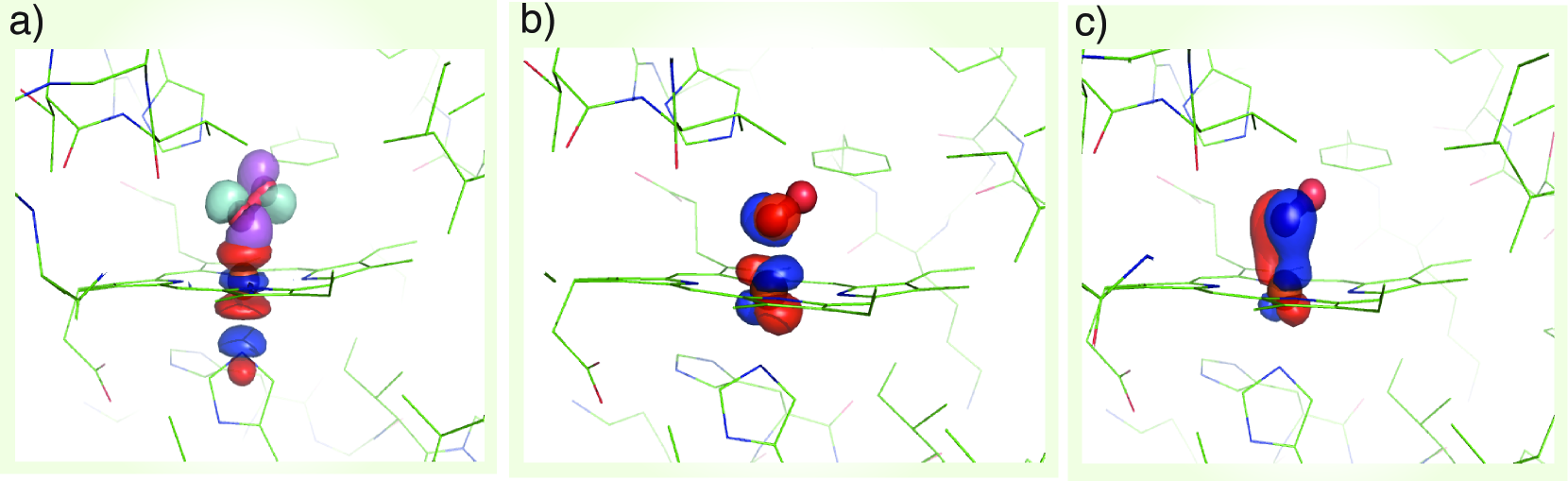
The net charge on the O2 molecule, from natural population analysis, is e, which is consistent with the Weiss picture of bonding in MbO2 [2]. It is worth noting that state-of-the-art CASSCF/MM calculations point toward a smaller O2 charge of e [4]. Metal-to-ligand charge transfer is expected to occur via -bonding interactions between Fe orbitals and O2 [4, 10]. Indeed, Fig. 2b,c are characteristic of the multi-configurational CASSCF orbitals that make up the proposed -type bonding in a previous study [4]. A notable difference between these calculations and the CASSCF/MM study is that the -bonding is much stronger than previously reported. Here, e are involved in -bonding, as opposed to approximately e in CASSCF/MM. Our calculations yield a hole character of 19 %. This compares extremely favorably with recent Fe L-edge X-ray absorption spectroscopy measurements of a small biomimetic heme model, which estimates the hole character to be % [10]. We, therefore, find that the -bonding character in MbO2 is similar to that in isolated porphyrins. We note that the stronger -bonding interaction between the iron and O2 also suggests that spin polarization of the electrons is less likely [10], suggesting that a broken spin symmetry description of MbO2 might not be entirely reliable.
| Protein | d | d | dxy | dxz | dyz |
| MbO2 ( eV) | 0.82 | 0.38 | 1.99 | 1.90 | 1.92 |
| MbO2 ( eV) | 0.96 | 0.96 | 1.10 | 1.87 | 1.96 |
| MbCO ( eV) | 0.95 | 1.13 | 1.99 | 1.88 | 1.89 |
| MbCO ( eV) | 1.00 | 1.15 | 1.99 | 1.84 | 1.84 |
| MbO2(CASSCF/MM) | 0.44 | 0.59 | 1.93 | 1.86 | 1.13 |
Next, we show, in Table 1, a comparison of the computed Fe orbital density with and without the explicit inclusion of Hund’s coupling in the Hamiltonian. We find that the effect of in MbO2 is to bring the d, dxy, and d orbitals closer to single-electron occupation, so that the Hund’s coupling enhances the spin magnetic moment on the Fe atom. Indeed, we find, in our calculations, a build up of a magnetic moment in the d, d and dxy orbitals, with a concomitant electron occupation of absent from our eV calculation and from the CASSCF/MM approach. The latter discrepancy may be due to the fact that CASSCF does not include dynamical correlation effects and may also be dependent on the chosen active space. In MbCO, unlike MbO2, we observe that the doublet on the dxy orbital is not emptied as the Hund’s coupling is increased. We next show, in Fig. 3a, the dependence of the Fe charge, computed using Mulliken analysis in the NGWF basis, on the Hund’s coupling for MbO2 and MbCO, respectively. For MbO2, we find that the charge of the Fe is transferred to the porphyrin ring and protein as the Hund’s coupling is increased. In contrast, for MbCO we find a very weak dependence of the Fe charge on the Hund’s coupling parameter (see Table 1). In our view, the latter indicates a very strong Fe-CO covalent bond, which remains stable against the Hund’s coupling. We find that the charge transferred from the Fe to O2 is e at the physical value of the Hund’s coupling eV (see Fig. 3b), confirming the estimation obtained using natural population analysis.
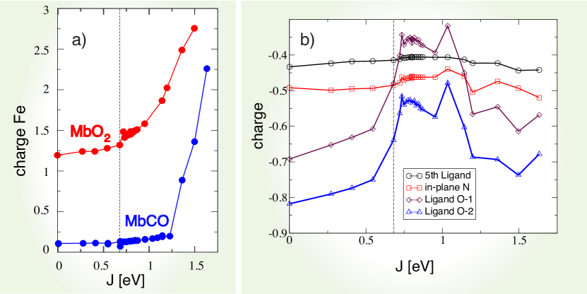
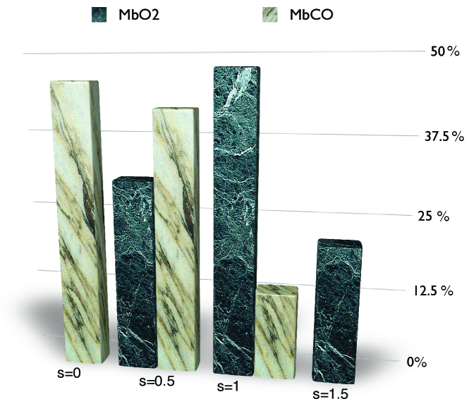
Fig. 4 depicts the computed spin fluctuations in MbO2 and MbCO, specifically, a histogram of the spin quantum number distribution obtained by looking at the 16 dominant states in the reduced ( subspace) density-matrix, obtained by tracing the atomic DMFT problem over the bath degrees of freedom. This gives an effective representation of the quantum states of the Fe atom. The ground-state wave-function is not a pure state with a single allowed value for the magnetic moment (singlet, doublet, triplet, etc.), yet we can describe the fluctuating magnetic moment of the Fe atom by analyzing the distribution of the magnetic moments obtained from the dominant configurations. In particular, we find that the reduced density-matrix of MbCO has states with dominant configurations, and MbO2 has dominant contributions from , with higher spin contributions at . This is consistent with our general observation that MbO2 has larger valence fluctuations (entanglement in the ground-state) than MbCO.
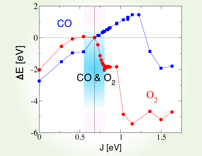
Having shown how the Hund’s coupling affects the orbital occupancy of the Fe site in Mb, and the associated charge transfer to the O2 molecule, we investigate, in what follows, how the energetics of ligand binding to Mb are determined by these effects, and how the protein uses quantum fluctuations to discriminate between O2 and CO. Fig. 5 shows the binding energies of O2 and CO to Mb (to within a constant shift), calculated using DFT+DMFT, as a function of the Hund’s exchange coupling . At eV, the binding energy of MbCO is approximately eV more favorable than the binding energy of MbO2, yielding an unphysical energetic imbalance. For eV, we find, on the contrary, that the binding energy of MbO2 is dramatically reduced. We attribute this to the enhancement of the spin magnetic moment in the Fe atom. In the intermediate regime, close to eV, we find that the imbalance between MbO2 and MbCO is thereby also dramatically reduced. In fact, the experimental binding free energy difference, between the two ligands, of kcal/mol, is recovered from our calculations when is near eV, a typical value used for iron-based materials [32]. In this case, the effect of on the binding energy of O2 may be regarded as a balance between two competing effects. The charge analysis (Fig.3) reveals that metal-to-ligand charge transfer is higher for eV, which is expected to enhance ligand–protein interactions for small values of . However, NBO analysis reveals a larger ligand-to-metal back charge transfer for eV, which is consistent with the increased occupancy of the Fe d orbital (Table 1), and is expected to cause variational energetic lowering at higher values of due to electronic delocalization.
Compared to previously reported DFT studies, including our own DFT+ study of the same system [13], the present study predicts a significantly larger charge on the O2 molecule ( e versus e). This charge is expected to stabilize the O2 molecule in the Mb protein via hydrogen bond interactions with His 64. Hence, we propose that both dynamical and multi-reference quantum effects, and large system sizes, must be accounted for in order to correctly determine the energetics of ligand binding in proteins with strongly correlated subspaces.
4 Conclusions
We have presented the application of a newly-developed methodology, designed to treat strong electronic interaction and multi-reference effects in systems of relatively very large numbers of atoms, to a molecule of important biological function. In particular, we have found that the Hund’s coupling is the crucial ingredient necessary to increase the multi-reference high-spin character of the ground-state, and so to bring the binding energetics into qualitative agreement with experiment. This provides a route to the solution of a long-standing problem in the density-functional theory based simulation of heme proteins, which often underestimates the Hund’s coupling and incorrectly-describes multi-reference effects, namely an unphysically large imbalance of CO and O2 binding energies. Our many-body description of the ligated myoglobin ground-state is further supported by quantitative agreement with experimental findings on both the ligand-dependence of the optical absorption spectra and the nature of the -bonding in Fe–O2. Our approach, optimized to describe molecules and nano-particles involving transition metal ions, supports a large range of applications, e.g., to strongly correlated oxide nano-particles [33] or to enzymes [34].
5 Methodology
In this work, we have carried out a detailed theoretical study of the electronic structure of the myoglobin molecule by means of a combination [22, 18] of linear-scaling density-functional theory (DFT) and the dynamical mean-field theory approximation (DFT+DMFT) [20, 35], a model which treats subspace-local dynamical, finite-temperature and multi-determinantal effects, for given Hamiltonian parameters.
The ONETEP linear-scaling DFT code [21, 18, 37] was used to obtain the DFT ground-state. The ONETEP method is particularly advanced in terms of its accuracy, equivalent to that of a plane-wave method, which is arrived at by means of an in situ variational optimization of the expansion coefficients of a minimal set of spatially-truncated Nonorthogonal Generalized Wannier Functions [38] (NGWFs), and is based on direct minimization of the total-energy with respect to the single-particle density-matrix. The use of a minimal, optimized Wannier function representation of the density-matrix allows for the DFT ground state to be solved with relative ease in large systems, particularly in molecules where their explicit truncation implies that the addition of vacuum does not increase the computational cost.
Preparation of the structures for DFT+DMFT analysis have been described in detail elsewhere [13]. Briefly, the computational models are based on the X-ray crystal structures of sperm whale Mb in oxygenated and carbonmonoxygenated ligation states (PDB: 1A6M, 1A6G) [9]. The heme group, ligand and 53 closest residues (1007 atoms in total) were extracted from the MbO2 crystal structure and optimized using spin-polarized DFT, with the PBE [39] gradient-corrected exchange-correlation functional augmented by damped London potentials to describe van der Waals interactions [36]. Following optimization, the heme group and three closest residues were replaced by their positions in the MbCO crystal structure and re-optimized. This scheme ensures that energy differences are directly attributable to local changes in the binding site, while accounting for long-ranged polarization and constraints of the protein scaffold. The DFT binding energy was converged to better than eV with respect to changes in the plane-wave energy cutoff and NGWF cutoff radii, and no additional restrictions on the variational freedom, such as the density kernel truncation, were invoked.
We refined our DFT calculations using the DFT+DMFT method [20, 35] in order to obtain a more accurate treatment of strong electronic correlation effects. In particular, DMFT introduces both quantum and thermal fluctuations, which are multi-reference effects not captured at the level of the Kohn-Sham DFT. In this, the Mb molecule was mapped, within DMFT, to an Anderson impurity model (AIM) Hamiltonian [40], and we used a recently developed extended Lanczos solver [41] to obtain the DMFT self energy. Since only a single impurity site ( orbital subspace) is present, the system becomes crystal momentum independent in the molecular limit, and since the Kohn-Sham Green’s function is computed in full, by inversion, before projection onto the impurity subspace, the Anderson impurity mapping is effectively exact, and the necessity of invoking the DMFT self-consistency is not required. However, in DFT+DMFT there is also a charge self-consistency cycle, albeit not routinely invoked at present due to computational cost, where the DFT+DMFT density kernel is used to generate a new Kohn-Sham Hamiltonian, which in turn provides a new input to the DMFT; the procedure being repeated until convergence is achieved. In this work, our data are obtained in the absence of charge self-consistency, however we checked that the corrections are small. Indeed, for MbO2 at eV, the changes obtained by converging the charge self-consistent DFT+DMFT induce a change in the energy of eV, which corresponds to the energy of MbO2 at eV when the charge self-consistency is absent. Other changes are also small, for example, the chemical potential changes by eV, and we find a change in the Fe charge of e. All these variations are consistent with a renormalized ( increased by 3% at eV).
To obtain the Kohn-Sham Green’s function, we performed the matrix inversion, as well as all matrix multiplications involved in the DMFT algorithm, on graphical computational units (GPUs) using a tailor-made parallel implementation of the Cholesky decomposition written in the CUDA programming language.
Electronic correlation effects are described within the localized subspace by the Slater-Kanamori form of the Anderson impurity Hamiltonian [42, 43], specifically:
| (1) | ||||
where are orbital indices, () annihilates (creates) an electron with spin in the orbital , is the orbital occupation operator. The first term describes the effect of intra-orbital Coulomb repulsion, parametrized by , and the second term describes the inter-orbital repulsion, proportional to , which is renormalized by the Hund’s exchange coupling parameter in order to ensure a fully rotationally invariant Hamiltonian (for further information on this topic, we refer the reader to Ref. [44]). The third term is the Hund’s rule exchange coupling, described by a spin exchange coupling of amplitude . denotes the spin corresponding to orbital , so that , where is the vector of Pauli matrices indexed by and . In this work, we used eV for the screened Coulomb interaction [16], and we explored the dependence of several observables on the Hund’s coupling (in the range eV). Our DMFT calculations were carried out at room temperature, K. In this work, we used the canonical form of the double-counting potential , given by:
| (2) |
assuming paramagnetic occupancy of the orbitals. Here, the parameter is the intra- and inter-orbital averaged repulsion [45]. In our calculations we found that the DMFT solution remains paramagnetic, although the possibility of spontaneous formation of a magnetic moment (spin symmetry broken state) was allowed for. However, the low energy states are in a quantum superposition of polarized states, giving a fluctuating magnetic moment at the iron site. The theoretical optical absorption was obtained in DFT+DMFT within the linear-response regime (Kubo formalism), in the no-vertex-corrections approximation [46], where it is given by:
| (3) | ||||
and the factor of two accounts for spin-degeneracy, is the simulation-cell volume, is the electron charge, is the reduced Planck constant, is the Fermi-Dirac distribution, and is the density-matrix given by the frequency-integral of the interacting DFT+DMFT Green’s function. The matrix elements of the velocity operator, , noting that we do not invoke the Peierls substitution [46], are given by:
| (4) |
This expression is general to the NGWF representation [47], used in this work, where the contribution to the non-interacting Hamiltonian due to the non-local part of the norm-conserving pseudopotentials [48, 49], represented by , is included. Once the self energy matrix is obtained, it can be used to correct the DFT total energy with the DMFT correction [50, 51]:
| (5) |
where indicates the many body interaction vertex of the DMFT, and the primed sum is over the occupied states. The symbol “Tr” indicates the one-electron trace for a generic representation and the sum over the Matsubara frequencies of the finite-temperature many-body formalism. The interaction term is obtained with the Galitskii-Migdal formula [52]:
| (6) |
() is the self-energy (Green’s function) matrix in the NGWF representation. We note that both the self energy and the Green’s function are slowly decaying functions, hence the trace over Matsubara frequencies has to be done with care [50, 51]. Finally, the double-counting correction must be introduced, since the contribution of interactions between the correlated orbitals to the total energy is already partially included in the exchange-correlation potential derived from DFT. The most commonly used form of the double-counting term is [45]:
| (7) |
A new approach, developed in this work, is the generation of natural bond orbitals based on the many-body Green’s function provided by DFT+DMFT, in order to obtain greater chemical insight into the ligand binding process. Natural bond orbitals (NBOs) [29] are post-processed linear-combinations of the basis functions in which the density-matrix is expanded, such that the projection of the density-matrix onto the subspace formed by atom-based and atom-pair based subsets of basis-functions is maximally diagonal. In the current calculations the basis-functions in question are NGWFs [38], transformed to NBOs using the NBO 5 programme [30], recently interfaced to ONETEP, as described in Ref. [31]. This procedure is carried out in such a manner that the final NBOs are then categorized into largely-occupied bonding and lone-pair orbitals, and largely-vacant anti-bonding and Rydberg orbitals. While normally applied to Kohn-Sham density-functional theory, to date, the NBO generation procedure is independent of the model (and so the Hamiltonian and self-energy) generating the density-matrix, and so we may apply it to the density-matrix integrated from the DFT+DMFT full Green’s function, for the first time. The resulting many-body NBOs largely retain the familiar profile of DFT-based NBOs, in this study, but their occupancies may be expected to deviate further from integer values due to quantum-mechanical and finite-temperature multi-reference effects captured within DFT+DMFT.
We computed the energy of MbCO and MbO2 as a function of the Hund’s exchange coupling . Defining and , the binding energy difference is given by . When , the MbO2 and MbCO binding energies are identical. In comparisons with the experimental relative free energy of binding, we have assumed that the relative change in entropy of the two ligands upon binding is zero, which is a reasonable approximation for two sterically similar diatomic ligands.
Acknowledgements.
We are grateful to Tanusri Saha-Dasgupta, Nicholas Hine, Gabriel Kotliar, Louis Lee, Peter Littlewood, and Andy Millis for helpful discussions. DJC is supported by a Marie Curie International Outgoing Fellowship within the 7th European Community Framework Programme. Calculations were performed on BlueGene/Q at the STFC Hartree Centre under project HCBG005 and on the Cambridge HPC Service, funded by EPSRC grants EP/J017639/1 and EP/F032773/1. We gratefully acknowledge the support of NVIDIA Corporation with the donation of Tesla K20 GPU used for this research.References
- [1] L. Pauling. Nature of the iron–oxygen bond in oxyhaemoglobin. Nature, 203:182–183, 1964.
- [2] J. J. Weiss. Nature of the iron–oxygen bond in oxyhaemoglobin. Nature, 202:83–84, 1964.
- [3] W. A. Goddard and B. D. Olafson. Ozone model for bonding of an O2 to heme in oxyhemoglobin. Proc. Natl. Acad. Sci. USA, 72:2335–2339, 1975.
- [4] H. Chen, M. Ikeda-Saito, and S. Shaik. Nature of the Fe-O2 bonding in oxy-myoglobin: Effect of the protein. J. Am. Chem. Soc., 130:14778–14790, 2008.
- [5] J. Ribas-Arino and J. J. Novoa. The mechanism for the reversible oxygen addition to heme: A theoretical CASPT2 study. Chem. Commun., pages 3160–3162, 2007.
- [6] M. Radón and K. Pierloot. Binding of CO, NO, and O2 to heme by density functional and multireference ab initio calculations. J. Phys. Chem. A, 112:11824–11832, 2008.
- [7] C. A. Reed and S. K. Cheung. On the bonding of FeO2 in hemoglobin and related dioxygen complexes. Proc. Natl. Acad. Sci. USA, 74:1780–1784, 1977.
- [8] M. Momenteau and C. A. Reed. Synthetic heme dioxygen complexes. Chem. Rev., 94:659–698, 1994.
- [9] J. Vojtěchovský, K. Chu, J. Berendzen, R. M. Sweet, and I. Schlichting. Crystal structures of myoglobin-ligand complexes at near-atomic resolution. Biophys. J., 77:2153–2174, 1999.
- [10] S. A. Wilson, T. Kroll, R. A. Decreau, R. K. Hocking, M. Lundberg, B. Hedman, K. O. Hodgson, and E. I. Solomon. Iron L-edge x-ray absorption spectroscopy of oxy-picket fence porphyrin: Experimental insight into FeO2 bonding. J. Am. Chem. Soc., 135:1124–1136, 2013.
- [11] R. D. Jones, D. A. Summerville, and F. Basolo. Synthetic oxygen carriers related to biological systems. Chem. Rev., 79:139–179, 1979.
- [12] J. S. Olson and G. N. Phillips Jr. Myoglobin discriminates between O2, NO, and CO by electrostatic interactions with the bound ligand. J. Biol. Inorg. Chem., 2:544–522, 1997.
- [13] D. J. Cole, D. D. O’Regan, and M. C. Payne. Ligand discrimination in myoglobin from linear-scaling DFT+. J. Phys. Chem. Lett., 3:1448–1452, 2012.
- [14] E. Sigfridsson and U. Ryde. Theoretical study of the discrimination between O2 and CO by myoglobin. J. Inorg. Biochem, 91:101–115, 2002.
- [15] F. De Angelis, A. A. Jarzȩcki, R. Car, and T. G. Spiro. Quantum chemical evaluation of protein control over heme ligation: CO/O2 discrimination in myoglobin. J. Phys. Chem. B, 109:3065–3070, 2005.
- [16] D. A. Scherlis, M. Cococcion, P. Sit, and N. Marzari. Simulation of heme using DFT+: A step toward accurate spin-state energetics. J. Phys. Chem. B, 111:7384–7391, 2007.
- [17] D. D. O’Regan, M. C. Payne, and A. A. Mostofi. Subspace representations in ab initio methods for strongly correlated systems. Phys. Rev. B, 83:245124, 2011.
- [18] D. D. O’Regan, N. D. M. Hine, M. C. Payne, and A. A. Mostofi. Linear-scaling DFT+ with full local orbital optimization. Phys. Rev. B, 85:085107, 2012.
- [19] H. J. Kulik, M. Cococcioni, D. A. Scherlis, and N. Marzari. Density functional theory in transition-metal chemistry: A self-consistent Hubbard approach. Phys. Rev. Lett., 97:103001, 2006.
- [20] A. Georges, Gabriel Kotliar, Werner Krauth, and Marcelo J. Rozenberg. Dynamical mean-field theory of strongly correlated fermion systems and the limit of infinite dimensions. Rev. Mod. Phys., 68:13, 1996.
- [21] N. D. M. Hine, P. D. Haynes, A. A. Mostofi, C.-K. Skylaris, and M. C. Payne. Linear-scaling density-functional theory with tens of thousands of atoms: Expanding the scope and scale of calculations with ONETEP. Comp. Phys. Commun., 180:1041–1053, 2009.
- [22] C. Weber, D. D. O’Regan, N. D. M. Hine, M. C. Payne, G. Kotliar, and P. B. Littlewood. Vanadium dioxide : A Peierls-Mott insulator stable against disorder. Phys. Rev. Lett., 108:256402, 2012.
- [23] N. Lin, C. A. Marianetti, A. J. Millis, and D. R. Reichman. Dynamical mean-field theory for quantum chemistry. Phys. Rev. Lett., 106:096402, 2011.
- [24] D. Jacob, K. Haule, and G. Kotliar. Dynamical mean-field theory for molecular electronics: Electronic structure and transport properties. Phys. Rev. B, 82:195115, 2010.
- [25] C. Weber, D. D. O’Regan, N. D. M. Hine, P. B. Littlewood, G. Kotliar, and M. C. Payne. Importance of many-body effects in the kernel of hemoglobin for ligand binding. Phys. Rev. Lett., 110:106402, 2013.
- [26] T. Nozawa, T. Yamamoto, and M. Hatano. Infrared magnetic circular dichroism of myoglobin derivatives. Biochim. Biophys. Acta, 427:28–37, 1976.
- [27] A. K. Churg and M. W. Makinen. The electronic structure and coordination geometry of the oxyheme complex in myoglobin. J. Chem. Phys., 68(4):1913–1925, 1978.
- [28] M. W. Makinen, A. K. Churg, and H. A. Glick. Fe-O2 bonding and oxyheme structure in myoglobin. Proc. Natl. Acad. Sci. USA, 75:2291–2295, 1978.
- [29] A. E. Reed, L. A. Curtiss, and F. Weinhold. Intermolecular interactions from a natural bond orbital, donor-acceptor viewpoint. Chem. Rev., 88:899–926, 1988.
- [30] E. D. Glendening, J. K. Badenhoop, A. E. Reed, J. E. Carpenter, J. A. Bohmann, C. M. Morales, and F. Weinhold. http://www.chem.wisc.edu/nbo5 & the NBO 5.9 manual, 2009.
- [31] L. P. Lee, D. J. Cole, M. C. Payne, and C.-K. Skylaris. Natural bond orbital analysis in the ONETEP code: Applications to large protein systems. J. Comput. Chem., 34(6):429–444, 2013.
- [32] J. M. Tomczak, K. Haule, and G. Kotliar. Signatures of electronic correlations in iron silicide. Proc. Natl. Acad. Sci. USA, 109(9):3243–3246, 2012.
- [33] O. Veiseh, J.W. Gunn, and M. Zhang. Design and fabrication of magnetic nanoparticles for targeted drug delivery and imaging. Adv. Drug Deliv. Rev., 62:284–304, 2010.
- [34] U. Ermler, W. Grabarse, S. Shima, M. Goubeaud, and R. K. Thauer. Active sites of transition-metal enzymes with a focus on nickel. Curr. Opin. Struct. Biol., 8(6):749–758, 1998.
- [35] T. A. Maier, T. Pruschke, and M. Jarrell. Angle-resolved photoemission spectra of the Hubbard model. Phys. Rev. B, 66(7):075102, 2002.
- [36] Q. Hill and C. K. Skylaris. Including dispersion interactions in the ONETEP program for linear-scaling density functional theory calculations. Proc. R. Soc. A, 465:669–683, 2009.
- [37] N. D. M. Hine, P. D. Haynes, A. A. Mostofi, and M. C. Payne. Linear-scaling density-functional simulations of charged point defects in Al2O3 using hierarchical sparse matrix algebra. J. Chem. Phys., 133(11):114111, 2010.
- [38] C.-K. Skylaris, A. A. Mostofi, P. D. Haynes, O. Diéguez, and M. C. Payne. Nonorthogonal generalized wannier function pseudopotential plane-wave method. Phys. Rev. B, 66:035119, 2002.
- [39] J. P. Perdew, K. Burke, and M. Ernzerhof. Generalized gradient approximation made simple. Phys. Rev. Lett., 77:3865–3868, 1996.
- [40] L. G. G. V. Dias da Silva, M. L. Tiago, S. E. Ulloa, F. A. Reboredo, and E. Dagotto. Many-body electronic structure and Kondo properties of cobalt-porphyrin molecules. Phys. Rev. B, 80:155443, 2009.
- [41] M. Aichhorn, M. Daghofer, H. G. Evertz, and W. von der Linden. Low-temperature Lanczos method for strongly correlated systems. Phys. Rev. B, 67:161103, 2003.
- [42] J. C. Slater. The ferromagnetism of nickel. Phys. Rev., 49:537–545, 1936.
- [43] J. Kanamori. Superexchange interaction and symmetry properties of electron orbitals. J. Phys. Chem. Solids, 10(2-3):87–98, 1959.
- [44] M. Imada, A. Fujimori, and Y. Tokura. Metal-insulator transitions. Rev. Mod. Phys., 70:1039, 1998.
- [45] T. Pruschke and M. Zölfl. Electronic structure and ordered phases in transition metal oxides: application of the dynamical mean-field theory. In Bernhard Kramer, editor, Advances in Solid State Physics 40, volume 40, pages 251–265. Springer Berlin / Heidelberg, 2000.
- [46] A. J. Millis. Optical conductivity and correlated electron physics. Strong Interactions in Low Dimensions, Physics and Chemistry of Materials with Low-Dimensional Structures., 25:195, 2004.
- [47] V. Halpern and A. Bergmann. Calculation of electronic green functions using nonorthogonal basis functions: application to crystals. J. Phys. C: Solid State Phys., 5(15):1953, 1972.
- [48] L. E. Ratcliff, N. D. M. Hine, and P. D. Haynes. Calculating optical absorption spectra for large systems using linear-scaling density functional theory. Phys. Rev. B, 84:165131, 2011.
- [49] A. J. Read and R. J. Needs. Calculation of optical matrix elements with nonlocal pseudopotentials. Phys. Rev. B, 44:13071–13073, 1991.
- [50] I. Di Marco, J. Minár, S. Chadov, M. I. Katsnelson, H. Ebert, and A. I. Lichtenstein. Correlation effects in the total energy, the bulk modulus, and the lattice constant of a transition metal: Combined local-density approximation and dynamical mean-field theory applied to Ni and Mn. Phys. Rev. B, 79:115111, 2009.
- [51] L. V. Pourovskii, B. Amadon, S. Biermann, and A. Georges. Self-consistency over the charge density in dynamical mean-field theory: A linear muffin-tin implementation and some physical implications. Phys. Rev. B, 76:235101, 2007.
- [52] V. M. Galitskii and A. B. Migdal. Application of quantum field theory methods to the many body problem. Sov. Phys. JETP, 7:96–104, 1958.