The Crystallization Behavior of Porous PLA Prepared by Modified Solvent Casting/Particulate Leaching Technique for Potential Use of Tissue Engineering Scaffold
Abstract
The porous PLA foams potential for tissue engineering usage are prepared by a modified solvent casting/particulate leaching method with different crystallinity. Since in typical method the porogens are solved in the solution and flow with the polymers during the casting and the crystallinity behavior of PLA chains in the limited space cannot be tracked, in this work the processing is modified by diffusing the PLA solution into a steady salt stack. With a thermal treatment before leaching while maintaining the stable structure of the porogens stack, the crystallinity of porous foams is made possible to control. The characterizations indicate the crystallization of porous foams is in a manner of lower crystallibility than the bulk materials. Pores and caves of around 250 size are obtained in samples with different crystallinity. The macro-structures are not much impaired by the crystallization nevertheless the morphological effect of the heating process is still obvious.
keywords:
Porous materials , Biomaterials , Poly(Lactic Acid) , Tissue Engineering Scaffold , Crystallization1 INTRODUCTION
Porous poly(lactic acid) (PLA) has been developed for tissue engineering scaffolds for decades [1-3]. Including poly(L-lactic acid) (PLLA), poly(D-lactic acid) (PDLA) and PLA-based copolymers like poly(lactic-co-glycolic) acid (PLGA), these bio-based resins have been proved to be a successful candidate of scaffold materials with excellent biocompatibility and biodegradablity. The tissue engineering requires sufficient interconnecting inner space in the scaffold for biofactor delivery, tissue growth, and the scaffold should be degradable after tissue’s growth meanwhile providing proper mechanical strength to support the tissue engineering system. Therefore the balance of growth space, degradation behavior and mechanical properties is the main concern of constructing a scaffold. With the respect to the particular requirements of certain tissue engineering, nowadays designed preparation and modification techniques of porous PLA scaffold materials become an intensive interests-drawing subject [4-13], which requires more understanding of the basic principles of physical and chemical properties, particular in the form of scaffold.
The crystallization of PLA plays an important role in its mechanical properties and degradability. Generally the crystallized polymers have higher strength and mechanical modulus [14]. In the case of PLA, the crystallinity also significantly affects the degradability, with the general behavior that the degradation time is longer with higher crystallinity, as the crystal segments are more stable than amorphous area and prevent water permeation into it. For example, it was reported that PLLA takes more than 5 years for total degradation, whereas only about 1 year for the amorphous PLA or PDLLA [15]. However unlike the crystallinity control for the inorganic components in the tissue engineering scaffold [16], very rare reports concerned the crystallinity of the polymer scaffold materials. One probable reason is that the preparation of porous scaffolds is a delicately process, where the control of crystallinity is usually difficult or unavailable. Except the solvent casting/particle leaching method, in other widely used preparation methods such as electrospun fiber, phase separation, membrane lamination and gas foaming, the polymers are not able to experience a thermal treating step, i.e. the most common way to control the crystallinity [1]. In some works the crystallinity is controlled by the raw materials itself, i.e. selecting raw materials of different molecular weight associated with different crystallization behaviors, or a particular processing procedure for chain cleavage to control the crystallinity [17]. And in some methods, even polymer with high crystallinity cannot be served as the raw materials to prepare the scaffold, for example the gas foaming technique reported by David J. Mooney et al. [18]. Nevertheless, the study of the crystallization of the scaffold should hold considerable practical merits in tissue engineering, for example to fit the scaffold degradation time with the expected tissue growth time by the control of crystallinity (if possible). Also, the scaffold structure may vary with crystallinity and influent the biological behavior of living tissue leaning on it. Park et al. reported a research on the sustained release of human growth hormone from semi-crystalline poly(l-lactic acid) and amorphous poly(d,l-lactic-co-glycolic acid) microspheres, which reveals that the morphological effect is important on protein release [19].
Crystallinity can be tailored in solvent casting/particulate leaching technique. However in this method the salt as porogens are solved in the solution and flow with the polymers during the casting, therefore without the immobilization of porogens the crystallinity behavior in the space-limited gap cannot be tracked [1]. In this work, we modified the solvent casting/particulate leaching technique by diffusing the PLA solution into a steady salt stack instead of solving the porogens. The control of crystallinity was made possible by inserting a thermal treating step before leaching, while maintaining the stable structure of salt stack. We have investigated the morphological effect of limited space on the crystallization of PLA, and the porous structure with different crystallinity under thermal treatments.
2 Experimental
2.1 Materials
The PLA of label 4032D is purchased from Natureworks®, with L/D ratios from : to :. The porogens is NaCl of analytical grade. The : mixture of dichloromethane and chloroform is served as solvent.
2.2 Porous sponge preparation
The PLA pellets are solved into dichloromethane and chloroform (:) with the concentration of . The NaCl powder is thoroughly grounded and sieved with then sieve to screen the particles of sizes in between, and paved onto a petri dish where it forms a ~ thick disc. The PLA solution is very slowly poured into the dish at the edge. The pouring is as slow as that the solution diffuses inside the salt stack instead of flowing over the surface, also the slow diffusing guarantees the salt particles are not considerably moved by the liquid flowing to keep the inner structure of the salt stack stable. After pouring, the salt-PLA solution composite is then placed in vacuum for for drying out the solvent. The product is a solid dry PLA-glued salt composite ready for thermal treatment. The composite is placed in water for to leach out the salts after the thermal treatment for recrystallization. The leached samples are freeze-dried for and stored in vacuum ready for characterizations.
2.3 Recrystallization
The recrystallization of composite is processed by the heating and cooling process. Four samples were made to have different crystallinity. One sample was kept as the original composite without thermal treatment for reference (sample R). The other three composites are heated in oven at for , then one composite (sample A) was immediately quenched in liquid nitrogen; sample B was linearly cooled down at the rate .; sample C was linearly cooled at till , kept at for , then cooled down with the same rate to room temperature. For comparison we also made two bulk PLA samples with the same crystallization process of sample B and C and they were labeled as sample B’ and C’.
2.4 Characterization
The crystallinity of samples were characterized by X-ray Diffraction (XRD) (D/max-2200/PC, Rigaku Corporation). The porous structure of samples were revealed by Scanning Electron Microscope (SEM) (Nova NanoSEM 450, FEI).
3 Results and Discussion
3.1 The crystallization behavior
The XRD results firstly dispel the doubt about the possibility of crystallization in confined geometry. Fig.1(1) shows the XRD of three thermal treated samples A, B and C (Note that the sample R is not included because its XRD curve almost overlap with the sample A, i.e. amorphous), and the bulk samples of B’ and C’ are shown in Fig.1(2). With the comparison of the bulk behavior, clear crystal peaks at and other minor peaks indicate that the heat treatment makes the crystallization possible but we can see significant impact of confined geometry, the crystallization of PLA chains in porogens slits is harder and the crystallinity is lower than the bulk sample under the same treatment condition. The crystallinity is calculated to be and in B and C, comparing to and in B’ and C’. It is clear that with the same thermal treatment the bulk samples are much easier to crystallize with higher crystallinity. This agrees with our expectation that the crystallization is more difficult in confined geometry, even the size of fibers and walls confining the pores is still in the magnitude of micrometers (see the SEM paragraph) which far overweighted the diameter of chain segments, the chain movement and rearrangement is obstructed by the limited space.
Another evidence that confined space impairs the crystallibility is the comparable crystallinity of B and C. The small difference is trivial due to many factors such as sample preparation or the baseline selection. It implies that linear cooling process may reach the upper limit crystallinity for the porogens confined environment, while for bulk sample, the effects of staying at for longer time is significant on crystallinity.
Although the samples are leached for there are unavoidable porogens residues left in the sample. The peaks on and are NaCl crystals [20], whereas these two peaks do not exist in the bulk samples B’ and C’.

3.2
The porous macro-structure
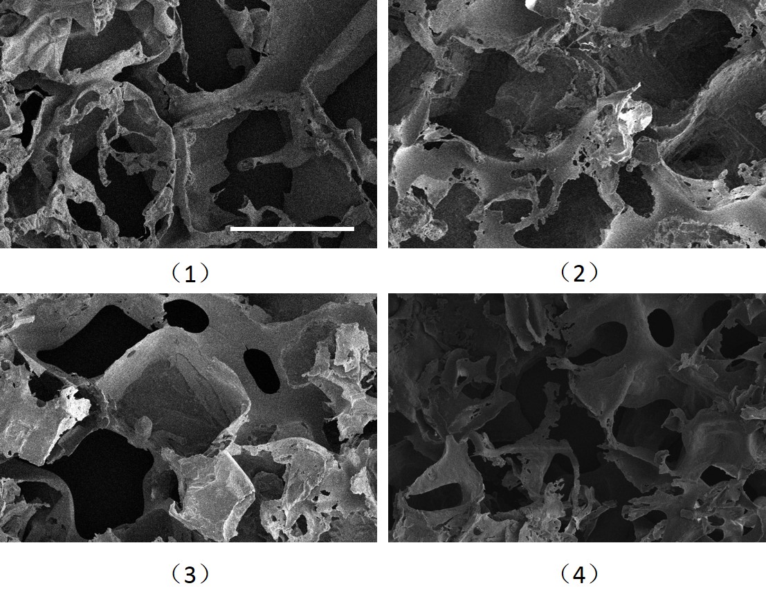
The SEM results in Fig.2 show the macro-structures of unheated original casting sample R and three samples with heat treatment A, B and C. A general observation of macro-structure confirms that the thermal treatment does not impressively affect the porous structure forming. The pores and caves structure in each sample can be clearly observed with the pore size of around , which accords to the sieving process. Nevertheless the morphological effect of heat treatment is obvious. In Fig.2(2), (3) and (4) the reheated samples present the features of thinner pore walls, branches and fragments, while in the reference sample the pore wall is thicker with rod-like branches. Regardless of the crystallinity, heating the samples to (the melting temperature of PLA) enables the polymer chains to remobilize and diffuse into the slits between salt particles where the solved chains had not diffused into and occupied. The thinner pore wall indicates that the chain remobilization also moves the porogens and makes narrower space among them. Although we employ the steady salt stack to confine the recrystallization within the limited space, the porogens are only relatively ”stable” comparing to typical solvent casting technique.
The quenching sample A has more fragmental structures than B and C, it is clear to understand the phenomenon that in sample A the polymer melt diffused into thinner slits is quenched to solidify its diffusing state of fragmental features. For sample B and C, the recrystallization process offers sufficient time for the diffused chains to mobilize and rearrange themselves to be more ordered, crystal structure. This rearrangement provides a less fragmental structure on the macro-scale. No obvious difference of macro-structures between sample B and C is observed, i.e. hours linear cooling is sufficient for recrystallization to achieve this structural effect, longer recrystallization time plays very little more effects on the crystallinity and the macro-structure, and this observation also agrees to the crystallinity results of B and C as indicated above.
4 Conclusion
The PLA porous matrix for potential use in tissue engineering have been prepared by modified salt casting and particulate leaching technique. The PLA solution is diffused into relative stable salt stack, instead of solving salts with polymers in typical method. Because the raw salt casting solidifies the salt-PLA composite, we are able to insert a step of thermal treatment to recrystallize the polymer matrix before leaching process. In this way we are able to: 1) investigate the crystallization behavior of PLA confined in limited space; 2) develop an available crystallinity control option in porous PLA scaffold preparation. The XRD results indicate the crystallization of porous foams, in a manner of lower crystallibility than the bulk materials. The marco-structure of porous samples are observed by SEM, by obtaining the pores of around , it is revealed that the polymer foam may crystallize without significant structure damage. The features of thinner pore walls, branches and fragments confirmed the effect of heating treatment. Both XRD and SEM results of sample B and C indicate that 7 hours linear cooling is sufficient to achieve certain crystallinity and marco-structure.
References
- [1] KF Leong, CM Cheah, CK Chua. Biomaterials 24 (2003) 2363–2378.
- [2] K Rezwana, QZ Chena, JJ Blakera, AR Boccaccinia. Biomaterials 27 (2006) 3413–3431.
- [3] I Armentano, M Dottori, E Fortunati, S Mattiolia, JM Kenny. Polymer Degradation and Stability 95 (2010) 2126-2146.
- [4] Vacanti JP, Morse MA, Saltzman WM, Domb AJ, Peter-Atayde A, Langer R. J Pediatr Surg 1988;23(1):3–9.
- [5] Mikos AG, Sarakinos G, Leite SM, Vacanti JP, Langer R. Biomaterials 1993;14(5):323–30.
- [6] Lu L, Mikos AG. MRS Bull 1996;21(11):28–32.
- [7] Thomson RC, Shung AK, Yaszemski MJ, Mikos AG. Polymer scaffold processing. In: Lanza RP, Langer R, Vacanti JP, editors. Principles of tissue engineering, 2nd ed. San Diego: Academic Press, 2000. p. 251–62 [Chapter 21].
- [8] Yang SF, Leong KF, Du ZH, Chua CK. Tissue Eng 2001;7(6):679–89.
- [9] Roether JA, Boccaccini AR, Hench LL, Maquet V, Gautier S, Jerome, R. Biomaterials 2002;23:3871–8.
- [10] Ma PX, Zhang R. J Biomed Mater Res 2001;56:469–77.
- [11] Xiong Z, Yan YN, Wang SG, Zhang RJ, Zhang C. Scr Mater 2002;46:771–6.
- [12] Taboas JM, Maddox RD, Krebsbach PH, Hollister SJ. Biomaterials 2003;24:181–94.
- [13] Lu HH, El-Amin SF, Scott KD, Laurencin CT. J Biomed Mater Res A 2003;64A:465–74.
- [14] M. Rubinstein, Ralph H. Colby. 2003, Polymer Physics, 1st Edition, Oxford University Press Inc., New York.
- [15] Rich J, Jaakkola T, Tirri T, Narhi T, Yli-Urpo A, Seppala J. In vitro evaluation of poly([var epsilon]-caprolactone-co-DL-lactide)/bioactive glass composites. Biomaterials 2002;23:2143–50.
- [16] Chihiro Mochizuki, Yuji Sasaki, Hiroki Hara, Mitsunobu Sato, Tohru Hayakawa, Fei Yang, Xixue Hu, Hong Shen, Shenguo Wang. Journal of Biomedical Materials Research Part B: Applied Biomaterials Volume 90B, Issue 1, pages 290–301, July 2009.
- [17] Sundararajan V. Madihally, Howard W.T. Matthew. Biomaterials 20 (1999) 1133-1142.
- [18] David J. Mooney, Daniel F. Baldwin, Nam P. Suh, Joseph P. Vacanti, Robert Larger. Biomaterials 17 (1996) 1417-1422.
- [19] Hong Kee Kim, Tae Gwan Park. Journal of Controlled Release 98 (2004) 115 – 125
- [20] NaCl powder X-ray diffraction pattern, Retrieved from http://www.uiowa.edu/~c004206/hand9.pdf (April 8, 2014).