Strength of the MeV resonance in the 40Ca(p,)41Sc reaction revisited
Abstract
The strength of the MeV resonance in the 40Ca(p,)41Sc reaction is determined with two different methods: First, by an absolute strength measurement using calcium hydroxide targets, and second, relative to the well-determined strength of the resonance triplet at = 4.5 MeV in the 40Ca(,)44Ti reaction. The present new value of eV is 37% (equivalent to ) higher than the evaluated literature value. In addition, the ratio of the strengths of the 1.842 MeV 40Ca(p,)41Sc and 4.5 MeV 40Ca(,)44Ti resonances has been determined to be . The newly corrected strength of the 1.842-MeV resonance can be used in the future as a normalization point for experiments with calcium targets.
pacs:
25.40.Lw, 25.40.Ny, 26.30.-kI Introduction
Precise values for selected resonance strengths may serve as normalization points for nuclear reaction experiments Iliadis2007 . This is particularly true for nuclei of astrophysical interest, where in several cases precision cross section data are needed in order to constrain astrophysical scenarios. One example are mirror nuclei such as 40Ca that are included in the -rich freezeout process believed to be responsible for the production of the supernova nuclide 44Ti Diehl2010 .
The MeV resonance in the 40Ca(p,)41Sc reaction provides a useful normalization point for experiments addressing the -rich freezeout, because it is relatively strong, easily accessible by a proton beam and generally in the astrophysically relevant energy range. This resonance populates the keV, 7/2+, seventh excited state in 41Sc, which, in turn, decays with 99.9% probability by -ray emission to the ground state Cameron2001 . 41Sc is unstable with a half life of 0.6 s and a positron endpoint energy of 5.473 MeV for the strongest decay branch.
The strength of the MeV resonance has been measured several times in the past (Table 1). In the framework of networks of (p,) resonance strengths involving several different nuclei, its value was first determined by Butler Butler1961 on calcium oxide targets using in-beam -ray spectroscopy with NaI detectors and detecting the positrons from the decay of 41Sc. Youngblood et al. Youngblood1968 devoted considerable effort to obtain a pure metallic calcium target and measured the resonance strength in an absolute way again by positron counting. A third absolute measurement was performed by Kozub et al. Kozub1977 , again on metallic calcium targets but using in-beam spectrometry with germanium detectors. Finally, as an ancillary result of an experiment aiming to study the 40Ca(,)44Ti reaction, Robertson et al. report an absolute value of the 40Ca(p,)41Sc resonance strength Robertson2012 . Relative resonance strengths measurements have been reported by Engelbertink and Endt Engelbertink1966 and by Paine and Sargood Paine1979 .
Here, a new measurement of the resonance strength is presented. To this end, data taken in the framework of a recent 40Ca(,)44Ti experiment Schmidt2013 are re-analyzed with a view to extract the strength of the MeV resonance in 40Ca(p,)41Sc. The sought for resonance strength is determined both absolutely and relative to the recently redetermined (,) strength.
| (eV) | Reference | Target | Technique | |
|---|---|---|---|---|
| ± | Butler Butler1961 | CaO | absolute; in-beam spectrometry and 41Sc -counting | |
| ± | Engelbertink and Endt Engelbertink1966 | Ca3(PO4)2, CaSO4 | relative to 31P(p,)32S, 32S(p,)33Cl resonances | |
| ± | Youngblood et al. Youngblood1968 | metallic Ca | absolute; 41Sc -counting | |
| ± | Kozub et al. Kozub1977 | metallic 40Ca | absolute; in-beam spectrometry | |
| ± | Paine and Sargood Paine1979 | CaO on Al | relative to 27Al(p,)28Si resonance | |
| ± | Robertson et al. Robertson2012 | metallic Ca | absolute; in-beam spectrometry | |
| ± | present work | Ca(OH)2 | both absolute and relative to 40Ca(,)44Ti | |
II Experiment
For the present purposes, the data from the scans of the MeV resonance for two different targets called #31 and #32 used for a study of the 40Ca(,)44Ti reaction Schmidt2013 are re-analyzed. The experiment has been performed at the 3 MV Tandetron accelerator of Helmholtz-Zentrum Dresden-Rossendorf (HZDR).
Targets consisting of calcium hydroxyde with natural isotopic composition on a tantalum backing were irradiated at an angle of 55∘ tilted to the beam. The rays from the reaction under study were detected by two escape-suppressed high-purity germanium (HPGe) detectors placed at angles of 55∘ and 90∘ to the beam direction, respectively. Further details on the experimental setup have been reported previously Schmidt2013 .
II.1 Analysis method
The resonance strength is related to the proton, photon, and total widths , , and of the resonance under study by the following equation:
| (1) | |||||
| (2) |
The statistical factor depends on the total angular momenta , , and of projectile, target, and resonance. In earlier works, commonly an alternative expression is used, i.e. = . For the reaction studied here, = 2. For a target of infinite thickness, the experimental yield as a function of is then given by the following relation Iliadis2007 :
| (3) |
where is the de Broglie wavelength at the resonance energy and is the effective stopping power for the hydrogen beam.
The yield critically depends on the stoichiometric composition of the target. Assuming the target to be of the stoichiometry CaOxHy, the stoichiometric parameters and affect the effective stopping power with {H, He} in the following way:
| (4) |
In this relation, is the stopping power of ion in calcium, in solid oxygen, and in solid hydrogen. The isotopic ratio of 40Ca in natural calcium is assumed to be % Coplen2002 . The correction factor MeV takes into account the slight deviations from Bragg’s stopping power summation rule. has been estimated using the so-called core and bond approach Ziegler1988 for stoichiometric Ca(OH)2. For helium ions at a laboratory energy of 4.5 MeV, MeV Schmidt2013 .
If one limits the experiment to just one target material, there are in principle two possible approaches to determine the resonance strength:
- 1.
-
2.
Two different resonances, e.g. a (p,) and an (,) resonance on the same target nucleus, are studied in the same target, determining the experimental yields for each of them separately. The ratio of resonance strengths is then determined as follows:
(5) As the ratio of effective stopping powers is usually only weakly dependent on the stoichiometry, this relative approach obviates the need to determine the target stoichiometry.
In the present work, both these approaches are used. Alternative approaches include relative measurements using either two resonances on different target nuclei both included in the same chemical compound Engelbertink1966 or two different chemical compounds deposited subsequently on the same target backing Paine1979 . However, different from approach (2) above these approaches still retain the dependence on the knowledge of the stoichiometric composition of each of the two compounds used.
II.2 Yield determination

The resonance under study decays by 99.9% by emission of a 2882 keV ray to the ground state in 41Sc Cameron2001 . The experimental yield can thus be determined as a function of beam energy over the entire target width by observing this ray. Two proton beam scans have been performed for target #32: before and after the -beam irradiation Schmidt2013 . For target #31, the -beam irradiation was interrupted by an additional scan, so that there are three scans (Fig. 1).
The targets used here are rather narrow, with an energetic thickness of just 7.5 keV for the proton beam. Therefore, the yield on the resonance plateau does not correspond to but must instead be extrapolated Iliadis2007 . The yield as a function of proton energy for a target of finite thickness Fowler1948 is
| (6) | |||||
This yield curve can then be used to fit the measured yields in Fig. 1. However, due to beam energy straggling inside the target Iliadis2007 , the slope of the right falling edge is less steep. In order to distinguish between the left and the right edge the two ’s in Eq. (6) have been replaced with and :
| (7) | |||||
The measured yield curves are very well described by Eq. (7) (solid lines in Fig. 1), and the resonance strength is obtained directly from these fits (Table 2). The statistical uncertainty is 6.2% (6.3%) for target #31 (#32). The combined value has a statistical uncertainty of 4.4%.
| Target scan | (eV) | ||
|---|---|---|---|
| #31 | before first -beam irradiation | ± | 0.012 |
| between -beam irradiations | ± | 0.008 | |
| after second -beam irradiation | ± | 0.008 | |
| Average #31 | ± | 0.012 | |
| #32 | before -irradiation | ± | 0.008 |
| after -irradiation | ± | 0.012 | |
| Average #32 | ± | 0.012 | |
| #31 and #32 combined | ± | 0.008 0.015 | |
III Results
III.1 Absolute determination of the strength of the 1.842-MeV resonance in 40Ca(p,)41Sc
For the absolute determination of the resonance strength, the stoichiometry of the targets has to be known. It has been determined previously for the two samples under study here Schmidt2013 in two different ways: First, with an elastic recoil detection (ERD) analysis for a sample target from the same production batch, and second, by the analysis of the primary rays from the 16O(p,)17F reaction. Both methods gave consistent results Schmidt2013 , and finally a stoichiometry of Ca(OH)1.88±0.21 is obtained, which is consistent with calcium hydroxyde Schmidt2013 . The stoichiometry contributes 5.9% to the uncertainty of the resonance strength, half of the total error budget (Table 3).
The -ray angular distribution of the 2882 keV rays detected here has been measured previously by three independent groups Youngblood1968 ; Rabin1973 ; Kozub1977 . Using the coefficients of Kozub et al. Kozub1977 , which agree with Youngblood et al. Youngblood1968 and Rabin Rabin1973 , the ratio of angular distribution corrections at 90∘ and 55∘ results in . The present experimental ratio of yields for the 90∘ and the 55∘ detectors is , which confirms the correctness of the literature angular distribution. For the determination of , the present yields are corrected with the literature Kozub1977 angular distribution, adding 3.8% uncertainty on .
The -ray detection efficiency has already been determined previously using calibrated radioactive sources and relative yields from the 27Al(p,)28Si reaction, with an uncertainty of 2.3% at 2882 keV Schmidt2013 . The normalization of the stopping power Ziegler2010 contributes another 1.4% uncertainty. The beam current was measured with a calibrated current integrator, and secondary electrons from the target were suppressed using a negatively charged tube just in front of the target, giving 1% uncertainty for the beam intensity Schmidt2013 .
Finally, the absolute resonance strength determined here is eV, with the error resulting from a quadratic combination of systematic and statistical uncertainties.
| Uncertainty | Contribution |
|---|---|
| Stoichiometry Schmidt2013 | |
| -ray angular distribution Kozub1977 | |
| -ray detection efficiency Schmidt2013 | |
| Stopping power Ziegler2010 | |
| Beam current Schmidt2013 | |
| Total systematic uncertainty |
III.2 Ratio of the strengths of the 1.842 MeV 40Ca(p,)41Sc and 4.5 MeV 40Ca(,)44Ti resonances
The present absolute strength (Sec. III.1) was determined on two targets which were also used to study the resonance triplet at = 4.5 MeV in the 40Ca(,)44Ti reaction. Therefore, as an alternative to the absolute resonance strength determination described in the previous section, the ratio of strengths of this resonance triplet and the resonance under study here has been determined.
To calculate a ratio of resonance strengths, according to Eq. (5) only the ratio of the two effective stopping powers has to be known, the absolute effective stopping power is not needed. The strength ratio depends only negligibly on the stoichiometric ratio, hence instead of 5.9% for the stoichiometry and 1.4% for the stopping power uncertainty, only 3.6% for the ratio of stopping powers between proton beam and beam Ziegler2010 have to be included in the error budget. Likewise, the beam current normalization cancels out.
For the present ratio, in order to simplify the error calculation, only the (,) resonance strength determined by the activation method Schmidt2013 is used. The (,) strength had also been determined previously using in-beam -ray spectrometry, but due to a poorly known angular distribution the results are less precise Schmidt2013 and not used here.
From the (,) resonance strength, the following contributions to the error budget have to be taken into account: 1.1% for the finite target thickness correction, 0.5% for the 44Ti half-life Ahmad2006 , and for the offline -ray counting 1.5% for the detection efficiency and 2.5% statistics Schmidt2013 . From the (p,) resonance strength, contributions of 2.3% for the efficiency in the in-beam -ray detection, 3.8% for the 2882-keV -ray angular distribution Kozub1977 , and 4.4% for the result of the fit procedures (Table 2) are included.
The total relative uncertainty for the resonance strength ratio is then 8%, and the value obtained here is:
Using eV Schmidt2013 , an absolute strength of eV is obtained, confirming the result of the absolute determination of the resonance strength in Sec. III.1.
IV Discussion
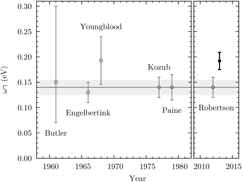
The present new value of the resonance strength is significantly (3.5 times the uncertainty of the evaluated value) higher than the evaluated strength of Endt90-NPA ; Cameron2001 . Only the values by Engelbertink and Endt Engelbertink1966 , Kozub et al. Kozub1977 , and Paine and Sargood Paine1979 had been included in the evaluation.
However, the present new value is consistent within 1 error bars with the previous results by Butler Butler1961 and Youngblood et al. Youngblood1968 , and consistent within 2 with all the other works when taken individually Engelbertink1966 ; Kozub1977 ; Paine1979 ; Robertson2012 (Fig. 2).
In order to understand the discrepancies, it is instructive to consider the target materials used. It has to be noted that metallic calcium targets may easily oxidize and transform to CaO or even Ca(OH)2. In case of an incorrectly determined stoichiometry for calcium, one would therefore expect an underestimate of the effective stopping power and, thus, an underestimate of for a given experimentally determined yield. Such an effect may well explain why several of the works using metallic calcium or calcium oxide Kozub1977 ; Paine1979 give lower strength values than the present one.
The early work of Engelbertink and Endt Engelbertink1966 was part of a network of intercomparison of various resonance strengths, including the = 588 keV resonance in the 32S(p,)33Cl reaction. The strength of this resonance has later been re-studied and seems to be a factor of 2 higher Iliadis92-NPA , which would bring the Engelbertink and Endt Engelbertink1966 result in agreement with the present value.
Two early works devoted particular attention to the target composition. Butler Butler1961 electrodeposited calcium on the backing and then deliberately oxidized it to form calcium oxide. Subsequently, the targets where ignited to white heat with a torch, removing impurities and ensuring that the target material remains burnt lime (CaO). Youngblood et al. Youngblood1968 used metallic calcium targets, and a detailed description of the procedures used is available Youngblood1965 . Calcium metal was scraped, removing surface oxidation, before the target was evaporated in situ. A proton elastic scattering experiment resulted in an oxygen content of less than 2%. The values of Butler Butler1961 and Youngblood et al. Youngblood1968 are in agreement with the present one.
Much less details on target preparation and handling are available in the works by Kozub et al. Kozub1977 , Paine and Sargood Paine1979 , and Robertson et al. Robertson2012 . Kozub et al. Kozub1977 state that they used the 16O(p,)17F reaction to determine the oxygen content of their targets, and that corrections to the effective stopping amounted to a few percent. However, no details on which of the three 16O(p,)17F transitions were actually used for the analysis and on the angular corrections (which are significant in this case) are given, hence it is possible that Kozub et al. underestimated the oxygen content of the target Kozub1977 . For the other two works by Paine and Sargood Paine1979 and by Robertson et al. Robertson2012 , even less details are available, so target oxidation cannot be excluded there, either.
The present target composition has been determined both by the elastic recoil detection (ERD) method and by nuclear reactions Schmidt2013 . It is consistent with fully oxydized and hydrogenated calcium.
The correctness of the present new result is further corroborated by the relative resonance strength determination, which to very good approximation is independent of stoichiometry. The strength of the 4.5 MeV resonance triplet in the 40Ca(,)44Ti reaction is now rather well-determined. The normalization value used here, eV Schmidt2013 is the most precise value available, but other previous resonance strengths Dixon1980 ; Nassar2006 ; Vockenhuber2007 that have been determined without reference to 40Ca(p,)41Sc are all close to it. An exception is the work by Robertson et al., where the 40Ca(,)44Ti resonance strength at 4.5 MeV has been determined relative to the 40Ca(p,)41Sc resonance strength under study here Robertson2012 .
V Summary
The strength of the = 1842 keV resonance in the 40Ca(p,)41Sc reaction has been re-measured. The result, eV, is higher than the value from a previous evaluation Endt90-NPA ; Cameron2001 . In addition, the ratio of strengths of the latter resonance and the 4.5 MeV resonance triplet in the 40Ca(,)44Ti reaction has been determined to be . The present value may be used in the future as a normalization point in coming precision experiments on 40Ca targets.
Acknowledgements.
The excellent support by the staff and operators of the HZDR ion beam center, by Andreas Hartmann (HZDR), and by Bettina Lommel and the GSI target laboratory is gratefully acknowledged. — This work was supported in part by DFG (BE4100/2-1), by HGF (NAVI, VH-VI-417), by EuroGENESIS, and by the European Union (FP7-SPIRIT, contract no. 227012).References
- (1) C. Iliadis, Nuclear Physics of Stars (Wiley-VCH Verlag, 2007)
- (2) R. Diehl, D. Hartmann, and N. Prantzos, Astronomy with Radioactivities, Lecture Notes in Physics (Springer, 2010) ISBN 9783642126970, http://books.google.de/books?id=F4fVUxDRvNAC
- (3) J. Cameron and B. Singh, Nuclear Data Sheets 94, 429 (2001), ISSN 0090-3752, http://www.sciencedirect.com/science/article/pii/S0090375201900213
- (4) J. Butler, Phys. Rev. 123, 873 (Aug 1961), http://link.aps.org/doi/10.1103/PhysRev.123.873
- (5) D. Youngblood, B. Wildenthal, and C. Class, Phys. Rev. 169, 859 (May 1968), http://link.aps.org/doi/10.1103/PhysRev.169.859
- (6) R. Kozub, B. Cooke, J. Leslie, and B. Robertson, Phys. Rev. C 16, 132 (Jul 1977), http://link.aps.org/doi/10.1103/PhysRevC.16.132
- (7) D. Robertson, J. Görres, P. Collon, M. Wiescher, and H.-W. Becker, Phys. Rev. C 85, 045810 (Apr 2012), http://link.aps.org/doi/10.1103/PhysRevC.85.045810
- (8) G. Engelbertink and P. Endt, Nucl. Phys. 88, 12 (1966), ISSN 0029-5582, http://www.sciencedirect.com/science/article/pii/0029558266904482
- (9) B. Paine and D. Sargood, Nucl. Phys. A 331, 389 (1979), ISSN 0375-9474, http://www.sciencedirect.com/science/article/pii/037594747990349X
- (10) K. Schmidt, S. Akhmadaliev, M. Anders, D. Bemmerer, K. Boretzky, A. Caciolli, D. Degering, M. Dietz, R. Dressler, Z. Elekes, Z. Fülöp, G. Gyürky, R. Hannaske, A. Junghans, M. Marta, M.-L. Menzel, F. Munnik, D. Schumann, R. Schwengner, T. Szücs, A. Wagner, D. Yakorev, and K. Zuber, Phys. Rev. C 88, 025803 (Aug 2013), http://link.aps.org/doi/10.1103/PhysRevC.88.025803
- (11) T. Coplen, J. Böhlke, P. De Biévre, T. Ding, N. Holden, J. Hopple, H. Krouse, A. Lamberty, H. Peiser, K. Revesz, S. Rieder, K. Rosman, E. Roth, P. Taylor, R. Vocke, Jr., and Y. Xiao, Pure Appl. Chem. 74, 1987 (2002)
- (12) J. Ziegler and J. Manoyan, Nucl. Instrum. Methods B 35, 215 (1988), ISSN 0168-583X, http://www.sciencedirect.com/science/article/pii/0168583X8890273X
- (13) W. Fowler, C. Lauritsen, and T. Lauritsen, Rev. Mod. Phys. 20, 236 (Jan 1948), http://link.aps.org/doi/10.1103/RevModPhys.20.236
- (14) B. Rabin, Contribution à l’étude spectroscopique du à l’aide des réactions , et , Ph.D. thesis, Université Louis Pasteur de Strasbourg (1973)
- (15) J. Ziegler, M. Ziegler, and J. Biersack, Nucl. Instrum. Methods B 268, 1818 (2010), ISSN 0168-583X, http://www.sciencedirect.com/science/article/pii/S0168583X10001862
- (16) I. Ahmad, J. Greene, E. Moore, S. Ghelberg, A. Ofan, M. Paul, and W. Kutschera, Phys. Rev. C 74, 065803 (2006)
- (17) P. Endt, Nucl. Phys. A 521, 1 (Dec. 1990)
- (18) C. Iliadis, U. Giesen, J. Görres, M. Wiescher, S. Graff, R. Azuma, and C. Barnes, Nucl. Phys. A 539, 97 (Mar. 1992)
- (19) D. H. Youngblood, The reaction, Ph.D. thesis, Rice University (1965), http://hdl.handle.net/1911/14250
- (20) W. Dixon, R. Storey, and J. Simpson, Can. J. Phys. 58, 1360 (1980), http://www.nrcresearchpress.com/doi/abs/10.1139/p80-175
- (21) H. Nassar, M. Paul, I. Ahmad, Y. Ben-Dov, J. Caggiano, S. Ghelberg, S. Goriely, J. P. Greene, M. Hass, A. Heger, A. Heinz, D. J. Henderson, R. V. F. Janssens, C. L. Jiang, Y. Kashiv, B. S. Nara Singh, A. Ofan, R. C. Pardo, T. Pennington, K. E. Rehm, G. Savard, R. Scott, and R. Vondrasek, Phys. Rev. Lett. 96, 041102 (Feb 2006), http://link.aps.org/doi/10.1103/PhysRevLett.96.041102
- (22) C. Vockenhuber, C. Ouellet, L. The, L. Buchmann, J. Caggiano, A. Chen, H. Crawford, J. D’Auria, B. Davids, L. Fogarty, D. Frekers, A. Hussein, D. Hutcheon, W. Kutschera, A. Laird, R. Lewis, E. O’Connor, D. Ottewell, M. Paul, M. Pavan, J. Pearson, C. Ruiz, G. Ruprecht, M. Trinczek, B. Wales, and A. Wallner, Phys. Rev. C 76, 035801 (Sep 2007), http://link.aps.org/doi/10.1103/PhysRevC.76.035801