Observation of strontium segregation in LaAlO3/SrTiO3 and NdGaO3/SrTiO3 oxide heterostructures by X-ray photoemission spectroscopy
Abstract
LaAlO3 and NdGaO3 thin films of different thickness have been grown by pulsed laser deposition on TiO2-terminated SrTiO3 single crystals and investigated by soft X-ray photoemission spectroscopy. The surface sensitivity of the measurements has been tuned by varying photon energy and emission angle . In contrast to the core levels of the other elements, the Sr line shows an unexpected splitting for higher surface sensitivity, signaling the presence of a second strontium component. From our quantitative analysis we conclude that during the growth process Sr atoms diffuse away from the substrate and segregate at the surface of the heterostructure, possibly forming strontium oxide.
I Introduction
In recent years pulsed laser deposition (PLD) has proved to be a powerful tool for stoichiometric epitaxial growth of a target material on top of a single crystal substrate. In combination with reflection high energy electron diffraction (RHEED) an atomically controlled layer-by-layer deposition is possible. This enables the manufacturing of epitaxial heterostructures exhibiting intriguing physical and electronic properties.Ohtomo et al. (2002); Brinkman et al. (2007); Aruta et al. (2010)
At the interface between two insulating metal oxides, LaAlO3 and SrTiO3 for example, a thickness threshold for an insulator to metal phase transition has attracted much interest. A highly mobile two dimensional electron gas (2DEG) is formed at the interface when 4 or more unit cells (uc) of epitaxial LaAlO3 are deposited on a single crystal, TiO2-terminated SrTiO3 substrate.Ohtomo and Hwang (2004); Thiel et al. (2006) The origin of this unexpected behavior has been so far the subject of passionate debates. The ”polar-catastrophe” scenario is believed by many scientists to catch most of the physics of this system. Within such model the electrostatic potential rises steadily with the growth of an increasing number of polar LaAlO3 layers, until the heterostructure accesses a new electrostatic ground state where a so-called “electronic reconstruction” process, transferring electronic charges from LaAlO3 to SrTiO3, takes place.Ohtomo and Hwang (2004); Nakagawa et al. (2006) However, it has been argued that oxygen vacancies may affect the interface conductivity,Kalabukhov et al. (2007); Siemons et al. (2007); Chen et al. (2011) which in fact depends on the oxygen partial pressure during growth and post-deposition annealing procedures. Finally, deviations from an abrupt interface due to cation intermixing at the LaAlO3/SrTiO3 interface has also been proposed as a possible source of chemical doping, giving rise to the observed interface conductivity. Such intermixing effects were found, e.g. by X-ray diffraction (XRD) Willmott et al. (2007), medium energy ion scattering Kalabukhov et al. (2009) and photoemission spectroscopy Qiao et al. (2010) for both the A-site (La Sr) and B-site sublattice (Al Ti). Chambers et al. (2010); Chambers (2011); Vonk et al. (2007, 2012) Recent scanning transmission electron microscopy and electron energy loss spectroscopy (STEM/EELS) data performed on samples similar to the ones employed in this work have posed nevertheless stringent upper limits to the amount of La cations crossing the interface. Cantoni et al. (2012)
X-ray photoemission spectroscopy (XPS) is a surface sensitive technique that enables in principle the investigation of all three scenarios mentioned above. Among the challenges of the method are the distinction of interface and bulk signal and the strong damping of the interface signal by the overlayer. We investigated the cation core level spectra of LaAlO3/SrTiO3 and NdGaO3/SrTiO3 samples by tuning the surface sensitivity. NdGaO3/SrTiO3 shares with LaAlO3/SrTiO3 a perovskite structure, an insulating nature of the single building blocks, a polar/non-polar character, and a critical thickness of four unit cells for the onset of conductivity. Furthermore, it also possesses transport properties that are similar to LaAlO3/SrTiO3. Di Gennaro et al. (2013) The electrical properties of the NdGaO3/SrTiO3 system have been investigated recently as a function of the growth conditions.Gunkel et al. (2013)
Here we show that an unexpected second strontium component, that we attribute to surface segregation of Sr cations, clearly emerges from collected spectra. Such phenomenon is frequently found in perovskites such as LaxSr1-xMnO3, Dulli et al. (2000) SrTiO3, Gunhold et al. (2002) SrTi1-xFexO3-δ Jung and Tuller (2012) and in the TiOSrTiO3 system. Radovic et al. (2011); Ciancio et al. (2012) Also the role of Sr-vacancies at the LaAlO3/SrTiO3 interface is under discussion. Gunkel et al. (2012) The Sr segregation effect occurs for all heterostructures and film thicknesses addressed in our investigation.
II Experimental
Room temperature soft X-ray photoemission spectroscopy measurements have been carried out at the UE52-PGM Beamline of the Berlin Synchrotron Facility (BESSY). Photon energies () varying from 200 eV to 1200 eV were chosen to control the surface sensitivity of the measurements. Spectra have been recorded with a Scienta SES 4000 energy analyzer and a total energy resolution smaller than meV, depending on . For quantitative interpretations the photon energy dependent inelastic mean free paths (IMFP) of the photoelectrons have been calculated by the semi-empirical TPP-2M modelTanuma et al. (2003) that considers the material specific density and band gap of the grown layer. The corresponding cross sections and asymmetry parameters for the core levels are given by Yeh and Lindau. Yeh and Lindau (1985)
LaAlO3/SrTiO3 and NdGaO3/SrTiO3 samples were grown by RHEED-assisted PLD technique on nominally single terminated SrTiO3 substrates that were purchased from TSST BV. The deposition conditions were as follows: substrate temperature C, oxygen partial pressure mbar, and laser fluence on the target. The target - substrate distance was 40 mm. A relatively high oxygen partial pressure was adopted in order to decrease the risk of incorporating oxygen vacancies into SrTiO3 during the growth process. This method has proved to yield high quality metallic samples, above threshold thickness, when proper growth conditions are adopted. Di Gennaro et al. (2013); Aruta et al. (2010, 2012) A slow cooldown to room temperature was performed after growth by keeping the oxygen pressure unchanged.
Samples were transported in air and no further cleaning steps were applied prior to experiment.
III Results and Discussion
Fig. 1 shows the background corrected and normalized Sr core levels for pure SrTiO3 (a) and 3 uc LaAlO3/SrTiO3 (b) measured with varying photon energy and emission angle. The Sr line consists of a doublet due to spin-orbit splitting (Sr , Sr ). Charging-related energy shifts have been encountered during the measurements. We will refer therefore in the following to the binding energy (BE) shifts with respect to the Sr peak maximum, rather than to absolute BE values. The surface sensitivity of the experiment can be tuned by changing the photon energy and the emission angle. Lower photon energies and higher emission angles increase the surface contribution to the overall signal and decrease the effective IMFP .
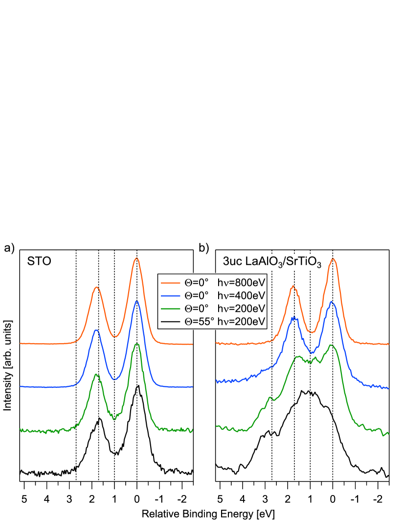
The SrTiO3 spectra show two single peaks with little dependence of the shape on measuring parameters, although in the most surface sensitive conditions (, eV) a slight broadening might be present. The spectra collected for LaAlO3/SrTiO3, on the contrary, feature a strong dependence on . Whereas the eV spectrum resembles the one of SrTiO3, at lower photon energies and higher emission angle an increasing high BE component is found, that dominates the spectrum for , eV.
Such behavior is observed on the Sr doublet of all the LaAlO3/SrTiO3 and NdGaO3/SrTiO3 samples, irrespective of polar film thickness. The core levels of the other cations do not show, instead, any comparable dependence on the measuring conditions.
The change of the Sr line in Fig. 1 is most naturally associated with the appearance of a second Sr component, which is chemically inequivalent with respect to the Sr2+ cations populating the perovskite A-site in SrTiO3. Alternative possibilities, as a strong band bending in SrTiO3 near the interface, would also cause a dependence of the spectra. Such hypothesis is nevertheless inconsistent with the absence of a comparable broadening for the other core levels. Furthermore, band bending would lead to a continuous shift of the peak positions of the Sr spectra as a function of between the two extreme BE values, for which no evidence was found in our data.
In order to extract quantitative information from the spectra we implemented a global fitting scheme. All the spectra for a given sample have been fitted with two doublets and the following constraints: The intensity ratio for the spin-orbit components has been set to 3 : 2, equal FWHM (full width half maximum) for doublet lines were imposed and the same energy separation between the two doublets was globally claimed for all measurements of a sample.
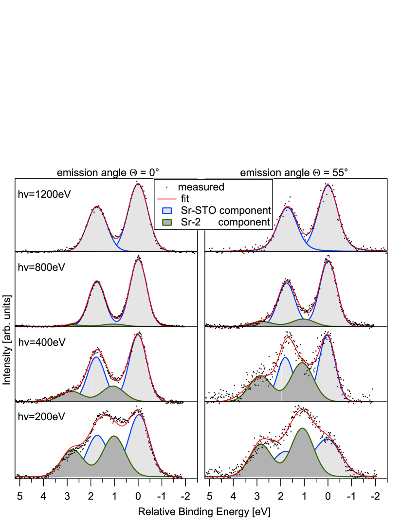
The results are shown in Figure 2 and Table 1. The fit is of good quality. For all three samples the same parameters satisfy (and therefore validate) the described procedure. The energy separation between the substrate and second Sr component is about 1 eV. The larger FWHM of the photoemission profile from the high BE component, called Sr-2 in the following, suggests a higher degree of disorder of Sr-2 cations with respect to the Sr-STO cations residing in crystalline SrTiO3.
| Parameter | 3uc LaAlO3 | 3uc NdGaO3 | 6uc NdGaO3 |
|---|---|---|---|
| Energy separation (eV) | 1.06 | 0.95 | 1.00 |
| FWHM(Sr-STO) (eV) | 0.9 | 0.9 | 0.9 |
| FWHM(Sr-2) (eV) | 1.2 | 1.3 | 1.3 |
It is obvious in Figure 2 that the integrated intensity of the Sr-2 component increases for lower and larger compared to the integrated intensity of Sr-STO, proving that Sr-2 cations are located above the SrTiO3 substrate.
Still, a number of more challenging issues about the Sr-2 component remain open: are the Sr-2 cations lying in-between LaAlO3 and SrTiO3, are they homogeneously intermixed in LaAlO3 or do they segregate above LaAlO3? Which is their amount? Which is their source and which is the origin of the chemical shift?
In Fig. 3(a) the integrated intensity ratios (IIRs) between the Sr and La profiles in LaAlO3/SrTiO3 are plotted vs for both Sr components. Each data point corresponds to a given photon energy and emission angle that is converted into . The data were corrected for core level and photon energy dependent cross sections and asymmetry parameters.Yeh and Lindau (1985) As expected, the Sr-STO IIR decreases exponentially for lower , due to the rising photoelectron damping by the LaAlO3 overlayer and the simultaneously increasing contribution of La. In contrast to this the Sr-2 IIR increases for lower . Once again, a similar behavior is observed for the NdGaO3/SrTiO3 samples shown in Fig. 3(b) and 3(c): the Sr-STO IIR (measured with respect to Ga ) increases with increasing , while Sr-2 clearly shows an opposite trend. If the Sr-2 cations were lying in between LaAlO3 (NdGaO3) and SrTiO3, their IIR would show the same trend as Sr-STO. A homogeneous intermixing of Sr-2 in LaAlO3 (NdGaO3) would cause a constant IIR vs . Only a top surface position of the Sr-2 cations entails the observed increase of IIR with decreasing .
The measured IIR profiles can be compared to a model that assumes an abrupt interface and an additional overlayer containing the Sr-2 species (see Fig. 3 inset). Within this scenario, the IIR values for Sr-STO and Sr-2 are expressed as a function of the thicknesses by Eq. (1) and (2):
| (1) |
| (2) |
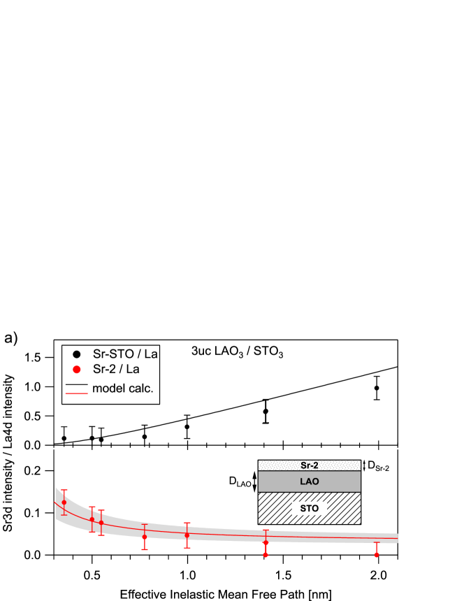
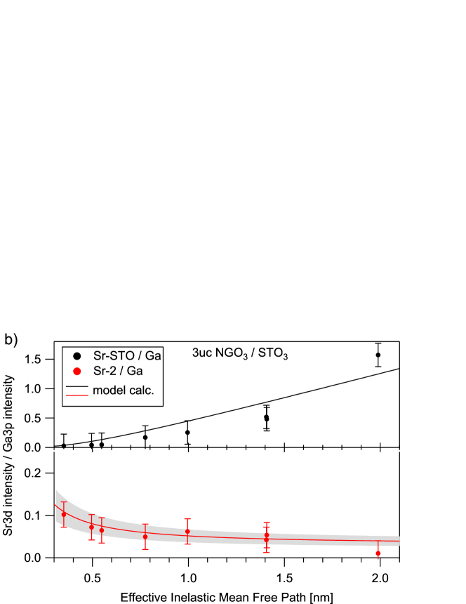
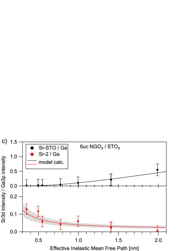
Here are the integrated intensities, directly extracted by fitting the data for Sr-STO and Sr-2 and La.Substrate related spurious Si intensity has been observed for low . The Si line partly superimposes the La line but can be removed by fitting with sufficient accuracy reflects the concentrations and cross sections of the different elements. In order to reduce the number of free parameters we set = , i.e. we assume the unknown atomic concentrations of Sr-2 to be equal to the Sr-STO case. D is the thickness of the layers (see Fig. 3 inset). The solid lines in Figure 3 represent the calculated profiles of Eq. (1) and (2) for a 3 uc LaAlO3 and 3 uc and 6 uc NdGaO3 layer ( nm, nm). The thickness of the Sr surface layer describing the datasets best is in the range of nm.
The model describes the data successfully and confirms that the Sr-2 cations lie predominantly at the surface of the heterostructures. appears to be much smaller than a SrTiO3 unit cell and all . For this situation the model employed in (1) and (2), which rests on the IMFP formalism, should be considered as an attempt to extract the order of magnitude of the top Sr concentration rather than exact quantization. The latter comes out as only a fraction of a single SrTiO3 unit cell, which hints to partial coverage or island formation. The model itself assumes a homogeneous Sr-2 layer, realized if the Sr is incorporated in the terminating LaAlO3 layer or by finely dispersed Sr-based molecules or clusters. However, to discriminate between these situations or other forms of island growth is not possible based on the data in Fig. 3 with certainty and remains, in essence, a task for future studies. Interestingly , i.e. the amount of Sr-2, appears to be independent of the chemical nature of the polar overlayer (LaAlO3 or NdGaO3) and of its thickness. Its observation requires low photon energies, rarely used for core level studies. This is probably the reason why this interesting phenomenon escaped the attention of previous investigations,Chambers et al. (2010); Takizawa et al. (2011) although some broadening of the Sr has been reported occasionally.Drera et al. (2013)
We now turn to the two final and most intriguing open questions: which is the origin of the extra Sr-2 component and what determines the shift in BE? The extra Sr might well migrate from the bulk of SrTiO3, that can be considered for our purposes as an “infinite Sr reservoir”. In this context, the driving force for Sr migration could either be an intrinsic non-stoichiometry of the single crystal or possibly the energy gain of a surface redox reaction of Sr in oxidizing conditions. As an alternative hypothesis, the excess Sr could lie initially on the nominally Ti-terminated SrTiO3 surface as clusters of residual atoms (possibly close to step edges) not removed by the surface treatment nominally guaranteeing the single TiO2 termination. This would suggest the existence of a driving force tending to maintain, even in the presence of a non-uniform SrTiO3 termination, a uniform SrO-TiO2-LaO-AlO2 sequence across the whole interface, by pushing the initial excess Sr to the top of the growing film. Finally, a finite amount of substrate surface Sr may be set free during the deposition process and experience an energy gain by floating at the surface of the heterostructure.
As for the BE shift between the two Sr components, it would be very tempting to attribute it to the electric potential foreseen to build-up across LaAlO3, within the polar catastrophe scenario. Nevertheless the analysis of current literature suggests that a BE shift of pure chemical nature, rather than of electrostatic nature, might well be at play. Chemical shifts very similar to the ones reported above have been in fact reported for bare, thermally treated SrTiO3 substrates and assigned to either SrOx, Szot et al. (2000) or Sr bonded to carbon (e.g. SrCO3). van der Heide et al. (2001); Vasquez (1991); Menou et al. (2009) A formation of Sr(OH)2, due to the reaction with water, can also cause a similar BE shift. Vasquez (1991) This hypothesis, if confirmed, could be consistent with the observation that BE shifts of electrostatic nature are hardly found in LaAlO3/SrTiO3. Yoshimatsu et al. (2008); Segal et al. (2009); Takizawa et al. (2011); Slooten et al. (2013)
At the end we address the general importance of the observed Sr-segregation for the physics of the oxide heterostructures. Any attempt to directly link this phenomenon to interface conductivity would be highly speculative at this stage. Migration of positively charged Sr2+ atoms from the interface to the upper LaAlO3 surface would certainly be an alternative or complementary means (with respect to electronic reconstruction) to alleviate the polar catastrophe. Nevertheless this argument would apply to any of the four cation species present in the system. Furthermore, this effect could be neutralized, if the migration involved neutral Sr2+ - O2- complexes rather than single ions. Finally, the very small amount of Sr-2 makes it insufficient to compensate a nominal polarity of 1/2 e- per in-plane uc. We will limit ourselves therefore to observe that the decade-long debate on the origin of the 2DEG in LaAlO3/SrTiO3 has taught us that the finest details in the atomic arrangement, including e.g. the SrTiO3 atomic termination, or submonolayer differences in LaAlO3 thickness above 3 uc, or LaAlO3 stoichiometry variations of the order of 1 Warusawithana et al. (2013), or undetectably low levels of oxygen vacancies, can dramatically alter the electronic properties of this system. Only a very accurate and complete understanding of the effective atomic configuration occurring in real systems will allow us to properly discern intrinsic and extrinsic effects.
IV Conclusions
By varying the surface sensitivity of X-ray photoemission we have unambiguously identified a previously elusive high binding energy Sr-component that we attribute to a submonolayer thick overlayer. The BE shift can be assigned to a purely chemical shift between the covering layer, presumably SrO, SrCO3 or Sr(OH)2, and SrTiO3, without any electrostatic contribution by the polar layer. The formation of such layer occurs both in LaAlO3/SrTiO3 and in NdGaO3/SrTiO3 for all thicknesses of the polar film. Our findings add further insight on the complex picture of oxide heterostructures, both in terms of their growth mechanisms and, possibly, of their electronic properties. They also confirm the tendency towards surface segregation of Sr in oxide systems with perovskite-related structure.
V Acknowledgement
E.D.G., U.S.d.U., and F.M.G. acknowledge financial support by the European Union (Programme No. FP7/2007-2013, Grant Agreement No. 264098 – MAMA), and by the Ministero dell’Istruzione, dell’Università e della Ricerca (Grant No. PRIN 2010-11 – OXIDE).
References
- Ohtomo et al. (2002) A. Ohtomo, D. A. Muller, J. L. Grazul, and H. Y. Hwang, Nature 419, 378 (2002).
- Brinkman et al. (2007) A. Brinkman, M. Huijben, M. van Zalk, J. Huijben, U. Zeitler, J. C. Maan, W. G. van der Wiel, G. Rijnders, D. H. A. Blank, and H. Hilgenkamp, Nat. Mater. 6, 493 (2007).
- Aruta et al. (2010) C. Aruta, S. Amoruso, R. Bruzzese, X. Wang, D. Maccariello, F. M. Granozio, and U. S. di Uccio, Appl. Phys. Lett. 97, 252105 (2010).
- Ohtomo and Hwang (2004) A. Ohtomo and H. Y. Hwang, Nature 427, 423 (2004).
- Thiel et al. (2006) S. Thiel, G. Hammerl, A. Schmehl, C. W. Schneider, and J. Mannhart, Science 313, 1942 (2006).
- Nakagawa et al. (2006) N. Nakagawa, H. Y. Hwang, and D. A. Muller, Nat. Mater. 5, 204 (2006).
- Kalabukhov et al. (2007) A. Kalabukhov, R. Gunnarsson, J. Börjesson, E. Olsson, T. Claeson, and D. Winkler, Phys. Rev. B 75, 121404 (2007).
- Siemons et al. (2007) W. Siemons, G. Koster, H. Yamamoto, W. Harrison, G. Lucovsky, T. Geballe, D. Blank, and M. Beasley, Phys. Rev. Lett. 98, 196802 (2007).
- Chen et al. (2011) Y. Chen, N. Pryds, J. E. Kleibeuker, G. Koster, J. Sun, E. Stamate, B. Shen, G. Rijnders, and S. Linderoth, Nano Lett. 11, 3774 (2011).
- Willmott et al. (2007) P. R. Willmott, S. A. Pauli, R. Herger, C. M. Schlepütz, D. Martoccia, B. D. Patterson, B. Delley, R. Clarke, D. Kumah, C. Cionca, and Y. Yacoby, Phys. Rev. Lett. 99, 155502 (2007).
- Kalabukhov et al. (2009) A. S. Kalabukhov, Y. A. Boikov, I. T. Serenkov, V. I. Sakharov, V. N. Popok, R. Gunnarsson, J. Börjesson, N. Ljustina, E. Olsson, D. Winkler, and T. Claeson, Phys. Rev. Lett. 103, 146101 (2009).
- Qiao et al. (2010) L. Qiao, T. C. Droubay, V. Shutthanandan, Z. Zhu, P. V. Sushko, and S. A. Chambers, J. Phys.: Condens. Matter 22, 312201 (2010).
- Chambers et al. (2010) S. A. Chambers, M. Engelhard, V. Shutthanandan, Z. Zhu, T. Droubay, L. Qiao, P. Sushko, T. Feng, H. Lee, T. Gustafsson, E. Garfunkel, A. Shah, J.-M. Zuo, and Q. Ramasse, Surf. Sci. Rep. 65, 317 (2010).
- Chambers (2011) S. A. Chambers, Surf. Sci. 605, 1133 (2011).
- Vonk et al. (2007) V. Vonk, M. Huijben, K. Driessen, P. Tinnemans, A. Brinkman, S. Harkema, and H. Graafsma, Phys. Rev. B 75, 235417 (2007).
- Vonk et al. (2012) V. Vonk, J. Huijben, D. Kukuruznyak, A. Stierle, H. Hilgenkamp, A. Brinkman, and S. Harkema, Phys. Rev. B 85, 045401 (2012).
- Cantoni et al. (2012) C. Cantoni, J. Gazquez, F. Miletto Granozio, M. P. Oxley, M. Varela, A. R. Lupini, S. J. Pennycook, C. Aruta, U. S. di Uccio, P. Perna, and D. Maccariello, Adv. Mater. 24, 3952 (2012).
- Di Gennaro et al. (2013) E. Di Gennaro, U. S. di Uccio, C. Aruta, C. Cantoni, A. Gadaleta, A. R. Lupini, D. Maccariello, D. Marré, I. Pallecchi, D. Paparo, P. Perna, M. Riaz, and F. M. Granozio, Adv. Opt. Mater. 1, 834 (2013).
- Gunkel et al. (2013) F. Gunkel, K. Skaja, A. Shkabko, R. Dittmann, S. Hoffmann-Eifert, and R. Waser, Appl. Phys. Lett. 102, 071601 (2013).
- Dulli et al. (2000) H. Dulli, P. A. Dowben, S.-H. Liou, and E. W. Plummer, Phys. Rev. B 62, R14629 (2000).
- Gunhold et al. (2002) A. Gunhold, K. Gömann, L. Beuermann, M. Frerichs, G. Borchardt, V. Kempter, and W. Maus-Friedrichs, Surf. Sci. 507–510, 447 (2002).
- Jung and Tuller (2012) W. Jung and H. L. Tuller, Energy Environ. Sci. 5, 5370 (2012).
- Radovic et al. (2011) M. Radovic, M. Salluzzo, Z. Ristic, R. Di Capua, N. Lampis, R. Vaglio, and F. Miletto Granozio, J. Chem. Phys. 135, 034705 (2011).
- Ciancio et al. (2012) R. Ciancio, E. Carlino, C. Aruta, D. Maccariello, F. M. Granozio, and U. Scotti di Uccio, Nanoscale 4, 91 (2012).
- Gunkel et al. (2012) F. Gunkel, P. Brinks, S. Hoffmann-Eifert, R. Dittmann, M. Huijben, J. E. Kleibeuker, G. Koster, G. Rijnders, and R. Waser, Appl. Phys. Lett. 100, 052103 (2012).
- Tanuma et al. (2003) S. Tanuma, C. J. Powell, and D. R. Penn, Surf. Interface Anal. 35, 268 (2003).
- Yeh and Lindau (1985) J. Yeh and I. Lindau, At. Data. Nucl. Data Tables 32, 1 (1985).
- Aruta et al. (2012) C. Aruta, S. Amoruso, G. Ausanio, R. Bruzzese, E. Di Gennaro, M. Lanzano, F. Miletto Granozio, M. Riaz, A. Sambri, U. Scotti di Uccio, and X. Wang, Appl. Phys. Lett. 101, 031602 (2012).
- (29) Substrate related spurious Si intensity has been observed for low . The Si line partly superimposes the La line but can be removed by fitting with sufficient accuracy, .
- Takizawa et al. (2011) M. Takizawa, S. Tsuda, T. Susaki, H. Y. Hwang, and A. Fujimori, Phys. Rev. B 84, 245124 (2011).
- Drera et al. (2013) G. Drera, G. Salvinelli, A. Brinkman, M. Huijben, G. Koster, H. Hilgenkamp, G. Rijnders, D. Visentin, and L. Sangaletti, Phys. Rev. B 87, 075435 (2013).
- Szot et al. (2000) K. Szot, W. Speier, U. Breuer, R. Meyer, J. Szade, and R. Waser, Surf. Sci. 460, 112 (2000).
- van der Heide et al. (2001) P. van der Heide, Q. Jiang, Y. Kim, and J. Rabalais, Surf. Sci. 473, 59 (2001).
- Vasquez (1991) R. Vasquez, J. Electron Spectrosc. Relat. Phenom. 56, 217 (1991).
- Menou et al. (2009) N. Menou, M. Popovici, S. Clima, K. Opsomer, W. Polspoel, B. Kaczer, G. Rampelberg, K. Tomida, M. A. Pawlak, C. Detavernier, D. Pierreux, J. Swerts, J. W. Maes, D. Manger, M. Badylevich, V. Afanasiev, T. Conard, P. Favia, H. Bender, B. Brijs, W. Vandervorst, S. V. Elshocht, G. Pourtois, D. J. Wouters, S. Biesemans, and J. A. Kittl, J. Appl. Phys. 106, 094101 (2009).
- Yoshimatsu et al. (2008) K. Yoshimatsu, R. Yasuhara, H. Kumigashira, and M. Oshima, Phys. Rev. Lett. 101, 026802 (2008).
- Segal et al. (2009) Y. Segal, J. H. Ngai, J. W. Reiner, F. J. Walker, and C. H. Ahn, Phys. Rev. B 80, 241107 (2009).
- Slooten et al. (2013) E. Slooten, Z. Zhong, H. J. A. Molegraaf, P. D. Eerkes, S. de Jong, F. Massee, E. van Heumen, M. K. Kruize, S. Wenderich, J. E. Kleibeuker, M. Gorgoi, H. Hilgenkamp, A. Brinkman, M. Huijben, G. Rijnders, D. H. A. Blank, G. Koster, P. J. Kelly, and M. S. Golden, Phys. Rev. B 87, 085128 (2013).
- Warusawithana et al. (2013) M. P. Warusawithana, C. Richter, J. A. Mundy, P. Roy, J. Ludwig, S. Paetel, T. Heeg, A. A. Pawlicki, L. F. Kourkoutis, M. Zheng, M. Lee, B. Mulcahy, W. Zander, Y. Zhu, J. Schubert, J. N. Eckstein, D. A. Muller, C. Stephen Hellberg, J. Mannhart, and D. G. Schlom, Nat. Commun. 4, 2351 (2013).