Shiga Toxin Detection Methods : A Short Review
Abstract
The Shiga toxins comprise a family of related protein toxins secreted by certain types of bacteria. Shigella dysenteriae, some strain of Escherichia coli and other bacterias can express toxins which caused serious complication during the infection. Shiga toxin and the closely related Shiga-like toxins represent a group of very similar cytotoxins that may play an important role in diarrheal disease and hemolytic-uremic syndrome. The outbreaks caused by this toxin raised serious public health crisis and caused economic losses. These toxins have the same biologic activities and according to recent studies also share the same binding receptor, globotriosyl ceramide (Gb3). Rapid detection of food contamination is therefore relevant for the containment of food-borne pathogens. The conventional methods to detect pathogens, such as microbiological and biochemical identification are time-consuming and laborious. The immunological or nucleic acid-based techniques require extensive sample preparation and are not amenable to miniaturization for on-site detection. In the present are necessary of techniques of rapid identification, simple and sensitive which can be employed in the countryside with minimally-sophisticated instrumentation. Biosensors have shown tremendous promise to overcome these limitations and are being aggressively studied to provide rapid, reliable and sensitive detection platforms for such applications.
∗INESC-Porto UOSE Optoelectronics and Sensors Unit, Faculty of Science University of Porto
——————————————————————————————
——————————————————————————————
1 Introduction
1.1 Preliminaries
Shiga toxin was isolated from Shigella dysenteriae serotype 1 by Kiyoshi Shiga in 1898, which named it Bacillus dysenterie [1]. In his work, Shiga described the production of the toxic factors actually known as Shiga toxin [2]. Shiga toxin-producing Escherichia coli (STEC) was discovered in 1977, and it was associated with the clinical hemolytic-uremic syndrome (HUS) in 1983, a life-threatening condition characterized by hemolytic anemia, thrombocytopenia, and renal failure [3]. This toxin has been also associated with hemorrhagic colitis and other severe disease conditions. Most of the work in the identification and characterization of Shiga toxin has been focused on E. coli O157:H7 strains, although many cases of Shiga toxin associated disease were caused by other serotypes of E. coli [1].
The main reservoirs of STEC are cattle and sheep, but other animals are recognized as a risk factor such as: deer, dogs, birds and horses. Nevertheless, new vehicles of infection resulting from environmental contamination and the intensive farming are continuously been identified [4]. Shiga toxin-producing Escherichia coli may cause diarrhea, bloody diarrhea and hemorrhagic colitis. The transmission of STEC may occurs through contaminated foods, such as ground beef, through contaminated water and by person-to-person interaction [5]. Fecal-oral transmission is also a common mode of transmission. Other ways to acquire that disease is the direct contact with animals on farms or at zoos. The leafy greens and unpasteurized apple cider are other recognized exposure sources. The transmission from person-to-person can occur directly (households, child care centers, institutions) or indirectly (contaminated drinking or recreational water) [6]. The ingestion of undercooked hamburgers in fast-food restaurants, was associate with the production of hemorrhagic colitis (HC), produced by STEC [7]. Stool cultures from patients infected in these episode, yielded a previously rarely isolated E. coli serotype, O157:H7. It has been reported that many E. coli strains isolated from these inpatients with diarrheal illness produce a Shiga-like toxin (SLT), including one of the strains that produce the Vero cytotoxin [8]. Afterward O’Brien et al., showed that Shiga-like toxin and the Vero cytotoxin were the same toxin [9]. Bacterial infections caused by Shiga toxin are responsible of many disease and the death of a great number of people. These infections have become a growing threat to the human health with worse manifestations in developed countries where the bacterias able to release Shiga-like toxins has been found in different food, including milk, apple juice and vegetables.
Surveillance data from the Center for Disease Control (CDC) of the U.S. for the 1998 calendar year showed an increase in the number of confirmed outbreaks of E. coli O157:H7 infection, from an average of 31 per year between 1994 and 1997 to 42 in 1998. In most of the Asian countries, STEC is not yet a major health problem, except in Japan, where 29 outbreaks were reported between 1991 to 1995 [9]. The first documentation of outbreak was produced by an episode involving strain O157:H7, in 1982 causing hemorrhagic colitis. Since then, the incidence of this strain in the diseases has increased annually [10]. An important massive outbreak of O157:H7 that gained particular attention happened in the western U.S. in 1993, having 501 cases, 151 hospitalizations and 45 cases of HUS. This outbreak was linked to consumption of undercooked hamburger meat at a fast food restaurants [11]. More recently, the transmission of foodborne bacteria remains an important public health threat because of the increased consumption of fresh vegetables and fruits, and the increased consumption of foods in public restaurants. The 80 % of cases of bacterial diarrhea that occurs every year in the United States are a result of foodborne transmission. Shiga toxin–producing E. coli is among the four most commonly reported bacterial enteropathogens and contributes to an estimated cost of $7 billion annually. STEC in particular, causes approximately 100,000 illnesses, 3,000 hospitalizations and 90 deaths annually in the United States [12].
Numerous outbreaks, as well as sporadic cases of STEC infections and HUS, have been documented worldwide. The largest outbreaks were recorded in industrialized countries. The modern industrialized large-scale food production might serve as a widespread vector in cases of food contamination. In Germany, STEC were commonly recognized as pathogens causing rare but severe disease almost exclusively in younger children. Before 2011, about 1,000 STEC infections per year and fewer than 100 cases of HUS were registered. The 2011 outbreak of a STEC O104:H4 in Northern Germany started at the beginning of May, reaching its peak on May 22. This an unusual outbreak of diarrhea with HUS affected Germany and was spread to other European countries, United States and Canada. Until July 4, when the end of the outbreak was officially declared 4075 cases: 3935 in Germany, with 48 dead, while in set of the other countries affected during this outbreak, were reported 140 cases [13].
The most comparable historical outbreak to the recent German epidemic, was took place in Japan in 1996 (Sakai city outbreak). About 6,000 persons were affected after white radish sprouts had been served at school canteens, exposing 47,000 children to the contaminated food. The incidence of HUS in the Sakai city outbreak was considerably lower (106 cases out of 6,000 infections). Another outbreak of a STEC O157:H7 have occurred in 1996, in Scotland, involving 512 persons, of whom 279 cases were microbiologically confirmed. One of the largest non-O157 STEC outbreaks was caused in Oklahoma in 2008, which reported 341 cases with gastroenteritis, 71 patients required hospitalization and 1 patient died [14].
The virulence of E. coli 0157:H7 can partially be attributed to its ability to establish infection at low doses in humans. The majority of STEC infections present with hemorrhagic colitis as 91% of patients give a history of bloody diarrhea at some point during their illness. Significant morbidity and mortality secondary to infection is attributed to the development of HUS [12]. Among the E. coli O157:H7 has received the most attention by the scientific and regulatory community because of its association with several large outbreaks of human illness with severe manifestations [15]. Even when the growing knowledge about toxins and its interaction with cells have allowed the production of molecules that help to treat this type of infection [16], this help some times comes out of time, as most of the above infections where confirmed several days after the initial episode.
It is known that available methods are not characterized by its speed. However, it is our intuition that biochemically-driven toxin detection can improve the speed of diagnosis kits for bacterial diseases. Currently, no extensive research on Stx identification and quantification is available. The purpose of this work is to review the state-of-the art techniques for Stx detection, trying to describe all the biological and (bio)chemical approaches for its diagnostics.
2 The Shiga family of toxins
2.1 Structural Characteristics
Shiga Toxin and Shiga-like Toxin belong to large family of plants and bacterial toxins. The real Shiga Toxin (Stx) is produced by Shigella dysenteriae and is almost identical to Shiga-like toxin 1 (Stx1). Shiga-like toxin is secreted by some strains of Escherichia coli (STEC), and also Citrobacter freundii, Aeromonas hidrofilas, Aeromonas caviae y Enterobacter cloacae have been reported able to express the toxin. Although the Shiga-like toxin is have a similar structure, do not have the same effect on cells [17] .
As said before, the virulence factor of Shiga toxin, also known as verotoxin, causes hemorrhagic colitis and diarrhea and in the most severe cases leads to the lethal hemolytic-uremic syndrome. The primary virulence factor in systemic host responses produced by clinical isolates of STEC is Shiga toxin type 2 (Stx2), but some isolates produce both Stx1 and Stx2, or more rarely only Stx1 [18].
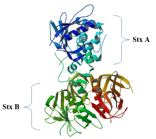
The two serological types of verotoxin, Stx1 and Stx2, are formed by a active enzymatic complex with a subunit A and five subunit B (Figure 1) [19]. The fragment A has an internal disulfide bond which when processed proteolytically generates two subunit: A1 and A2. The A1 fragment inhibits protein synthesis after it is released in the cytosol by the elimination of one adenine from the 28S RNA of the 60S ribosomal subunit [20]. The B pentamer, of 89 aminoacids, can join the terminal of disaccharide galabiose (gal-1,4-gal) in the surface of the host cells. This interaction carbohydrate-toxin can be used to STEC detection [21].
The molecular weight of the toxin is around 70kD, composed by subunit A of 32 kD and each subunit B of 7.7 kD. Stx1 is virtually identical to Stx, differing in only one aminoacid residue, whereas the Stx2 isoforms share less sequence similarity with Stx (60%). Toxins Stx1 and Stx2 have similar structure but differ in their sequences (Figure 2): the fragment A has 315 aminoacid, while for Stx2 the subunit A show 318 aminoacid [16]. Although their primary sequence of aminoacid are related, Stx1 and Stx2 are immunologically different: both are able to join the Gb3 receptor but they do not target same organs and tissues[22]. Gb3 is expressed in many cells of the body human, but the fact that amongst the more common complication hemorragic colitis and the HUS are prevalent, suggests that the infection is directed to specific organs. For example, Gb3 is expressed in the kidneys, mainly in the pediatric glomeruli, and when the kidneys becomes adult this expression decreases or is lost. This fact explains why HUS observed in children is mainly caused by Stx1 [23]. It has been reported that 90% of HUS cases occurs in children younger than three years old. Furthermore has been observed that Stx1 binds little or nothing in adults renal glomeruli[24].
The affinity of Stx1 by the receptor Gb3 is ten times bigger than with Stx2. The Shiga toxin genes are located in the bacteriophage (bacteria virus), which is associated with all pathogenic STEC [25]. The Stx1 and Stx2 are secreted by different ways and are translated in different ways across the outer membrane [26].

In eukaryotic cells the surface receptor for members of the Stx family is the neutral glycosphingolipid globotriaosylceramide (Gb3; gal- gal Glc-ceramide), with exception of Stxe variant that recognizes the globotetraosylceramide (Gb4; galNAc- gal gal Glc-ceramide). Several synthetic analogues of Gb3 exists and these are able to distinguish amongst Stx variants with a modification in the N-acetyl group. Complicating this picture is the fact that many cellular binding sites to Stx are available, and each of the subunits in the B pentamer have three Gb3 binding sites [27, 28].
Gb3 is synthesized in the Golgi of eukaryotic cells and is transported to the plasma membrane, having its trisaccharide residue out-membrane and the non covalent ceramide hydrocarbon in the plasma membrane. The joining subunit of Stx specifically recognizes the terminal alpha 1,4 trisaccharide digalactose. However, not only the part of carbohydrate is important in the toxin joining mechanism, also the lipid tail is important in the interaction of this toxin with its receptor. The sensitivity of the cell to the toxin and the linking procedure between the toxin and the cell can be regulated by many factors. For instance, changes in the transduction signals can be important for such regulations: an incubation prolonged with AMPc, or the exposition to butyric acid can induce the production of new receptors for the Shiga toxin. Cytokines (IL-1), tumoral necrosis factor (TNF) induced by Shiga toxin or LPS during the infection time could induce Gb3 synthesis in many types of cells. The induction of Gb3 production triggers the action of Shiga toxins and causes severe complication during the infection. The internalization involves formation of a clathrin-coated pit on the cell membrane. In some cells, vesicles-bound toxins are subjected to the fusion with the cellular lysosome, resulting in toxin degradation. In those cells Stx-sensitives, the endosomal vesicle contain a toxin-complex receptor which is transported to the endoplasmatic reticule.
During this process the subunit A is cleaved by a protease of membrane furin-like, generating a catalytically active 27 kD A1 N-terminal fragment, and a 4 kD A2 C-terminal fragment which remains attached by a disulphide bound. The active catalytically fragment for this dimer exerts its effect upon the ribosomes when is released in the cytoplasm [17, 16, 29].
Toxin can exerts its effect in eukaryotic cells by one of following
three mechanisms: firstly, the ribosomes inactivation and the inhibition
of synthesis of the cytoplasmic protein causing the cellular death.
Secondly, the depurination in the ribosomes generates a unique response
of signals translation named “ribotoxic stress response
” (RSR), that leads to the production of cytokines,
or others factors that resulting in many different events including
the apoptosis of the cells implicated. In the last site, the joint
of the holotoxin Stx or its subunit at the receptor, can start a cascade
of cytoplasmic signals translation different of the response RSR.
The end result of this events is the cellular death (apoptosis or
necrosis) or an response of inflammation in the cells that remain
viable, and maybe others immediate responses [22].
3 Detection
3.1 Biological Methods
It is considered that an appropriate response to bacterial outbreaks consist in its early detection and the use of adequate antibiotics to control them. The methods used to detect food pathogens have been grouped in four mainly categories. In the first group are the conventional microbiological methods, in which the food is mixed with selective medium enriched to increase the population of a target organism. Also, in this categories is located the agar plating in selective or differential media to isolate the pure culture, and the test of the culture by means of phenotype analysis or take of metabolic fingerprinting. These conventional microbiological methods, which are considered the gold standard, are reliable and accurate, but require much time and are very labor intensive to obtain the results [30]. Typically, the traditional methods involve a series of steps: pre-enrichment, selective enrichment, biochemical screening and serological confirmation. These step are laborious, requiring significant amount of time, expensive equipment and trained personal [19].
The detection of microorganisms which produce Stx has been difficult, due to the microorganism diversity and the detection limits of the samples in the environment [10]. The bacterial detection methods have to be rapid and very sensitive because, as explained before, the presence of even a single pathogenic organism in the body or food may become infectious. Extremely selective detection methodology is required, due to the big numbers of non-pathogenic organisms, which are often present in a complex biological environment, and coexist with the pathogenic bacterias. The traditional methods used for bacterial identification comprises the counting of cells with the aid of a optical microscope or by flow cytometry; measuring physical parameters by piezocrystals, impedimetry, redox reactions, optical methods, calorimetry, ultrasound techniques and detecting cellular compounds such as ATP, DNA, protein, lipid derivatives and radioactive isotopes [31].
The second group is composed by variants of the polymerase chain reaction (PCR) technique. Numerous articles has been published employing these methods in the determination of pathogens amongst them, E.coli in food . The methods to detect and identified the verotoxin produced by E.coli, based in these PCR genetic techniques, have been developed very recently. An advantage of this techniques is that with small amounts of samples make possible the accurate detection of the target species [32]. The PCR technique is very sensible for this task, but require hours to process the sample. Besides considerable bio-molecular skills are needed to prove the identification. The whole process could take from 48 hours to five days to obtain results, to this time is needed to sum the time expended in sample transportation, from the controlled site to the lab [33]. Moreover, the presence of certain genes has not, necessarily, a direct correlation with dangerous toxin levels [19].
The PCR in real time is the most used to the quantification of specific fragments of DNA. The amount of product synthesized during PCR is measured in real time by the detection of a fluorescent signal produced to result of a specific amplification. This methods is rapid and sensitive, but in some cases false positive and negative results are obtained, requiring an additional confirmation through to the hybridization probe and polymorphism of length of restriction fragment [30]. Several methods based in real-time PCR have been commercialized for Shiga toxin genes analysis in microbiologic food [34].
The third group includes the methods of immunosorbent assay (ELISA), its principle is based in the join of an antibody to the target antigen. It is a accurate and precise method, ideal for the quantitative and qualitative detection of many types of protein in complex matrices, when searching objectives are known. The sensitivity is low and up to 3 or 4 hours are is necessary in order to complete the analysis [30]. The ELISA for Stx1 and Stx2 can be used in stool sample. There are also commercial immunoassay for the detection of the Shiga-like toxin in milk and chopped meat [35, 36].
One of detection methods based in the capture of these toxins with antibodies was realized by Hattum et al. The presence of verotoxin in the sample is verified through a fluorescence signal coming from an antibody-antibiotin complex. The method is complementary to genetic methods, because it allow the detection of mRNA but does not provides information about if the toxin is only expressed or it is functional [19].
The researcher Ashkenazi et al, have evaluated the efficacy of a Gb3-based ELISA for detection of Shiga toxin, both from culture pure plates and, most importantly, directly from a mixed bacterial culture[37]. The glycodendrimers and glycol-conjugated nanoparticles has been used as anti-adhesive molecules for toxins and biosensors to monitor the protein-carbohydrate interactions. The glyconanoparticles show the best potential for studying these protein-carbohydrate interactions. The size distribution of these nanoparticles is reasonably narrow and is comparable with the size of the studied biomolecules, moreover, these structure have been well described and are easily manipulated chemical structures. The glycopolydiacetylene nanoparticle (GPDA) has been used to monitor receptor-ligand events junctions for viruses, toxins, bacteria and antibody-receptor interactions, due to its unique colorimetric transition when joining to these macromolecules. Therefore, due to the well-known affinity of Shiga toxin towards the final [gal-1,4-gal] disaccharide unit, GPDA nanoparticles containing this specific disaccharide sequence, has been used to identify E.coli Shiga toxin in in 96-well plates [21].
The fourth, and most recent group of detection methodologies is based on microarray techniques. These methods allow the simultaneous identification, in foods, of a large number of pathogens with a simple reaction. The basic idea is that many complementary probes are joined in a matrix shape on a solid surface, each one of these site contains several copies of specific probes. The matrix is hybridized with the DNA isolated from the sample of interest, producing a characteristic fluorescence. During this step of hybridization, the fragment is joined to the probe about the bases of DNA complementarity. However, this regular microarrays method needs an expensive equipment, both for exploring that matrix and for compiling the generated data [30].
3.2 Detection by Biosensors
Many of the reported methods for the detection of pathogenic bacteria are applied to Escherichia coli. Most E.coli-specific methods have a detection limit between and cells/mL and some rely on the amplification of specific genes of the E. coli genome for specific identification [38]. Structure-based, designed synthetic biosensors have been recently considered as attractive alternative to conventional platforms for pathogens detection. Moreover, these biosensors have important characteristics, such as high grade of sensitivity and detection specificity, minimum effort in sample preparation, profitability, miniaturization and portability to real-time monitoring while reduce the total time requested for detection. Therefore, a great effort has been allocated the development of these fast biosensors of diverse nature, as they are considered promising devices for pathogenic bacteria detection [39].
Ideal attributes of any element of recognition would be a great stability, facility of immobilization in the sensor platform and the specificity of recognition to the host with minimal cross-reactivity to other pathogens agents [40]. In the detection of biochemical and physiological processes, the biosensors have been converted in a fundamental tool.
According to the methods used for signal transduction, biosensors are divided in four groups: mass, optical, electrochemical, and thermal sensors. taking into account the way used for target identification, biosensors can be classified into two categories: sensors for direct detection and sensors with indirect detection. Direct detection biosensors are designed in such a way that the bio-specific reaction is directly determined in real time by measuring the physical changes induced by the complex formation. Whereas indirect detection biosensors are those in which a preliminary biochemical reaction takes place and then, one of some products of this reaction are detected by a sensor.
Very often, they have been grouped into the following categories:
biosensors based on direct detection of bacteria, flow-injection biosensors,
monitoring bacterial metabolism, detection of enzyme labels, genosensors
and the emerging artifical nose [31].
The mot popular bio-probes employed for these surface biosensors for
pathogens detection are nucleic acid, antibody, entire phages, phage
display peptide (PDP) and more recently the phage’s
receptor binding proteins. Biosensors does not suffer for a long
time of sample pre-enrichment, nor a step of secondary
enrichment, therefore, they can predict the level
and type of contamination of food. All this, faster than biological,
microbiological, immunological and conventional molecular methods
[40].
For long time nucleic acid lateral flow immunoassays (NALFIA) has been used as biosensors for the detection of nucleic acid. In these assays, nucleic acids can be captured on the lateral flow test strips by means of relation of independent or dependent antibodies. Amongst the advantages of lateral flow strips can be mentioned: one-step, simplicity, fast results, low cost, versatile and a prolonged shelf life. Moreover if compared with traditional electrophoresis detection, NALFIA has other advantages like shorter response time, and no needs hazardous reagents. NALFIA can be used as biosensor for bacterial detection due to its characteristics: test finished after 5 -10 min, species selectivity, direct measurement, portable and designed for field application. This is complemented with a simple manufacturing process. Finally, if NALFIA strips are used with coloured nanoparticles, an easy visual detection can be afforded. This combination overcomes the need of have expensive equipments which is a major shortcomings in the application of immunosensor techniques, a fact acknowledged by Noguera et al, whom have reported the use of a carbon nanoparticles-NALFIAs combination for rapid detection and identification of genes encoding various STEC virulence factors (vt1, vt2) [38].
The technique of Localized Surface Plasmon Resonance (LSPR) which is based in the analyzing intermolecular interactions of biomacromolecules have been used for detection of biological toxins. In the recently study developed by Nagatsuka et al, was used a portable LSPR detection system that uses glyco-chips containing Au nanoparticles coated with synthetic oligosaccharides that specifically bind toxins. The ricin, Shiga toxin, and cholera toxin were selected as the targets in this study. For every case, the LSPR detection was completed within 20 min and was highly specific to the target toxin. Because of its sensitivity, a LSPR system based on glyco-nanotechnology is competitive with other techniques and has the added advantage of being portable and of providing simple and rapid analysis in contaminated areas [41].
In an assay of rapid detection, efficient, accurate and of low cost, Bai et al, have developed methods to detect pathogens transmitted by food in the surface of and optical biosensor thin film. An advantage of this technology is that, due to the characteristic optic of the thin film to the biosensors chip, the experimental results can be visualized by the human eyes without specific instruments. Therefore, this technology avoids an initial investment on expensive instruments and can distribute to any laboratory of individual research with basic facilities for molecular biology. Generally, these methods are rapids and robust, has an excellent sensitivity and specificity, and are quite competitive, in terms of cost, when compared with existing technologies [30].
Tu et al in 2006, have developed a biosensor based on the immobilization of an antibody on a optic fiber for fast detection of low levels of E.coli O157: H7 and to Shiga-similar toxins in ground beef sample. The principle of the sensor is an type sandwich immunoassay using an antibody specific to E.coli O157: H7 or its toxins. A polyclonal antibody was first immobilized on polystyrene fiber waves guides through a reaction of biotin-streptavidin, which served as entity for capturing bacterias and toxins. The fluorescent molecules immobilized on the fiber were excited by the evanescent wave, and the portion of the emission light was transmitted by the fiber and finally collected in the photo-detector at 670-710 nm [42].
Another similar approach was used by Ngundi et al in 2006, which have studied an array-based technique that provides the capability to perform multiple analysis simultaneously. They report a technique for immobilizing sugars onto planar waveguides and employing the patterned arrays to analyze carbohydrate-binding protein toxins. In this study are used an array biosensor, and are employing two monosaccharide-derivatives: N-acetylneuraminic acid (Neu5Ac), and N-acetyl galactosamine (GalNAc) as receptors for protein toxins [43].
4 Conclusion
Large outbreaks of human illness with severe manifestations due to STEC infections have occurred in recent decades caused by Shiga toxin producers microorganisms. The high infectious capacity of these microorganism make dangerous even the presence of few individuals in a given medium, as a results these episodes has received great attention by the community scientific.
These outbreaks have taken place due to an historical little monitoring or surveillance in the food supply chain. This incomplete control on food or its precursors is attributable, in part, to the lack of standardized methods for the detection or enumeration of these bacteria in food matrices, but they are also due to the lack of consensus on which serotypes are most important. Still, other issues difficult the battle against pathogens, such as: the rapid urbanization, the increasing number of poor and hungry people around the world, the rapid development of transportation, environmental change and the human activity.
The war between humans and pathogens never ends. New diseases are continuously appearing, and these organism will find more chances to interact with humans. Therefore, we must take advantage of new advances in technology and the strengthen the search to develop new diagnostic mechanisms and design new drugs to fight against infectious diseases.
In this scenario, it is a challenge to create techniques to detect the bacterias involved in these outbreaks with high degree of efficiency. In recent times, the use of structure-based designed biosensors have emerged as an alternative, with the necessary properties for reliable and effective use in routine applications. Many researchers are in the search of biosensor systems with specificity to distinguish the target bacteria in a multi-organism matrix, the sensitivity to detect bacteria directly, the adaptability to detect different analytes without sample pre-enrichment and the capacity to give real-time results. At the same time, the biosensor must have relatively simple and inexpensive configurations. In the case of STx, little have been done in this field, no robust sensors for continuous water and food monitoring have been developed. Therefore, the necessity to explore new ways to develop robust, long-time of use sensors, exists, and its a challenge to be accomplished in the next years
References
- [1] James E. Lee, Junelina Reed, Malcolm S. Shields, Kathleen M. Spiegel, Larry D. Farrell, and Peter P. Sheridan. Phylogenetic analysis of shiga toxin 1 and shiga toxin 2 genes associated with disease outbreaks. BMC Microbiology, 7(1):109, December 2007. PMID: 18053224.
- [2] Andrew F. Trofa, Hannah Ueno-Olsen, Ruiko Oiwa, and Masanosuke Yoshikawa. Dr. kiyoshi shiga: Discoverer of the dysentery bacillus. Clinical Infectious Diseases, 29(5):1303–1306, November 1999. PMID: 10524979.
- [3] Cheleste M. Thorpe. Shiga toxin producing escherichia coli infection. Clinical Infectious Diseases, 38(9):1298–1303, May 2004. PMID: 15127344.
- [4] Asis Khan, S. C. Das, T. Ramamurthy, A. Sikdar, J. Khanam, S. Yamasaki, Y. Takeda, and G. Balakrish Nair. Antibiotic resistance, virulence gene, and molecular profiles of shiga toxin-producing escherichia coli isolates from diverse sources in calcutta, india. Journal of Clinical Microbiology, 40(6):2009–2015, June 2002. PMID: 12037056 PMCID: PMC130831.
- [5] Christina R. Hermos, Marcie Janineh, Linda L. Han, and Alexander J. McAdam. Shiga toxin producing escherichia coli in children: Diagnosis and clinical manifestations of O157:H7 and non-O157:H7 infection. Journal of Clinical Microbiology, 49(3):955–959, March 2011. PMID: 21177902.
- [6] Luis A. Ameal Maria F. Calvino Maria C. Martinez Luciano Miccio Osvaldo J. DegregorioO Adriana B, Betancour. Risk factors for shiga toxin-producing escherichia coli infections in preadolescent schoolchildren in buenos aires, argentina. J Infect Dev Ctries, 6(5), 2012. PMID: 12037056 pages = 378–386,.
- [7] Lali Growther and Niren Andrew.S. Shiga toxin producing e. coli an emerging pathogen a review. World Journal of Science and Technology, 1(7):20–27, 2011.
- [8] M A Karmali, B T Steele, M Petric, and C Lim. Sporadic cases of haemolytic-uraemic syndrome associated with faecal cytotoxin and cytotoxin-producing escherichia coli in stools. Lancet, 1(8325):619–620, March 1983. PMID: 6131302.
- [9] V. L. Tesh and A. D. O’Brien. The pathogenic mechanisms of shiga toxin and the shiga-like toxins. Molecular Microbiology, 5(8):1817–1822, 1991.
- [10] Steven A. Mauro and Gerald B. Koudelka. Shiga toxin: Expression, distribution, and its role in the environment. Toxins, 3(12):608–625, June 2011.
- [11] Susanne Hauswaldt, Martin Nitschke, Friedhelm Sayk, Werner Solbach, and Johannes K.-M. Knobloch. Lessons learned from outbreaks of shiga toxin producing escherichia coli. Current Infectious Disease Reports, 15(1):4–9, February 2013.
- [12] Clyde Collins and Jesse A. Green. A review of the pathophysiology and treatment of shiga toxin producing e. coli infection. Practical Gastroenterology, 2010.
- [13] YuJun Cui, DongFang Li, and RuiFu Yang. Shiga toxin-producing escherichia coli O104:H4: an emerging important pathogen in food safety. Chinese Science Bulletin, 58(14):1625–1631, May 2013.
- [14] Piercefield EW, Bradley KK, Coffman RL, and Mallonee SM. HEmolytic uremic syndrome after an escherichia coli o111 outbreak. Archives of Internal Medicine, 170(18):1656–1663, October 2010.
- [15] Roger Johnson Janet Harris Catherine M. Logue Jianghong Meng John N. Sofos Michael A. Grant, Craig Hedberg and James S. Dickson. The significance of non-o157 shiga toxin producing escherichia coli in food. Food Protection Trends, 2011.
- [16] K. Sandvig. Shiga toxins. Toxicon, 39(11):1629–1635, November 2001.
- [17] James C. Paton and Adrienne W. Paton. Pathogenesis and diagnosis of shiga toxin-producing escherichia coli infections. Clinical Microbiology Reviews, 11(3):450–479, July 1998. PMID: 9665978.
- [18] Xiaoping Zhang, Aaron D. McDaniel, Lucas E. Wolf, Gerald T. Keusch, Matthew K. Waldor, and David W. K. Acheson. Quinolone antibiotics induce shiga toxin-encoding bacteriophages, toxin production, and death in mice. Journal of Infectious Diseases, 181(2):664–670, February 2000. PMID: 10669353.
- [19] Hilde van Hattum, Kim van der Zwaluw, Gerben M. Visser, Jos van Putten, Rob Ruijtenbeek, and Roland J. Pieters. Functional assay for shiga-like toxin via detection by antibody capture and multivalent galabiose binding. Bioorganic & Medicinal Chemistry Letters, 22(24):7448–7450, December 2012.
- [20] Yaeta Endo, Kunio Tsurugi, Takashi Yutsudo, Yoshifumi Takeda, Toshihiro Ogasawara, and Kazuei Igarashi. Site of action of a vero toxin (VT2) from escherichia coli O157:H7 and of shiga toxin on eukaryotic ribosomes. European Journal of Biochemistry, 171(1-2):45–50, 1988.
- [21] Jon O. Nagy, Yalong Zhang, Wen Yi, Xianwei Liu, Edwin Motari, Jing Catherine Song, Jeffrey T. Lejeune, and Peng George Wang. Glycopolydiacetylene nanoparticles as a chromatic biosensor to detect shiga-like toxin producing escherichia coli O157:H7. Bioorganic & medicinal chemistry letters, 18(2):700–703, January 2008. PMID: 18086524 PMCID: PMC2839895.
- [22] Tom G. Obrig. Escherichia coli shiga toxin mechanisms of action in renal disease. Toxins, 2(12):2769–2794, December 2010.
- [23] Niels W. P. Rutjes, Beth A. Binnington, Charles R. Smith, Mark D. Maloney, and Clifford A. Lingwood. Differential tissue targeting and pathogenesis of verotoxins 1 and 2 in the mouse animal model. Kidney International, 62(3):832–845, September 2002.
- [24] Davin Chark, Anita Nutikka, Natasha Trusevych, Julia Kuzmina, and Clifford Lingwood. Differential carbohydrate epitope recognition of globotriaosyl ceramide by verotoxins and a monoclonal antibody. European journal of biochemistry / FEBS, 271(2):405–417, January 2004. PMID: 14717708.
- [25] Nikolai Engedal, Tore Skotland, Maria L Torgersen, and Kirsten Sandvig. Shiga toxin and its use in targeted cancer therapy and imaging. Microbial Biotechnology, 4(1):32–46, January 2011. PMID: 21255370 PMCID: PMC3023029.
- [26] Takeshi Shimizu, Satomi Kawakami, Toshio Sato, Terumi Sasaki, Masato Higashide, Takashi Hamabata, Toshiko Ohta, and Masatoshi Noda. The serine 31 residue of the b subunit of shiga toxin 2 is essential for secretion in enterohemorrhagic escherichia coli. Infection and Immunity, 75(5):2189–2200, May 2007. PMID: 17325057 PMCID: PMC1865754.
- [27] Luis A. Ameal Maria F. Calvino Maria C. Martinez Luciano Miccio Osvaldo J. DegregorioO Adriana B, Betancour, Ramesh R. Kale, Colleen M. McGannon, Cynthia Fuller-Schaefer, Duane M. Hatch, Michael J. Flagler, Shantini D. Gamage, Alison A. Weiss, and Suri S. Iyer. Differentiation between structurally homologous shiga 1 and shiga 2 toxins by using synthetic glycoconjugates. J Infect Dev Ctries, 47(7):1265––1268, January. PMID: 12037056 pages = 378–386,.
- [28] Tetsuya Okuda, Noriyo Tokuda, Shin-ichiro Numata, Masafumi Ito, Michio Ohta, Kumiko Kawamura, Joelle Wiels, Takeshi Urano, Orie Tajima, Keiko Furukawa, and Koichi Furukawa. Targeted disruption of Gb3/CD77 synthase gene resulted in the complete deletion of globo-series glycosphingolipids and loss of sensitivity to verotoxins. Journal of Biological Chemistry, 281(15):10230–10235, April 2006. PMID: 16476743.
- [29] Mitsumasa Saito, Murugespillai Mylvaganum, Patty Tam, Anton Novak, Beth Binnington, and Clifford Lingwood. Structure-dependent pseudoreceptor intracellular traffic of adamantyl globotriaosyl ceramide mimics. Journal of Biological Chemistry, 287(20):16073–16087, May 2012. PMID: 22418442.
- [30] Jinyi Zhao Sulan Bai. Rapid and reliable detection of 11 food-borne pathogens using thin-film biosensor chips. Applied microbiology and biotechnology, 86(3):983–90, 2010.
- [31] Ihab Abdel-Hamid Dmitri Ivnitski. Biosensors for detection of pathogenic bacteria. Biosensors and Bioelectronics, (7):599–624, 1999.
- [32] Rambabu Naravaneni and Kaiser Jamil. Rapid detection of food-borne pathogens by using molecular techniques. Journal of Medical Microbiology, 54(1):51–54, January 2005. PMID: 15591255.
- [33] Sowmya Subramanian, Konrad H. Aschenbach, Jennifer P. Evangelista, Mohamed Badaoui Najjar, Wenxia Song, and Romel D. Gomez. Rapid, sensitive and label-free detection of shiga-toxin producing escherichia coli o157 using carbon nanotube biosensors. Biosensors and Bioelectronics, 32(1):69–75, February 2012.
- [34] Heike Margot, Nicole Cernela, Carol Iversen, Claudio Zweifel, and Roger Stephan. Evaluation of seven different commercially available real-time PCR assays for detection of shiga toxin 1 and 2 gene subtypes. Journal of food protection, 76(5):871–873, May 2013. PMID: 23643131.
- [35] K S Kehl, P Havens, C E Behnke, and D W Acheson. Evaluation of the premier EHEC assay for detection of shiga toxin-producing escherichia coli. Journal of Clinical Microbiology, 35(8):2051–2054, August 1997. PMID: 9230380 PMCID: PMC229901.
- [36] Louise D. Teel, Judy A. Daly, Robert C. Jerris, Diana Maul, Gregory Svanas, Alison D. O’Brien, and Choong H. Park. Rapid detection of shiga toxin-producing escherichia coli by optical immunoassay. Journal of Clinical Microbiology, 45(10):3377–3380, October 2007. PMID: 17670920 PMCID: PMC2045338.
- [37] S Ashkenazi and T G Cleary. Rapid method to detect shiga toxin and shiga-like toxin i based on binding to globotriosyl ceramide (gb3), their natural receptor. Journal of clinical microbiology, 27(6):1145–1150, June 1989. PMID: 2666433.
- [38] P. Noguera, G. A. Posthuma-Trumpie, M. van Tuil, F. J. van der Wal, A. de Boer, A. P. H. A. Moers, and A. van Amerongen. Carbon nanoparticles in lateral flow methods to detect genes encoding virulence factors of shiga toxin-producing escherichia coli. Analytical and Bioanalytical Chemistry, 399(2):831–838, January 2011.
- [39] Jianling Wang, Guihua Chen, Hui Jiang, Zhiyong Li, and Xuemei Wang. Advances in nano-scaled biosensors for biomedical applications. Analyst, 138(16):4427–4435, July 2013.
- [40] Amit Singh, Somayyeh Poshtiban, and Stephane Evoy. Recent advances in bacteriophage based biosensors for food-borne pathogen detection. Sensors, 13(2):1763–1786, 2013.
- [41] Takehiro Nagatsuka, Hirotaka Uzawa, Keita Sato, Satoshi Kondo, Masayuki Izumi, Kenji Yokoyama, Isaac Ohsawa, Yasuo Seto, Paola Neri, Hiroshi Mori, Yoshihiro Nishida, Masato Saito, and Eiichi Tamiya. Localized surface plasmon resonance detection of biological toxins using cell surface oligosaccharides on glyco chips. ACS Applied Materials & Interfaces, 5(10):4173–4180, May 2013.
- [42] Shu-I Tu, Tao Geng, Joe Uknalis, and Arun Bhunia. Fiber-optic biosensor employing alexa-fluor conjugated antibodies for detection of escherichia coli O157:H7 and shiga-like toxins. volume 6381, pages 638106–638106–6, 2006.
- [43] Miriam M. Ngundi, Chris R. Taitt, Scott A. McMurry, Daniel Kahne, and Frances S. Ligler. Detection of bacterial toxins with monosaccharide arrays. Biosensors and Bioelectronics, 21(7):1195–1201, January 2006.