Room Temperature In-plane Magnetic Easy Axis for Fe3O4/SrTiO3(001):Nb Grown by Infrared PLD
Abstract
We examine the magnetic easy-axis directions of stoichiometric magnetite films grown on SrTiO3:Nb by infrared pulsed-laser deposition. Spin-polarized low-energy electron microscopy reveals that the individual magnetic domains are magnetized along the in-plane film directions. Magneto-optical Kerr effect measurements show that the maxima of the remanence and coercivity are also along in-plane film directions. This easy-axis orientation differs from bulk magnetite and films prepared by other techniques, establishing that the magnetic anisotropy can be tuned by film growth.
Magnetite (Fe3O4)Cornell and Schwertmann (1997), a ferrimagnet, is the oldest magnetic material knownMills (2004). It is a highly correlated electron material that presents a prototypical metal-insulator transition close to 120 K (the Verwey transitionWalz (2002); García and Subías (2004)). At low temperature it becomes ferroelectric, and thus, multiferroicKato and Iida (1982); Alexe et al. (2009). A bad metal at room temperature (RT), but predicted to be a half-metal with only the minority-spin band crossing the Fermi levelKatsnelson et al. (2008), it has been considered a promising material for spintronic applications as an spin-injectorWada et al. (2010) or as part of a spin-valveBibes et al. (2011). For such purposes, it is often desired to obtain highly perfect magnetite films on different oxide substrates. In particular, SrTiO3 is a very attractive material in the microelectronics industry and can be doped to provide either an insulating or metallic substrate. In consequence, there is interest in the magnetic and transport properties of magnetite films grown on SrTiO3, both Nb-dopedCarvello and Ranno (2004); Ziese et al. (2005); Kundaliya et al. (2006); Satoh et al. (2008); Wei et al. (2010) and undopedKale et al. (2001); Zheng et al. (2007); Chen et al. (2008); Cheng et al. (2008); Lee and Chern (2008); Leung et al. (2008); Hamie et al. (2012), by using techniques such as molecular beam epitaxy or pulsed-laser deposition (PLD).
The magnetization bulk easy-axis directions of Fe3O4 at RT are the cubic ones. The first order anisotropy constant changes sign upon cooling to 130 K, temperature below which the easy axis are the directionsBickford (1950); Abe et al. (1976); Jackson et al. (2011), down to Verwey transition at 120 K where the structure changes from cubic to monoclinic. Thus, in the (001) surface of bulk samples the magnetization is expected to lie along the projection of the bulk on the (001) surface, i.e., the in-plane directionsWilliams and Wright (1998), an expectation confirmed by spin-polarized low-energy electron microscopy observations (SPLEEM)de la Figuera et al. (2013). Most magnetic studies of thin films on SrTiO3 are performed by techniques such as magneto-optical Kerr effect (MOKE), and SQUID or vibrating-sample magnetometry (VSM), all of which average over the full thickness of the magnetite filmKale et al. (2001); Cheng et al. (2008); Chen et al. (2008); Wei et al. (2010); Hamie et al. (2012). In most cases, in-plane directions are reported for the easy-axisKale et al. (2001); Brandlmaier et al. (2008); Fonin et al. (2011), although some works indicate in-plane isotropic filmsCheng et al. (2008). There are several reports of real-space imaging of the surface domains by magnetic-force microscopy (MFM)Chen et al. (2008); Wei et al. (2010); Hamie et al. (2012), showing domains of about 60–100 nm in size, similar to the observed grain size, but they do not identify the local domain magnetization direction. On other (100) substrates, easy-axis directions are also usually reportedvan der Heijden et al. (1998). Although attempts have been made to modify the easy axis orientation by the use of piezoelectric substratesBrandlmaier et al. (2008) or through growth on stepped substratesMcGuigan et al. (2008), to our knowledge no four-fold magnetization axis have been obtained in magnetite films.
In this work we report on the growth by infrared pulsed-laser deposition (PLD)Sanz et al. (2013) of highly perfect magnetite films on SrTiO3:Nb and their characterization by a variety of techniques. The films present robust in-plane four-fold easy axes at RT but, in contrast with precedent results, they are oriented along the directions as detected locally by SPLEEM and averaged by MOKE.
As for many complex oxidesMoussy (2013), one of the preferred growth methods for magnetite on SrTiO3 has been PLD. In contrast to previous reported work using ultraviolet light, we have grown magnetite films by infrared PLD at 1064 nm using a hematite targetSanz et al. (2013). The Q-switched Nd:YAG laser had a full width at half-maximum of 15 ns with a 10 Hz repetition rate at a typical fluence of 4 J/cm2. SrTiO3(100) substrates doped with 0.1% Nb from Crystek were heated to 780 K during deposition. Data reported in this work is from films 160 nm thick, although similar results have been obtained in 50 nm thick ones.
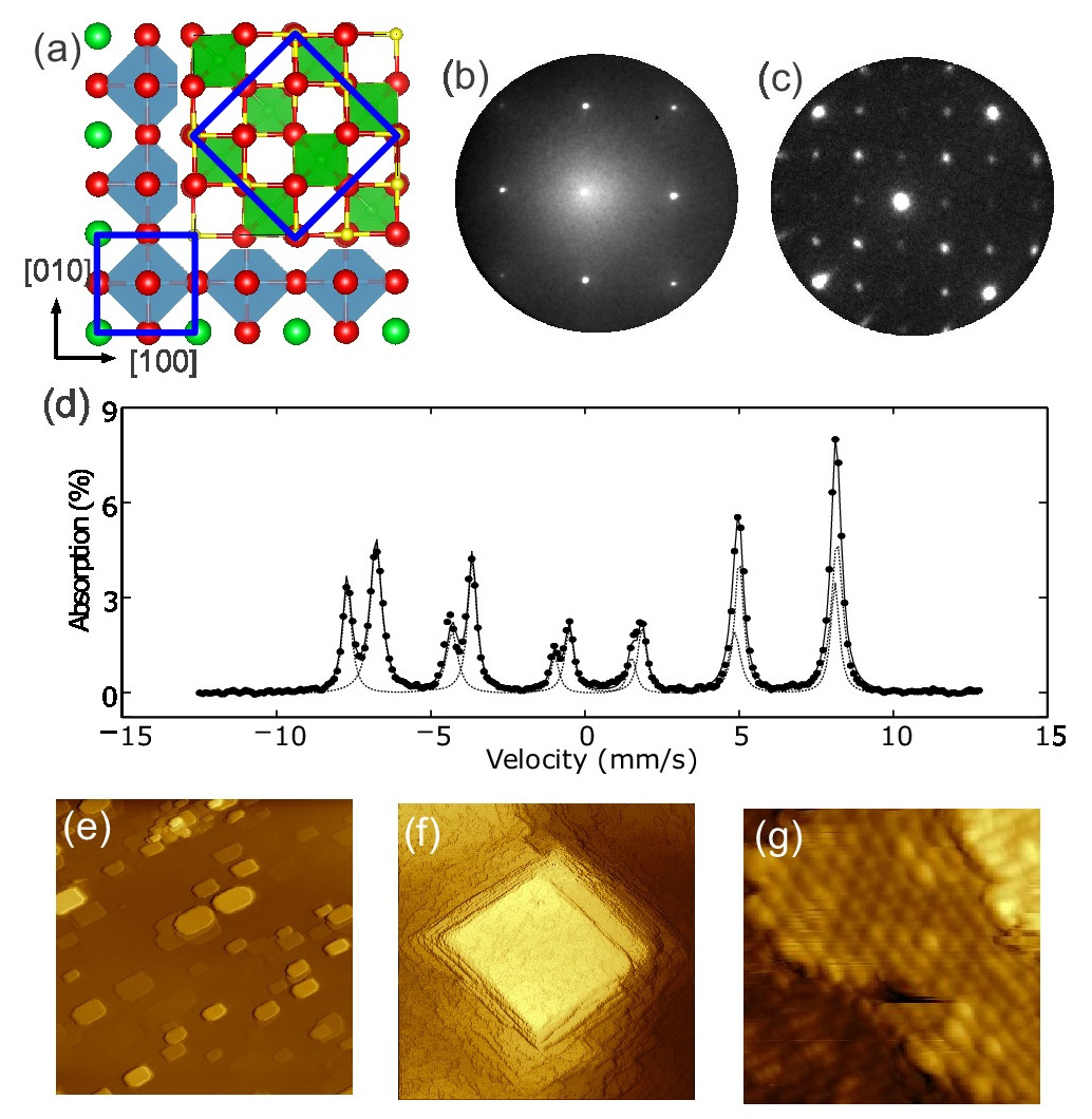
The epitaxial relationship between the perovskite substrate and the spinel film is expected to be cube-on-cube, , and depicted in Figure 1(a). The low-energy electron diffraction (LEED) pattern for SrTiO3:Nb (after annealing the substrate in 10-6 Torr of O2 at 800 K for several hours) is shown in Figure 1(b). The surface showed parallel steps nm apart in LEEM and AFM (not shown). The magnetite LEED pattern measured after several cycles of cleaning by Ar+ sputtering and annealing in 10-6 Torr O2, typical for preparing a clean magnetite surface in ultra-high vacuumParkinson et al. (2011), is shown in Figure 1(c). The strongest spots correspond to the first order and second order unit vectors of the surface primitive cell, which is rotated by 45∘ relative to the fcc cubic-cell unit vectors. The additional diffracted beams correspond to the reconstruction of magnetitePentcheva et al. (2008); Parkinson et al. (2011). Thus, the LEED pattern confirms the cube-of-cube epitaxial relationship, and it further shows that the film is monocrystalline.
The films have been characterized by integral conversion electron Mössbauer spectroscopy (ICEMS), x-ray diffraction (XRD), x-ray photoemission spectroscopy (XPS) and SQUID magnetometry. In an ICEMS RT spectrum of stoichiometric magnetite, two sextet components are detected corresponding to iron in the octahedral and tetrahedral positions, respectivelyVandenberghe et al. (2000). In such spectrum the component corresponding to octahedral iron presents parameters intermediate of those of Fe2+ and Fe3+. The magnetite film spectrum shown in Figure 1(d) has been fitted with two components that have the expected values for magnetite, in isomer shift (0.23 mm/s and 0.69 mm/s), quadrupole shift (-0.04 mm/s and 0.01 mm/s) and hyperfine magnetic fields (49.0 T and 46.4 T). The ratio of the two components is 1.8–1.9 depending on the particular sample indicating that the film is of stoichiometric composition. The out-of-plane lattice spacing from XRD is 0.840 nm, indicating that the magnetite film is mostly relaxed. This is expected given the 7.5% mismatch between magnetite and SrTiO3. Our films thickness (50-160 nm) is well above the limit for pseudomorhic growthKale et al. (2001) as detected by TEMZheng et al. (2007); Hamie et al. (2012). No contamination was detected by XPS, which showed only Fe and O, the former corresponding to a typical magnetite spectraFujii et al. (1999) with a mixture of Fe2+ and Fe3+. The Verwey temperature was measured to be 114 K with a SQUID magnetometer.
Typical microscopy images of the films are presented in Figure 1(e–g). AFM images of the surface show square features (“mesas”), with heights of up to 30 nm and lateral sizes in the 100-200 nm range, emerging from a flat film, as shown in Figure 1(e). In STM, both the areas between the mesas and their tops are confirmed to be quite flat, with small terraces tens of nanometers wide separated by atomic steps [0.21 nm high, see Figure 1(f) where the contrast has been enhanced so individual steps can be located]. While the orientation of the atomic steps, both on top of the mesas and on the areas between them, is not well defined, the mesas themselves are remarkably well aligned with the in-plane directions. On the individual atomic terraces, atomic rows 0.6 nm apart run along the direction in one terrace, and along the direction of the next atomic terrace [Figure 1(g)]. These rows correspond to the octahedral rows of iron of the magnetite unit cell, see Figure 1(a)Parkinson et al. (2011). Along the rows there is also an additional 0.6 nm periodicity, out-of-phase between consecutive rows. These periodicities corresponds to the reconstruction observed by LEED shown in Figure 1(c). This reconstruction, typical of magnetite cleaned by cycles of Ar+ sputtering and annealing in vacuum, has been interpreted as a Jahn-Teller distortion of the topmost octahedral iron atom positions along the rows of the surfacePentcheva et al. (2008).
Cleaning the sample for ultra-high vacuum experiments (i.e., for the STM and LEEM observations), which involve mild sputtering and annealing, changes slightly the as-grown surface morphology. While it is obvious that individual atomic step positions are changed, we remark that the AFM measurements were done on the “as-grown” films. The agreement between the STM, LEEM and AFM results indicates that no large morphological changes have occurred during UHV cleaning.
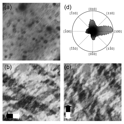
We have imaged the magnetic domains by means of SPLEEMRougemaille and Schmid (2010). After cleaning the samples with Ar+ sputtering and annealing, the film was heated above the Curie temperature and slowly cooled back to RT. A low-energy electron micrograph of the film is shown in Figure 2(a). Faint squares are observed, which correspond to the mesas detected in the AFM and STM images in Figure 1. The squares are oriented along in-plane providing an internal direction reference. The two magnetic contrast images are obtained by calculating the pixel-by-pixel asymmetry between LEEM images acquired illuminating the sample with beams of electrons with opposite spin polarization: bright (dark) areas indicate that the local surface magnetization has a component parallel (anti-parallel) to the spin-polarization direction of the electron beam. Grey areas indicate the absence of a magnetization component along the spin-polarization direction. The SPLEEM images thus indicate the local surface magnetization along a given direction. We note that SPLEEM is extremely surface sensitive, detecting the magnetization of the topmost atomic layers of the film. As the electron beam spin-polarization can be changed with respect to the sample, the magnetization vector can be determined in real space with nanometer resolutionRamchal et al. (2004). More details on the SPLEEM instrumentGrzelakowski et al. (1994), the spin-polarization control methodDuden and Bauer (1995) or the vector magnetometric application of SPLEEM can be found in the literatureRamchal et al. (2004); El Gabaly et al. (2006, 2008). SPLEEM images acquired (not shown) with out-of-plane spin direction presented negligible contrast, indicating that the magnetization lies mostly within the film plane. In Figures 2(b) and (c) white, black and grey regions are easily resolved. As the domains are not very large, it is difficult to determine by visual inspection whether the magnetization lies along some preferred axis. A plot of the magnetization-vector histogram vs. angle is shown in Figure 2(d) obtained by combining the images pixel by pixel to calculate the in-plane magnetization vector. It indicates that the magnetization lies mostly along the four in-plane directions, i.e. the histogram shows peaks at angles corresponding to the [100], [010], and orientations. The domain walls also show a preferred orientation, but along [110] and directions, i.e. along the sides of the 3D mesas on the film. The domains are up to one micrometer in size.
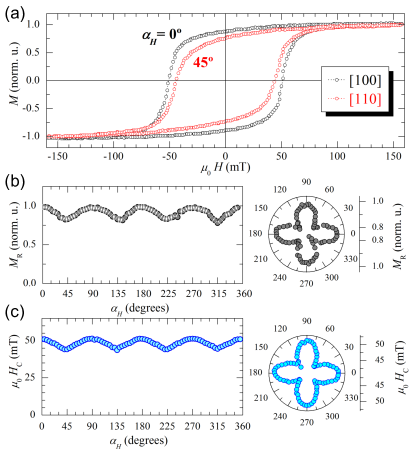
The unexpected magnetization easy-axis orientation of the film is confirmed by angular-dependent averaged magnetometry measurements. MOKE hysteresis loops have been systematically recorded by changing the in-plane orientation of the applied magnetic field in the angular range = 0–360∘ (Figure 3). Two representative plots ( = 0∘ and 45∘) are shown in Figure 3(a). The film coercivity is 45–50 mT, in line with published values for magnetite on SrTiO3Chen et al. (2008); Cheng et al. (2008); Wei et al. (2010). The low saturation fields around 150 mT evidence the high structural quality of our films, although often much higher saturation fields are reported for magnetite films on 100 substrates, probably due to magnetic domain pinning at defectsMargulies et al. (1997). Hysteresis loops displayed in Figure 3(a) show different remanence and coercivity values as a function of the in-plane orientation of the applied magnetic field. In particular, larger remanence and coercivity values are found for , i.e., along the [100] direction. Moreover, The angular dependence of the remanence [Figure 3(b)] and the coercivity [Figure 3(c)] show the fourfold symmetry of the magnetic anisotropy with the highest values found at 0, 90, 180 and 270∘. The fourfold symmetry can be easily identified in the corresponding polar plots of the remanence and the coercivity [right-hand side of Figures 3(b) and (c)]. As the maxima of both remanence and coercivity correspond to to easy-axis directions, the angular MOKE indicates that for the full film the easy-axes are the in-plane directions, in agreement with the microscopic SPLEEM observations of the surface magnetization.
In summary, we have grown pure stoichiometric magnetite films on SrTiO3:Nb by infrared PLD. Unlike films reported to date, these films have a robust well defined easy-axis along the in-plane directions. The individual magnetic domains at the surface of the films have been imaged in remanence by SPLEEM. The magnetic domains present magnetization vectors along the in-plane directions, while the domain walls are aligned with the in-plane directions. Hysteresis cycles have been measured by MOKE, obtaining the angular dependence of remanence and coercivity which both have maxima at the directions and thus confirm the local determination of the easy-axis directions by an averaging technique. Our films prove that modifying the growth parameters in magnetite films allows tunning the easy-axis directions.
Acknowledgments
Authors acknowledge fruitful discussions with Prof. M. Ziese. This research was supported by Projects CTQ2010-15680, MAT2009-14578-C03-01 (MICINN), MAT2012-38045-C04-01 (MINECO), MAT2011-27470-C02-02 (MICINN), MAT2011-25598 (MINECO), the EU-FP7 NANOPYME Project (No. 310516) and by the Office of Basic Energy Sciences, Division of Materials and Engineering Sciences, U. S. Department of Energy under Contract No. DE-AC04-94AL85000 (Sandia National Laboratories). Experiments performed at the National Center for Electron Microscopy, Lawrence Berkeley National Laboratory, were supported by the Office of Science, Office of Basic Energy Sciences, Scientific User Facilities Division, of the U.S. Department of Energy under Contract No. DE-AC02—05CH11231. E.R, M.O., M.S., A.T.N. and M.M. gratefully thank financial support from the Ramón y Cajal Programme (MINECO), a CSIC contract, a Geomateriales (CAM, S2009/Mat-1629) contract, a Feodor Lynen Postdoctoral Fellowship from the Alexander von Humboldt Foundation and a contract through the MICINN FPI Programme, respectively. We are grateful to Prof. T. Ezquerra (IEM, CSIC) for the use of the AFM system and M. Juanco (ICA, CSIC) for XRD measurements.
References
- Cornell and Schwertmann (1997) R. M. Cornell and U. Schwertmann, The Iron Oxides (John Wiley & Sons Ltd, 1997) p. 604.
- Mills (2004) A. A. Mills, Ann. Sci. 61, 273 (2004).
- Walz (2002) F. Walz, J. Phys. Cond. Mat. 14, R285 (2002).
- García and Subías (2004) J. García and G. Subías, J. Phys. Cond. Mat. 16, R145 (2004).
- Kato and Iida (1982) K. Kato and S. Iida, J. Phys. Soc. Jap. 51, 1335 (1982).
- Alexe et al. (2009) M. Alexe, M. Ziese, D. Hesse, P. Esquinazi, K. Yamauchi, T. Fukushima, S. Picozzi, and U. Gösele, Adv. Mat. 21, 4452 (2009).
- Katsnelson et al. (2008) M. I. Katsnelson, V. Y. Irkhin, L. Chioncel, A. I. Lichtenstein, and R. A. de Groot, Rev. Mod. Phys. 80, 315 (2008).
- Wada et al. (2010) E. Wada, K. Watanabe, Y. Shirahata, M. Itoh, M. Yamaguchi, and T. Taniyama, Appl. Phys. Lett. 96, 102510 (2010).
- Bibes et al. (2011) M. Bibes, J. E. Villegas, and A. Barthélémy, Adv. Phys. 60, 5 (2011).
- Carvello and Ranno (2004) B. Carvello and L. Ranno, J. Mag. Magn. Mat. 272-–276, 1926 (2004).
- Ziese et al. (2005) M. Ziese, U. Köhler, A. Bollero, R. Höhne, and P. Esquinazi, Phys. Rev. B 71, 180406 (2005).
- Kundaliya et al. (2006) D. C. Kundaliya, S. B. Ogale, L. F. Fu, S. J. Welz, J. S. Higgins, G. Langham, S. Dhar, N. D. Browning, and T. Venkatesan, J. Appl. Phys. 99, 08K304 (2006).
- Satoh et al. (2008) I. Satoh, J. Takaobushi, H. Tanaka, and T. Kawai, Sol. Stat. Comm. 147, 397 (2008).
- Wei et al. (2010) A. D. Wei, J. R. Sun, Y. Z. Chen, W. M. Lue, and B. G. Shen, J. Phys. D-App. Phys. 43, 205004 (2010).
- Kale et al. (2001) S. Kale, S. M. Bhagat, S. E. Lofland, T. Scabarozi, S. B. Ogale, A. Orozco, S. R. Shinde, T. Venkatesan, B. Hannoyer, B. Mercey, and W. Prellier, Phys. Rev. B 64, 205413 (2001).
- Zheng et al. (2007) J. G. Zheng, G. E. Sterbinsky, J. Cheng, and B. W. Wessels, J. Vac. Sci. Tech. B 25, 1520 (2007).
- Chen et al. (2008) Y. Z. Chen, J. R. Sun, Y. N. Han, X. Y. Xie, J. Shen, C. B. Rong, S. L. He, and B. G. Shen, J. Appl. Phys. 103, 07D703 (2008).
- Cheng et al. (2008) J. Cheng, G. Sterbinsky, and B. Wessels, J. Cryst. Growth 310, 3730 (2008).
- Lee and Chern (2008) D. Lee and G. Chern, Sol. Stat. Comm. 148, 353 (2008).
- Leung et al. (2008) G. Leung, M. Vickers, R. Yu, and M. Blamire, J. Cryst. Growth 310, 5282 (2008).
- Hamie et al. (2012) A. Hamie, Y. Dumont, E. Popova, A. Fouchet, B. Warot-Fonrose, C. Gatel, E. Chikoidze, J. Scola, B. Berini, and N. Keller, Thin Sol. Films 525, 115 (2012).
- Bickford (1950) L. R. Bickford, Phys. Rev. 78, 449 (1950).
- Abe et al. (1976) K. Abe, Y. Miyamoto, and S. Chikazumi, J. Phys. Soc. Japan 41, 1894 (1976).
- Jackson et al. (2011) M. Jackson, J. Bowles, and S. Banerjee, IRM Quaterly 21, 2 (2011).
- Williams and Wright (1998) W. Williams and T. M. Wright, J. Geophys. Res. 103, PP. 30,537 (1998).
- de la Figuera et al. (2013) J. de la Figuera, L. Vergara, A. T. N’Diaye, A. Quesada, and A. K. Schmid, Ultramicroscopy (2013), 10.1016/j.ultramic.2013.02.020, arXiv:1301.4350 [cond-mat] .
- Brandlmaier et al. (2008) A. Brandlmaier, S. Geprägs, M. Weiler, A. Boger, M. Opel, H. Huebl, C. Bihler, M. S. Brandt, B. Botters, D. Grundler, R. Gross, and S. T. B. Goennenwein, Phys. Rev. B 77, 104445 (2008).
- Fonin et al. (2011) M. Fonin, C. Hartung, U. Rüdiger, D. Backes, L. Heyderman, F. Nolting, A. F. Rodríguez, and M. Kläui, J. Appl. Phys. 109, 07D315 (2011).
- van der Heijden et al. (1998) P. van der Heijden, M. van Opstal, C. Swüste, P. Bloemen, J. Gaines, and W. de Jonge, J. Magn. Magn. Mat. 182, 71 (1998).
- McGuigan et al. (2008) L. McGuigan, R. C. Barklie, R. G. S. Sofin, S. K. Arora, and I. V. Shvets, Phys. Rev. B 77 (2008), 10.1103/PhysRevB.77.174424, WOS:000256763800078.
- Momma and Izumi (2011) K. Momma and F. Izumi, J. Appl. Crystallogr. 44, 1272 (2011).
- Sanz et al. (2013) M. Sanz, M. Oujja, E. Rebollar, J. F. Marco, J. de la Figuera, M. Monti, A. Bollero, J. Camarero, F. J. Pedrosa, M. García-Hernández, and M. Castillejo, Appl. Surf. Sci. (2013), 10.1016/j.apsusc.2013.06.026.
- Moussy (2013) J.-B. Moussy, J. Phys. D: Appl. Phys. 46, 143001 (2013).
- Parkinson et al. (2011) G. S. Parkinson, Z. Novotný, P. Jacobson, M. Schmid, and U. Diebold, Surf. Sci. 605, L42 (2011).
- Pentcheva et al. (2008) R. Pentcheva, W. Moritz, J. Rundgren, S. Frank, D. Schrupp, and M. Scheffler, Surf. Sci. 602, 1299 (2008).
- Vandenberghe et al. (2000) R. Vandenberghe, C. A. Barrero, G. M. da Costa, E. Van San, and E. De Grave, Hyperfine Interactions 126, 247 (2000).
- Fujii et al. (1999) T. Fujii, F. M. F. de Groot, G. A. Sawatzky, F. C. Voogt, T. Hibma, and K. Okada, Phys. Rev. B 59, 3195 (1999).
- Rougemaille and Schmid (2010) N. Rougemaille and A. K. Schmid, Eur. Phys. J. App. Phys. 50, 20101 (2010).
- Ramchal et al. (2004) R. Ramchal, A. K. Schmid, M. Farle, and H. Poppa, Phys. Rev. B 69, 214401 (2004).
- Grzelakowski et al. (1994) K. Grzelakowski, T. Duden, E. Bauer, H. Poppa, and S. Chiang, IEEE Trans. Mag. 30, 4500 (1994).
- Duden and Bauer (1995) T. Duden and E. Bauer, Rev. Sci. Inst. 66, 2861 (1995).
- El Gabaly et al. (2006) F. El Gabaly, S. Gallego, M. C. Muñoz, L. Szunyogh, P. Weinberger, C. Klein, A. K. Schmid, K. F. McCarty, and J. de la Figuera, Phys. Rev. Lett. 96, 147202 (2006).
- El Gabaly et al. (2008) F. El Gabaly, K. F. McCarty, A. K. Schmid, J. de la Figuera, M. C. Muñoz, L. Szunyogh, P. Weinberger, and S. Gallego, New J. Phys. 10, 073024 (2008).
- Margulies et al. (1997) D. T. Margulies, F. T. Parker, M. L. Rudee, F. E. Spada, J. N. Chapman, P. R. Aitchison, and A. E. Berkowitz, Phys. Rev. Lett. 79, 5162 (1997).