Random Raman lasing
Propagation of light in a highly scattering medium is among the most fascinating optical effect that everyone experiences on an everyday basis and possesses a number of fundamental problems which have yet to be solved [1, 2, 3, 4, 5, 6, 7]. Conventional wisdom suggests that non-linear effects do not play a significant role because the diffusive nature of scattering acts to spread the intensity, dramatically weakening these effects. We demonstrate the first experimental evidence of lasing on a Raman transition in a bulk three-dimensional random media. From a practical standpoint, Raman transitions allow for spectroscopic analysis of the chemical makeup of the sample [8, 9]. A random Raman laser could serve as a bright Raman source allowing for remote, chemically specific, identification of powders and aerosols. Fundamentally, the first demonstration of this new light source opens up an entire new field of study into non-linear light propagation in turbid media [10], with the most notable application related to non-invasive biomedical imaging [11, 12, 13].
Raman scattering is the inelastic scattering of a photon off a molecule. The frequency of the scattered photon is determined by the frequency of a vibrational level of the molecule, making Raman spectroscopy a valuable tool for molecular and structural identification. In the context of Raman lasing, spontaneous Raman scattering and stimulated Raman scattering (SRS) are analogous to fluorescence and stimulated emission, respectively. However, the nonlinearity associated with SRS distinguishes it from stimulated emission [14]. In other words, the gain in the Raman scattered field is proportional to the intensity of the pump. Hence, Raman lasers typically require high optical pump intensities in order to attain sufficient gain to achieve lasing [15, 16, 17, 18]. Unlike a traditional laser, whose wavelength is determined by the electronic structure of the gain medium, the wavelength of a Raman laser is determined not only by the vibrational spectrum of the gain medium, but also by the wavelength of the pump laser used.
Random lasing was first predicted by V. S. Letokhov in 1968 [19], but was not experimentally observed until 1994 [20, 21]. Random lasers operate on many of the same principles as traditional lasers. The exception is that in random lasers multiple elastic scattering provides feedback in place of mirrors. Analogous to traditional lasers, random lasers have a threshold where gain from electronic transitions characteristic of the material exceeds losses and exponential amplification occurs at the lasing frequency [22]. Random lasers can be divided into two categories, coherent and incoherent, based on the feedback that drives them [23]. Coherent feedback occurs with the generation of unstable periodic trajectories by multiple elastic scattering events [24]. These trajectories are analogous to many randomly oriented ring cavities existing in the gain medium. When multiple scattering acts only to return energy back into the gain medium, but phase information is lost in the process, the system is driven by incoherent feedback [25]. Incoherent feedback is diffusive in nature, meaning the wave properties of light are negligible in such a system. Thus, such a system can be accurately described with Monte Carlo simulations.
A random Raman laser has a primary gain mechanism of SRS, and relies on elastic scattering to provide feedback into the gain medium. To date, random Raman lasing has only been observed in a one-dimensional fiber system [26]. While many properties of the random Raman laser closely mimic those of a traditional random laser there are some notable differences. First, the gain bandwidth of a Raman transition is quite narrow. This makes it very unlikely for the resonant frequency of a closed cavity to lie within this small gain window and observe gain. Thus, the dynamics of a random Raman laser will be dominated by incoherent feedback. Second, Raman transitions are much faster than electronic transitions, thus the transient (picosecond) dynamics of pulse propagation through the medium will play a pivotal role. The complicated dynamics of nonlinear pulse propagation in a turbid medium make an analytical approach to describing this problem very difficult. In order to better understand these processes, we employed a Monte Carlo model developed previously [27]. These simulations were used to guide our experiments and offer the chance to illuminate aspects of the dynamics that cannot be easily observed experimentally.
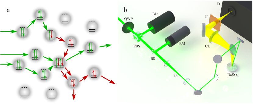
The random Raman laser (illustrated in Fig. 1a) was made of barium sulfate () powder with particle diameters of . plays the role of both the Raman gain medium and the scattering centers, and was chosen because of its low absorption and high scattering cross section throughout the visible spectrum. Micron-sized particles were chosen to ensure that the Raman gain in any one particle is not too large, thus requiring the feedback provided via elastic scattering for sufficient amplification of the Raman signal. To pump the random Raman laser, laser radiation with a wavelength of and a pulse duration of was employed. The incident radiation was gently focused onto the sample using a slightly offset telescope as a compound lens, allowing for adjustments in the beam diameter incident on the sample (see Fig. 1b). The generated Raman output light was collected at near normal incidence and passed into an energy meter, a CCD camera, or a spectrometer via a lens for the appropriate measurements.
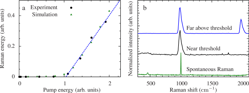
At low pump pulse energies, spontaneous Raman scattering dominates our detected Raman signal. However, once the pump energy increases beyond the threshold, ( in our experiment) gain exceeds losses and SRS dominates the conversion process, which is the onset of random Raman lasing (see Fig. 2a). At a maximum of of pump energy, as much as of Raman signal was measured by the energy meter. Assuming a homogeneous angular distribution of emitted Raman photons, our conversion efficiency of pump photons into Raman photons was approximately . This demonstrates that the random Raman laser is remarkably efficient considering typical conversion efficiencies for spontaneous Raman scattering are on the order of [28].
Above the lasing threshold, the Raman spectrum narrows to a single Raman peak with a frequency shift of as shown in Fig 2b. At higher pump energies, a second peak was observed corresponding to a second order SRS signal. This signal was generated by a second shifted Raman response. The primary Raman light served as the pump for a second Raman process which corresponded to a total Raman shift of . The presence of a second-order process further illustrates the substantial efficiency of this random Raman laser.
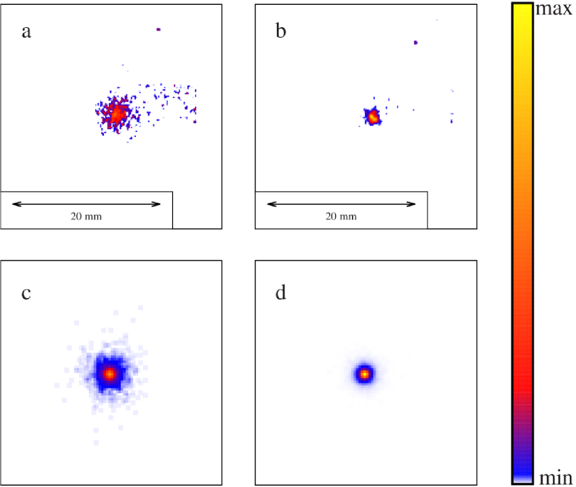
Below the lasing threshold, the spatial distribution of the emitted Raman light on the random Raman laser is very broad and predominately due to spontaneous Raman scattering (see Fig. 3). This is the result of deep penetrating photons that spend a long time in the medium, providing a greater chance to be converted into a Raman photon [27]. These deep penetrating photons have a larger probability of exiting the sample with large radial offsets. However, above the threshold, the majority of the energy is emitted from a highly localized area on the surface (see Fig. 3). This is primarily due to the fact that a large amount of the SRS light is generated near the surface, resulting in much smaller radial offsets. Strong SRS generation near the surface can be attributed to the slower speed of light diffusion in turbid media compared to the speed of light in a vacuum [29]. Thus, photons initially arriving in the medium do not have time to leave the front surface of the sample before photons from the back of the pulse enter, causing the pulse to effectively be compressed. This light compression raises the intensity, generating a large SRS signal close to the surface (See the supplimental material for a video of the simulation showing this effect).
We have demonstrated the first random Raman laser in a three-dimensional random medium. Above the threshold, SRS gain exceeds losses due to diffusion and absorption, resulting in Raman lasing. While many of the features are similar to traditional random lasers, the dynamics of random Raman lasing have a few notable differences which we have elucidated with the help of Monte Carlo simulations. These devices open up an entire new frontier on which to test the understanding of nonlinear optics, and offer a possible mechanism that could provide a luminously bright source of Raman light making remote sensing of powders or aerosols a natural extension.
References
- [1] Mie, G. Articles on the optical characteristics of turbid tubes, especially colloidal metal solutions. Ann. Phys. 25, 377–445 (1908).
- [2] Pasternack, R. F. & Collings, P. J. Resonance light scattering: a new technique for studying chromophore aggregation. Science 269, 935–939 (1995).
- [3] Licht, J. et al. Interactions between electrical activity and cortical microcirculation revealed by imaging spectroscopy: implications for functional brain mapping. Science 272, 551 (1996).
- [4] Tegen, I., Lacis, A. A. & Fung, I. The influence on climate forcing of mineral aerosols from disturbed soils. Nature 380, 419–422 (1996).
- [5] Skipetrov, S. & Van Tiggelen, B. Dynamics of Anderson localization in open 3D media. Phys. Rev. Lett. 96, 043902 (2006).
- [6] Yakovlev, V. V. et al. Stimulated Raman photoacoustic imaging. Proc. Natl. Acad. Sci. U.S.A. 107, 20335–20339 (2010).
- [7] Bertolotti, J. et al. Non-invasive imaging through opaque scattering layers. Nature 491, 232–234 (2012).
- [8] Freudiger, C. W. et al. Label-free biomedical imaging with high sensitivity by stimulated Raman scattering microscopy. Science 322, 1857–1861 (2008).
- [9] Schopf, J. W., Kudryavtsev, A. B., Agresti, D. G., Wdowiak, T. J. & Czaja, A. D. Laser–Raman imagery of Earth’s earliest fossils. Nature 416, 73–76 (2002).
- [10] Wiersma, D. S., Bartolini, P., Lagendijk, A. & Righini, R. Localization of light in a disordered medium. Nature 390, 671–673 (1997).
- [11] Siddique, M., Yang, L., Wang, Q. & Alfano, R. Mirrorless laser action from optically pumped dye-treated animal tissues. Opt. Commun. 117, 475–479 (1995).
- [12] Song, Q. et al. Random lasing in bone tissue. Opt. Lett. 35, 1425–1427 (2010).
- [13] Redding, B., Choma, M. A. & Cao, H. Speckle-free laser imaging using random laser illumination. Nat. Photonics 6, 355–359 (2012).
- [14] Shen, Y. & Bloembergen, N. Theory of stimulated Brillouin and Raman scattering. Phys. Rev. 137, A1787 (1965).
- [15] Spillane, S., Kippenberg, T. & Vahala, K. Ultralow-threshold Raman laser using a spherical dielectric microcavity. Nature 415, 621–623 (2002).
- [16] Rong, H. et al. An all-silicon Raman laser. Nature 433, 292–294 (2005).
- [17] Rong, H. et al. A continuous-wave Raman silicon laser. Nature 433, 725–728 (2005).
- [18] Troccoli, M. et al. Raman injection laser. Nature 433, 845–848 (2005).
- [19] Letokhov, V. S. Generation of light by a scattering medium with negative resonance absorption. Soviet Physics JETP 26, 835–840 (1968).
- [20] Genack, A. Z. & Drake, J. M. Scattering for super-radiation. Nature 368, 400–401 (1994).
- [21] Lawandy, N. M., Balachandran, R. M., Gomes, A. S. L. & Sauvain, E. Laser action in strongly scattering media. Nature 368, 436–438 (1994).
- [22] Wiersma, D. S. The physics and applications of random lasers. Nat. Phys. 4, 359–367 (2008).
- [23] Cao, H., Ling, Y., Xu, J. Y. & Cao, C. Q. Photon statistics of random lasers with resonant feedback. Phys. Rev. Lett. 86, 4524–4527 (2001).
- [24] Cao, H. et al. Spatial confinement of laser light in active random media. Phys. Rev. Lett. 84, 5584–5587 (2000).
- [25] Cao, H. et al. Random laser action in semiconductor powder. Phys. Rev. Lett. 82, 2278–2281 (1999).
- [26] Turitsyn, S. K. et al. Random distributed feedback fibre laser. Nat. Photonics 4, 231–235 (2010).
- [27] Hokr, B. H. & Yakovlev, V. V. Raman signal enhancement via elastic light scattering. Opt. Express 21, 11757–11762 (2013).
- [28] Boyd, R. W. Nonlinear Optics (Academic Press, 2003), 2 edn.
- [29] van Albada, M. P., van Tiggelen, B. A., Lagendijk, A. & Tip, A. Speed of propagation of classical waves in strongly scattering media. Phys. Rev. Lett. 66, 3132–3135 (1991).
Supplementary Information is available in the online version of the paper.
Acknowledgments The authors acknowledge the Texas A&M Supercomputing Facility (http://sc.tamu.edu/) for providing computing resources useful in conducting the research reported in this paper. This work was partially supported by National Institutes of Health Grant R21EB011703 and National Science Foundation Grants ECCS-1250360, DBI-1250361, and CBET-125036.
Author Contributions BHH, MC, JDM, HTB, BAR, GN, GIP, and VVY conducted experiments. BHH and RJT conducted simulations. BHH, BAR, GIP, LAG, and VVY designed research. All authors contributed to the writing of the paper.
Author Information Reprints and permissions information is available at
www.nature.com/reprints. The authors declare no competing financial interests. Readers are welcome to comment on the online version of the paper. Correspondence and requests for materials should be addressed to VVY (yakovlev@bme.tamu.edu).